aResearch Assistant, Department of Orthodontics, Faculty of Dentistry, Kocaeli University, Kocaeli, Turkey.
bResearch Assistant, dAssociate Professor, Department of Orthodontics, Faculty of Dentistry, Dicle University, Diyarbakir, Turkey.
cResearch Assistant, Department of Orthodontics, Faculty of Dentistry, Erciyes University, Kayseri, Turkey.
eProfessor and Chair, Department of Orthodontics, Faculty of Dentistry, Izmir Katip Celebi University, Izmir, Turkey.
fProfessor, Department of Pediatric Dentistry and Orthodontics, College of Dentistry, King Saud University, Riyadh, Saudi Arabia.
Corresponding author: Tancan Uysal.
İzmir Katip Çelebi Üniversitesi Dişhekimliği Fakültesi, Ortodonti A.D. Çiğli, İzmir, Turkey.
+902323293999; e-mail, [email protected].
Received February 12, 2011; Last Revision April 1, 2011;
Accepted April 5, 2011.
DOI:10.4041/kjod.2011.41.3.200
*King Saud University, Visiting Professor Project Unit (Grant No:
KSU-VPP-112).
Changes in mandibular transversal arch dimensions after rapid maxillary expansion procedure assessed through
cone-beam computed tomography
Asli Baysal, DDS, PhD,a Ilknur Veli, DDS,b Faruk Izzet Ucar, DDS,c Murat Eruz, DDS,c Torun Ozer, DDS, PhD,d Tancan Uysal, DDS, PhDe,f
Objective: This study aimed at evaluating the changes in mandibular arch widths and buccolingual in- clinations of mandibular posterior teeth after rapid maxillary expansion (RME). Methods: Baseline and post-expansion cone-beam computed tomographic (CBCT) images of patients who initially had bilateral posterior cross-bite and underwent RME with a banded-type expander were assessed in this study. The patients included 9 boys (mean age: 13.97 ± 1.17 years) and 11 girls (mean age: 13.53 ± 2.12 years).
Images obtained 6 months after retention were available for 10 of these patients. Eighteen angular and 43 linear measurements were performed for the maxilla and mandible. The measurements were performed on frontally clipped images at the following time points; before expansion (T1), after expansion (T2), and after retention (T3). Statistical significance was assessed with paired sample t-test at p < 0.05. Results:
T1-T2 comparisons showed statistically significant post-RME increases for all measurements; similarly, T2-T1 and T3-T1 comparisons showed statistically significant changes. The maxillary linear and angular measurements showed decreases after expansion, and mandibular linear and angular measurements in- creased after retention. Conclusion: All mandibular arch widths increased and mandibular posterior teeth were uprighted after RME procedure. (Korean J Orthod 2011;41(3):200-210)
Key words: CT, Arch form, Expansion
INTRODUCTION
Rapid maxillary expansion (RME) was introduced in 1860 by Angell.1 This procedure gained popularity in the 1960s and has currently become a common ortho- dontic procedure.2 Briefly, the effects of RME are in- creased nasal cavity width,3-6 separation of the maxil- lary halves,7 lowering of the palatal processes,3,4 bend- ing of the alveolar processes,7 and tipping-extrusion of the posterior teeth.8,9
Mandibular teeth become upright after RME.4,5 Haas3 stated that RME results in changes of the man- dibular arch. These changes are believed to be the re- sult of alterations in the balance between the tongue and the buccinator muscles. Another explanation for
these changes is that RME is accompanied by changes in the orientation of the inclined planes of the teeth.2 Brodie10 had previously observed that “the interaction of the forces of these 2 antagonistic muscle masses would dictate the size and form of the arches as well as the axial inclination of the teeth.” When the maxilla is expanded, the pressure from the buccinator muscle is decreased. This causes the mandibular teeth to ex- pand in a buccal direction owing to pressure from the tongue.3 Gryson11 found no change in or an increase of up to 1 mm in the mandibular intermolar width. Addi- tionally, no correlation was found between the increase in mandibular intermolar and intercanine width with re- spect to the increase in maxillary intercanine and inter- molar width. Lima et al.12 stated that the mandibular intermolar width increased during RME and remained stable thereafter. The intercanine width, on the other hand, was also found to be stable during all ob- servation periods.12 However, Miller2 found no change in both intercanine and intermolar widths after RME.
Previous studies have shown the existence of a rela- tionship between RME and changes in mandibular arch width. However, all studies cited above were based on- ly on the dental cast measurements. To the best of our knowledge, none of the studies conducted thus far have evaluated the post-RME changes in the axial in- clinations of mandibular teeth.
With the introduction of cone-beam computed tomo- graphy (CBCT), it is now possible to obtain high-reso- lution images (isotropic resolution: 0.4 - 0.125 mm) within a very short period (scanning time: 10 - 70 s) and with minimal radiation exposure (up to 15 times lower than that of conventional CT scans).13 CBCT al- so enables multiplanar imaging and provides 3D infor- mation.
The aim of this study was to evaluate the post-RME changes in mandibular arch widths and buccolingual inclinations of mandibular posterior teeth by using CBCT images.
MATERIAL AND METHODS
The CBCT images of patients who underwent RME with banded-type expander for the correction of maxil- lary constriction were retrieved from the archives of
the Oral and Maxillofacial Radiology Department of our hospital. The images were obtained for 20 subjects, including 9 boys (mean age: 13.97 ± 1.17 years) and 11 girls (mean age: 13.53 ± 2.12 years). For 10 of these patients, CBCT images obtained at the 6-month follow-up examination were also available, and these records were also examined in this study.
The parents of the patients provided informed con- sent after they and the patients were explained about the CBCT scanning procedure. Ethical approval for this study was obtained from the Ethical Committee of the Dicle University, Faculty of Dentistry.
RME was performed using a banded-type expander.
The screw was turned twice a day, and active ex- pansion was terminated when the palatal cusps of the upper posterior teeth came in contact with the buccal cusps of the lower posterior teeth. During the retention period, the expander was retained in the mouth for the first 3 months and then replaced with a transpalatal arch.
Images were obtained by using a CBCT device (iCATⓇ, Model 17 - 19, Imaging Sciences Interna- tional, Hatfield, PA, USA) set at the following parame- ters: exposure, 5.0 mA and 120 kV; exposure time, 9.6 s; and axial slice thickness, 0.3 mm. The CBCT im- ages were obtained with the iCATⓇ scanner (Imaging Sciences International) at a single 360o rotation. All the transversal linear and angular measurements were per- formed using the Dolphin Imaging 11.0 Premium soft- ware (Dolphin Imaging & Management Solutions, Chats- worth, CA, USA). Patient information was recorded in the database, and DICOM data sets of each patient were imported into the program. The software program automatically segments the tissues according to density values, i.e., into Hounsfield Units. All measurements were made on a 4-Equal layout screen to display all the measurements on the same screen.
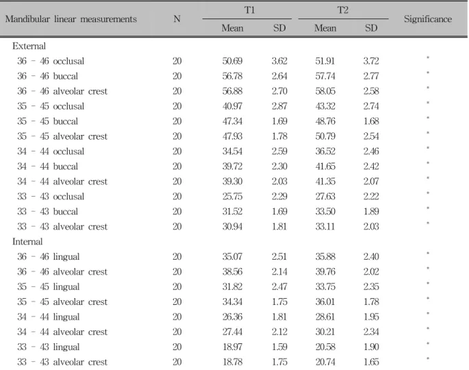
Measurements were performed for cross-sectional images obtained at the following time points: before expansion (T1), after expansion (T2) and after re- tention (T3). In all, 20 linear and 6 angular measure- ments were performed on mandibular sections (Fig 1).
The linear measurements include both internal and ex- ternal measurements. The internal measurements com- prise intermolar, interpremolar, and intercanine widths
Fig 1. Mandibular linear and angular measurements. A, External mandibular measurements performed at 3 different levels: alveolar crest, most prominent point of the crown, and buccal cusp tip; B, internal mandibular measurements were performed at 2 different levels: alveolar crest and most prominent point of the crown; C, mandibular long axis measurement performed between the long axis of the mesial root and to the reference plane, which passes through the most prominent point of the lower borders (right and left) of the mandible.
Fig 2. Maxillary linear and angular measurements. A, Nasal and palatal floor measurements; B, external maxillary measurements performed at 4 different levels: apical, alveolar crest, most prominent point of the crown, and buccal cusp tip; C, internal maxillary measurements were performed at 3 different levels: apical, alveolar crest, and most prominent point of the crown; D, maxillary long axis measurement performed between the long axis of the palatal root and to the palatal plane.
measured at 2 levels: the alveolar crest and the most prominent point of the crowns at the lingual aspect.
The external measurements were performed between
the alveolar crests, the most prominent point of the crowns at the buccal aspect, and the buccal cusp tips.
The angular measurements of mandibular teeth were
Table 1. Comparison of mandibular linear measurements before and after rapid maxillary expansion (unit, mm)
Mandibular linear measurements N T1 T2
Significance
Mean SD Mean SD
External
36 - 46 occlusal 20 50.69 3.62 51.91 3.72 *
36 - 46 buccal 20 56.78 2.64 57.74 2.77 *
36 - 46 alveolar crest 20 56.88 2.70 58.05 2.58 *
35 - 45 occlusal 20 40.97 2.87 43.32 2.74 *
35 - 45 buccal 20 47.34 1.69 48.76 1.68 *
35 - 45 alveolar crest 20 47.93 1.78 50.79 2.54 *
34 - 44 occlusal 20 34.54 2.59 36.52 2.46 *
34 - 44 buccal 20 39.72 2.30 41.65 2.42 *
34 - 44 alveolar crest 20 39.30 2.03 41.35 2.07 *
33 - 43 occlusal 20 25.75 2.29 27.63 2.22 *
33 - 43 buccal 20 31.52 1.69 33.50 1.89 *
33 - 43 alveolar crest 20 30.94 1.81 33.11 2.03 *
Internal
36 - 46 lingual 20 35.07 2.51 35.88 2.40 *
36 - 46 alveolar crest 20 38.56 2.14 39.76 2.02 *
35 - 45 lingual 20 31.82 2.47 33.75 2.35 *
35 - 45 alveolar crest 20 34.34 1.75 36.01 1.78 *
34 - 44 lingual 20 26.36 1.81 28.61 1.95 *
34 - 44 alveolar crest 20 27.44 2.12 30.21 2.34 *
33 - 43 lingual 20 18.97 1.59 20.58 1.90 *
33 - 43 alveolar crest 20 18.78 1.75 20.74 1.65 *
SD, Standard deviation. *p < 0.001.
made by considering a plane passing through the lower borders of the mandible as the reference plane. The an- gle between the long axis of the teeth and the refer- ence plane was measured for the left and right sides and recorded as the long axis angle for each tooth.
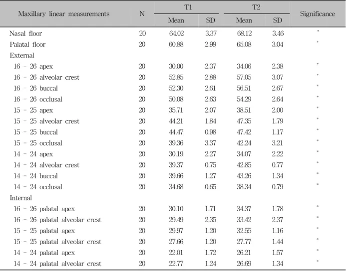
The linear and angular measurements for the maxilla and maxillary teeth were performed using the method described by Kartalian et al.14 For the maxilla, 18 line- ar and 6 angular measurements were performed (Fig 2). The nasal floor was measured tangent to the nasal floor at its most superior level and parallel to the low- er border of the hard palate. The line parallel to the lower border of the hard palate and tangent to the hard palate was recorded as the palatal plate. External max- illary measurements include the intermolar and inter-
premolar widths measured between the apex of palatal root, alveolar crest, most prominent point of the crown, and the buccal cusp tip levels. The internal maxillary measurements were performed at the apex of the pala- tal root and the alveolar crest levels on the palatal side. The angle between the long axis of the palatal root and the palatal plane was also measured.
Statistical analysis
All statistical analyses were performed using the stat- istical package for social sciences (SPSS), 16.0 (SPSS for Windows; SPSS Inc, Chicago, IL, USA). The nor- mality test of Shapiro-Wilks and Levene’s variance ho- mogeneity tests were applied to the data. Baseline
Table 2. Comparison of the changes in mandibular linear measurements between the active expansion and retention periods (unit, mm)
Mandibular linear measurements N T2-T1 T3-T1
Significance
Mean SD Mean SD
External
36 - 46 occlusal 10 0.90 0.39 1.32 0.38 †
36 - 46 buccal 10 0.73 0.39 1.46 0.49 ‡
36 - 46 alveolar crest 10 0.88 0.41 1.41 0.64 †
35 - 45 occlusal 10 2.24 1.22 2.81 1.37 †
35 - 45 buccal 10 1.27 1.22 1.48 1.29 *
35 - 45 alveolar crest 10 1.69 0.77 2.00 0.92 *
34 - 44 occlusal 10 1.59 2.20 2.35 2.08 ‡
34 - 44 buccal 10 1.77 0.67 2.23 0.67 †
34 - 44 alveolar crest 10 1.84 0.95 2.53 0.90 ‡
33 - 43 occlusal 10 1.50 0.90 2.24 0.91 ‡
33 - 43 buccal 10 1.68 0.68 2.02 0.83 ‡
33 - 43 alveolar crest 10 2.02 0.76 2.38 0.99 *
Internal
36 - 46 lingual 10 0.89 0.44 1.08 0.48 *
36 - 46 alveolar crest 10 1.25 0.68 1.37 0.77 *
35 - 45 lingual 10 2.02 0.76 2.55 0.82 ‡
35 - 45 alveolar crest 10 1.59 0.88 1.92 1.01 ‡
34 - 44 lingual 10 1.84 1.21 2.58 1.13 ‡
34 - 44 alveolar crest 10 2.49 0.39 3.02 0.34 ‡
33 - 43 lingual 10 1.56 0.55 2.26 0.64 ‡
33 - 43 alveolar crest 10 2.04 0.98 2.46 1.16 †
SD, Standard deviation. *p < 0.05; †p < 0.01; ‡p < 0.001.
(T1), post-expansion (T2) and 6-month follow-up (T3) data were found to be normally distributed, and homo- geneity of variance was noted among the groups.
Therefore, the statistical evaluations of these data were performed using parametric tests.
Arithmetic mean and standard deviation values were calculated for all measurements. Paired samples t-test was used to compare the mean values. Statistical sig- nificance was set at p < 0.05.
To determine the errors associated with CBCT measurements, 15 CBCTs were selected randomly.
Their measurements were repeated 5 weeks after the first measurements. A paired samples t-test was applied
to the first and second measurements, and the differ- ences between the measurements were found to be in- significant. Correlation analysis applied to the same measurements showed the highest r value (0.982) for internal 34 - 44 alveolar crest level measurement and the lowest r value (0.711) for 16 - 26 CEJ measure- ment.
RESULTS
The mandibular linear measurements before and after expansion are shown in Table 1. All the transversal linear measurements increased after RME. According
Table 4. Comparison of the changes in mandibular angular measurements between the active expansion and re- tention periods (unit, mm)
Mandibular angular
measurements N T2-T1 T3-T1
Significance
Mean SD Mean SD
46 10 0.67 0.24 1.10 0.29 *
36 10 1.00 0.67 1.93 0.94 *
45 10 1.24 0.77 2.58 1.90 *
35 10 0.78 0.44 1.65 0.52 *
44 10 1.18 0.41 2.04 0.38 *
34 10 1.90 0.63 2.77 0.57 *
SD, Standard deviation. *p < 0.001.
Table 3. Comparison of mandibular angular measurements before and after rapid maxillary expansion (unit, mm) Mandibular angular
measurements N T1 T2
Significance
Mean SD Mean SD
46 long axis 20 75.20 1.11 75.88 1.03 *
36 long axis 20 75.39 0.64 76.33 0.98 *
45 long axis 20 75.67 1.99 76.94 2.28 *
35 long axis 20 79.40 1.87 80.26 1.95 *
44 long axis 20 87.12 0.92 88.43 0.98 *
34 long axis 20 88.07 1.16 89.85 1.38 *
SD, Standard deviation. *p < 0.001.
Table 5. Comparison of maxillary angular measurements before and after rapid maxillary expansion (unit, mm) Maxillary angular
measurements N T1 T2
Significance
Mean SD Mean SD
16 long axis 20 98.05 1.79 101.66 2.10 *
26 long axis 20 98.34 1.40 101.97 2.12 *
15 long axis 20 95.32 2.05 96.52 2.08 *
25 long axis 20 85.81 1.48 87.49 1.64 *
14 long axis 20 90.77 1.00 96.36 1.10 *
24 long axis 20 91.71 1.19 96.16 2.75 *
SD, Standard deviation. *p < 0.001.
to the results of the paired samples t-test, these incre- ments were statistically significant (p < 0.001). Since the number of subjects was not equal for the active ex- pansion and retention periods, the comparisons be- tween these stages have been provided in separate tables. The T3-T1 and T2-T1 differences showed stat-
istical significance for all measurements (Table 2).
Increase in linear measurements was found to continue during the retention period, and these increases were statistically significant.
The mandibular angular measurements for all 20 pa- tients before and after RME procedure have been
Table 6. Comparison of the changes of maxillary angular measurements in the active expansion and retention periods (unit, mm)
Maxillary angular
mesurements N T2-T1 T3-T1
Significance
Mean SD Mean SD
16 long axis 10 3.52 0.74 2.21 0.55 *
26 long axis 10 3.26 0.86 2.53 0.72 *
15 long axis 10 1.17 0.43 0.88 0.45 *
25 long axis 10 1.58 0.60 1.13 0.64 *
14 long axis 10 5.43 0.72 3.59 0.58 *
24 long axis 10 5.07 0.61 3.10 0.80 *
SD, Standard deviation. *p < 0.001.
Table 7. Comparison of maxillary linear measurements before and after rapid maxillary expansion (unit, mm)
Maxillary linear measurements N T1 T2
Significance
Mean SD Mean SD
Nasal floor 20 64.02 3.37 68.12 3.46 *
Palatal floor 20 60.88 2.99 65.08 3.04 *
External
16 - 26 apex 20 30.00 2.37 34.06 2.38 *
16 - 26 alveolar crest 20 52.85 2.88 57.05 3.07 *
16 - 26 buccal 20 52.30 2.61 56.51 2.67 *
16 - 26 occlusal 20 50.08 2.63 54.29 2.64 *
15 - 25 apex 20 35.71 2.07 38.51 2.00 *
15 - 25 alveolar crest 20 44.21 1.84 47.35 1.79 *
15 - 25 buccal 20 44.47 0.98 47.42 1.17 *
15 - 25 occlusal 20 39.36 3.37 42.24 3.21 *
14 - 24 apex 20 30.19 2.27 34.07 2.22 *
14 - 24 alveolar crest 20 39.37 0.75 42.85 0.77 *
14 - 24 buccal 20 39.66 1.27 43.26 1.34 *
14 - 24 occlusal 20 34.68 0.65 38.34 0.79 *
Internal
16 - 26 palatal apex 20 30.10 1.71 34.37 1.78 *
16 - 26 palatal alveolar crest 20 29.49 2.35 33.42 2.37 *
15 - 25 palatal apex 20 29.97 1.20 32.55 1.16 *
15 - 25 palatal alveolar crest 20 27.66 1.20 27.77 1.44 *
14 - 24 palatal apex 20 22.01 1.72 26.21 1.57 *
14 - 24 palatal alveolar crest 20 22.77 1.24 26.69 1.34 *
SD, Standard deviation. *p < 0.001.
shown in Table 3. Statistically significant increases were found in long axis measurements for all inves-
tigated teeth (p < 0.001). The T3-T1 and T2-T1 com- parisons also showed significant differences for all
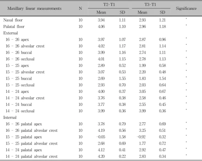
Table 8. Comparison of the changes of maxillary linear measurements between the active expansion and retention periods (unit, mm)
Maxillary linear measurements N T2-T1 T3-T1
Significance
Mean SD Mean SD
Nasal floor 10 3.94 1.11 2.93 1.21 *
Palatal floor 10 4.06 1.10 2.96 1.18 *
External
16 - 26 apex 10 3.97 1.07 2.87 0.96 *
16 - 26 alveolar crest 10 4.02 1.17 2.81 1.14 *
16 - 26 buccal 10 3.99 1.16 2.74 1.11 *
16 - 26 occlusal 10 4.01 1.15 2.78 1.13 *
15 - 25 apex 10 2.89 0.52 1.99 0.58 *
15 - 25 alveolar crest 10 3.07 0.53 2.20 0.48 *
15 - 25 buccal 10 2.69 1.55 1.83 1.54 *
15 - 25 occlusal 10 2.93 0.70 2.03 0.64 *
14 - 24 apex 10 4.00 0.37 3.05 0.67 *
14 - 24 alveolar crest 10 3.76 0.38 2.58 0.46 *
14 - 24 buccal 10 3.77 0.38 2.55 0.45 *
14 - 24 occlusal 10 3.99 0.36 3.99 0.36 *
Internal
16 - 26 palatal apex 10 3.78 0.79 2.77 0.69 *
16 - 26 palatal alveolar crest 10 4.19 0.56 3.25 0.51 *
15 - 25 palatal apex 10 -0.05 1.58 -0.92 0.32 *
15 - 25 palatal alveolar crest 10 2.68 0.69 1.77 0.72 *
14 - 24 palatal apex 10 4.12 0.41 2.92 0.47 *
14 - 24 palatal alveolar crest 10 4.20 0.22 2.83 0.34 *
SD, Standard deviation. *p < 0.001.
measurements (Table 4). The increases in the man- dibular angular measurements for all the investigated posterior teeth were found to continue during the re- tention period (p < 0.001).
Statistical analyses indicated that all the maxillary angular measurements increased after the RME proce- dure (Table 5) and that the increments from the base- line to the post-expansion period were statistically sig- nificant (p < 0.001). However, the actual increment after the retention period was lower (Table 6).
All maxillary linear measurements increased after RME and were found to be statistically significant (Table 7) (p < 0.001). These increments were reduced after the retention period (Table 8). The decreases in
the angular measurements were statistically significant (p < 0.001).
DISCUSSION
Changes in the maxillary dimensions during the RME procedure have been studied extensively. Although spontaneous expansion of the mandibular arch has been reported nearly 50 years ago,3 information about the changes in the mandibular arch widths is limited.
Additionally, the studies concerning the changes in mandibular arch during and after RME were based on- ly on dental cast measurements. According to the liter- ature, the mandibular teeth become upright after the
expansion of the upper jaw.4,5 This inference may not be entirely true since the roots of the teeth may not be taken into account when only dental cast measurements are used. CBCT imaging enables the detailed assess- ment of the changes in the long axis of all posterior teeth. In this study, the changes in maxillary and man- dibular alveoli were measured at different levels along with the changes in the inclination of maxillary and mandibular teeth. CBCT images facilitate the accurate evaluation of the changes at any level of the maxilla or mandible. To the best of our knowledge, this is the first study that investigated the post-RME changes in the buccolingual inclinations of mandibular posterior teeth.
In this study, the axial inclinations of all mandibular posterior teeth were found to have increased after RME. These increases tended to continued even after the retention period. Patients who require RME as a part of their orthodontic treatment generally have con- stricted maxillary arches and a compensatory narrow- ing of the mandibular arch.15 In 1961, Haas3 stated that since the maxilla expanded buccally, the mandibular dentition also expanded and tilted in the same direc- tion. The initial expansion of the mandibular arch im- mediately after RME may be interpreted as decom- pansation after the widening of the maxillary arch.
However, the continued uprighting of mandibular teeth is an important finding. This may be attributed to the position of the tongue, which may be lowered as a consequence of the lowering of the palatal halves (vault).7 Another influencing factor may be the bulky screw attachment of the device. This device was re- tained for 3 months in order to achieve retention, after which it was replaced by a transpalatal arch. Both de- vices may have forced the tongue to shift downwards.
Additionally, the elimination of the pressure of the buccinator muscles with the expansion of the maxilla3 may cause the mandibular dental arch to widen.
Similarly, the mandibular linear measurements showed gradual increase from the termination of active ex- pansion to the end of the retention period.
The banded-type expander was chosen. Bonded-type appliances have an occlusal coverage that may elimi- nate occlusal interference. In general, RME results in the tipping and extrusion of maxillary posterior teeth.7
The use of a banded-type expander may increase oc- clusal interference, and this may result in greater ex- pansion of the lower dental arch. This notion is sup- ported by the findings of Miller2 who reported that banded expanders afforded greater width gain.
All linear maxillary measurements increased after RME. Similar results were reported by Kartalian et al.14 In the current study, after 6 months of retention the actual increase in the linear maxillary measure- ments were found to be lower than those observed im- mediately after RME. This may be attributed to the re- bound phenomenon occurring on the maxillary halves.
According to Bishara and Staley,7 alveolar processes bend during the early phases of RME. After 5 - 6 weeks of retention, the active forces dissipate, and any residual force in the displaced tissues act on the alveo- lar processes causing them to rebound.16 This phenom- enon was also noted in this study dental tipping was found to be reverted after the retention period.
Haas3 reported the findings for 10 patients aged (9 - 18 years) who underwent mid-palatal suture expansion.
He found increases of 0.5 - 2.0 mm in the lower inter- molar width. While the intercanine distance was in- creased in 4 cases, it remained the same in 5 cases, and increased in 1 case.3 Later, Wertz6 evaluated 48 patients and recorded the distance between the perma- nent first molars. In 35 patients, the distance remained the same, increased in 12 of the patients, and de- creased in 1 patient. He concluded that a long-term study should be performed in order to evaluate the changes as “the over-expanded maxillary buccal seg- ments would tend to upright mandibular antagonists”.6 Gryson11 evaluated the mandibular arch dimensions 7 months after RME. He found a small gain in the man- dibular intermolar width, but the mandibular inter- canine width did not increase significantly. Sandstrom et al.17 reported the long-term results after 2 years of retention. After RME and fixed appliance therapy, they reported statistically significant increases for inter- canine and intermolar widths. Geran et al.18 performed a long-term follow-up study and reported favorable re- sults on arch dimensions at short-term evaluation. At long-term evaluation, they found that the molar width continued to increase, while the intercanine width decreased. In this study, the banded rapid maxillary ex-
pander was used, and the treatment was followed with fixed appliances. Compared to our study, other studies had very different ages of the patients, treatment regi- men (RME followed by fixed appliances or not), and evaluation periods, making comparisons impossible.
According to Moyers et al.,19 maxillary and man- dibular intercanine width increases mildly up to the age of 6 years. Following the eruption of all permanent teeth, the intercanine width decreases slightly (approxi- mately at 12 years). According to Moorrees and Reed,20 the intercanine width does not change from the age of 8 - 10 years. It was determined that the inter- molar width increases 5 - 6 mm for the maxilla and 3 - 4 mm for the mandible from 6 - 17 years of age.
Thus, the increase in the width of the molar region should be distinguished from normal growth. In a ma- jority of the studies, increase in the molar region was statistically significant, whereas the intercanine width was shown to be stable. These results may be attrib- uted to normal growth.
Haas21 observed lower intercanine expansion in his patients. This increase in the intercanine distance was attributed to the changing forces of occlusion against the lower arch and the elimination of the crushing ef- fect of the buccinator muscle on the lower arch. These factors were suggested to cause permanent expansion across the canines. He concluded that the lower inter- canine expansion might be absolutely stable even in non-growers. However, if concomitant apical base ex- pansion is performed, care should be taken with an- chorage and during long-term retention. In 2 long-term studies, Moussa et al.22 and Glenn et al.23 evaluated the stability of intercanine width after RME and reported good stability of the upper intercanine and lower and upper intermolar widths. However, the lower inter- canine distance stability was shown to be poor since it was found to closely approximate the pretreatment width. The results should be interpreted cautiously be- cause in both studies, the patients underwent fixed or- thodontic treatment. This may camouflage the effects of RME alone.
In 2004 Lima et al.12 performed a longitudinal study. The patients did not undergo fixed appliance therapy, and the mean ages of the subjects at baseline was 11.3 years. To eliminate growth changes, the oc-
clusal width measurements for each patient were sub- tracted from Moorrees’ mean width changes for each antimere and for each child’s age and sex. On the ba- sis of these criteria, these results may be considered comparable to those of our study. On a short-term ba- sis (at expander removal or close to 1 year after initial images were obtained) the mandibular intermolar widths were increased, and the increments were stat- istically significant, but the intercanine width was found to be the same. On a long-term basis, the 2 widths were found to be slightly decreased. In a meta-analysis conducted by Lagravère, et al.24 in 2006, it was reported that the majority of the mandibular in- termolar increments noted immediately after RME was not statistically significant.
From the ethical point of view, the main limitation of this study is the small sample size. To overcome this limitation, the patients’ age and gender were al- most homogenized, and the same author carefully per- formed all the measurements. The high precision of quantitative analyses of CBCT images contributes to the reliability of the outcomes and makes the small sample size acceptable. Future studies with large sam- ple size are needed for further evaluation.
Within the limitations of this study, short-term changes following RME were addressed. Future studies should be performed to evaluate long-term changes af- ter RME since relapse can occur over a period of time.
CONCLUSION
According to the results of this retrospective study, the following conclusions may be drawn:
1. All maxillary and mandibular arch widths increased immediately after RME.
2. Maxillary and mandibular posterior teeth tipped buc- cally immediately after RME.
3. The linear maxillary arch width measurements de- creased during follow-up, while the linear man- dibular arch width measurements increased.
4. The angular measurements showing the buccolingual tipping of maxillary posterior teeth decreased at fol- low-up, while these were increased for the man- dibular posterior teeth.
REFERENCES
1. Angell EC. Treatment of irregularity of the permanent or adult teeth. Dent Cosmos 1860;1:540-4.
2. Miller CL. Concomitant changes in mandibular arch dimen- sions during bonded and banded rapid maxillary expansion (dissertation). Missouri: Saint Louis University, 2010.
3. Haas AJ. Rapid expansion of the maxillary dental arch and na- sal cavity by opening the mid-palatal suture. Angle Orthod 1961;31:73-90.
4. Haas AJ. The treatment of maxillary deficiency by opening the midpalatal suture. Angle Orthod 1965;35:200-17.
5. Haas AJ. Palatal expansion: just the beginning of dentofacial orthopedics. Am J Orthod 1970;57:219-55.
6. Wertz RA. Skeletal and dental changes accompanying rapid midpalatal suture opening. Am J Orthod 1970;58:41-66.
7. Bishara SE, Staley RN. Maxillary expansion: clinical impli- cations. Am J Orthod Dentofacial Orthop 1987;91:3-14.
8. Byrum AG Jr. Evaluation of anterior-posterior and vertical skeletal change vs. dental change in rapid palatal expansion cases as studied by lateral cephalograms. Am J Orthod 1971;
60:419.
9. Hicks EP. Slow maxillary expansion. A clinical study of the skeletal versus dental response to low magnitude force. Am J Orthod 1978;73:121-41.
10. Brodie AG. The fourth dimension in orthodontia. Angle Orthod 1954;24:15-30.
11. Gryson JA. Changes in mandibular interdental distance con- current with rapid maxillary expansion. Angle Orthod 1977;
47:186-92.
12. Lima AC, Lima AL, Filho RM, Oyen OJ. Spontaneous man- dibular arch response after rapid palatal expansion: a long-term study on Class I malocclusion. Am J Orthod Dentofacial Orthop 2004;126:576-82.
13. Scarfe WC, Farman AG, Sukovic P. Clinical applications of
cone-beam computed tomography in dental practice. J Can Dent Assoc 2006;72:75-80.
14. Kartalian A, Gohl E, Adamian M, Enciso R. Cone-beam com- puterized tomography evaluation of the maxillary dentoskeletal complex after rapid palatal expansion. Am J Orthod Dentofa- cial Orthop 2010;138:486-92.
15. Baker LW. The influence of the formative dental organs on the growth of the bones of the face. Am J Orthod 1941;27:
489-506.
16. Isaacson RJ, Wood JL, Ingram AH. Forces produced by rapid maxillary expansion. Angle Orthod 1964;34:256-70.
17. Sandstrom RA, Klapper L, Papaconstantinou S. Expansion of the lower arch concurrent with rapid maxillary expansion. Am J Orthod Dentofacial Orthop 1988;94:296-302.
18. Geran RG, McNamara JA Jr, Baccetti T, Franchi L, Shapiro LM. A prospective long-term study on the effects of rapid maxillary expansion in the early mixed dentition. Am J Orthod Dentofacial Orthop 2006;129:631-40.
19. Moyers RE, van der Linden FPGM, Riolo ML, McNamara JA Jr. Standards of Human Occlusal Development. Monograph 5.
Craniofacial Growth Series. Ann Arbor: Center for Human Growth and Development, University of Michigan; 1976. p.
49-157.
20. Moorrees CF, Reed RB. Changes in dental arch dimensions expressed on the basis of tooth eruption as a measure of bio- logic age. J Dent Res 1965;44:129-41.
21. Haas AJ. Long-term posttreatment evaluation of rapid palatal expansion. Angle Orthod 1980;50:189-217.
22. Moussa R, O’Reilly MT, Close JM. Long-term stability of rap- id palatal expander treatment and edgewise mechanotherapy.
Am J Orthod Dentofacial Orthop 1995;108:478-88.
23. Glenn G, Sinclair PM, Alexander RG. Nonextraction ortho- dontic therapy: posttreatment dental and skeletal stability. Am J Orthod Dentofacial Orthop 1987;92:321-8.
24. Lagravère MO, Heo G, Major PW, Flores-Mir C. Meta-analy- sis of immediate changes with rapid maxillary expansion treatment. J Am Dent Assoc 2006;137:44-53.