Broussonetia kazinoki Siebold stimulates immune response in ovalbumin-immunized mice
Da Young Jung
1, Hyekyung Ha
1, Hoyoung Lee
1,2, Jin-Ah Lee
1, Seung-Il Jeong
3, Young-Jae Choi
4, Hyeun-Kyoo Shin
1*1Herbal Medicine EBM Research center, Korea Institute of Oriental Medicine, 483 Exporo, Yuseong-gu, Daejeon, 305-811, Republic of Korea
2Hongcheon Institute of Medicinal Herb, 101 Yeonbong-ri, Hongcheon-eup, Hongcheon, Gangwon, 250-930, Republic of Korea
3Jeonju Biomaterials Institute, Jeonju, 561-360, Republic of Korea
4ChunYang Paper Co., Ltd., 16-12, 3 Ga, Pungnam-Dong, Wansan-Gu, Jeonju, Jeonbuk, Republic of Korea Original Article
⋅Received:11 February 2011 ⋅Revised:21 April 2011 ⋅Accepted:22 April 2011
⋅Correspondence to:Hyeun-Kyoo Shin
Herbal Medicine EBM Research center, Korea Institute of Oriental Medicine, 483 Exporo, Yuseong-gu, Daejeon, 305-811, Republic of Korea
Tel:+82-42-868-9464, Fax:+82-42-864-2120, Email:[email protected]
Objective: To evaluate the immune-stimulatory potential of extracts of Broussonetia kazinoki Siebold (BK) on specific cellular and humoral immune responses in ovalbumin (OVA)-immunized mice.
Material and Methods: C57BL/6 mice were immunized intraperitoneally with OVA/alum (100 μg/200 μg) on days 1, 8, and 15. BK (100, 300 or 1000 mg/kg) was given to mice orally for 21 days (from day 1 to day 21). At day 22, OVA-, lipopolysaccharide (LPS)- and concanavalin A (Con A)-stimulated splenocyte proliferation and OVA-specific and total antibodies were measured in plasma. Further, the effects of BK on expression of cytokine mRNA in OVA-immunized mice splenocytes were evaluated by RT-PCR analysis.
Results: BK significantly enhanced OVA-, LPS-, and Con A-induced splenocyte proliferation in OVA-immunized mice (p<0.01). BK also significantly enhanced total IgM and OVA-specific IgG1 levels in plasma compared with the OVA control group. Moreover, BK up-regulated significantly the expression of mRNA level of IL-2 and IFN-γ in splenocytes.
Conclusions: BK has immune-stimulating activity in an OVA-immunized mouse model system, enhancing the Th1 immune response. BK showed no cytotoxicity in this system, suggesting that BK may be a safe and effective adjuvant in humans.
Key Words : Broussonetia kazinoki Siebold, OVA, splenocyte proliferation, antibody, adjuvant, immune response
Introduction
Adjuvants significantly influence immune responses and can tilt the immune system to favor Th1- or Th2-type responses
1,2). The Th1 immune response, mediated by Th1 helper cells, is characterized by production of the cytokines IL-2, TNF-β and IFN-γ, and enhanced synthesis of IgG2a, IgG2b and IgG3.
Moreover, the Th1 immune response is a prerequisite for the production of cytotoxic T lymphocytes (CTLs).
In contrast, the Th2 response is characterized by production of the cytokines IL-4, IL-5 and IL-10, and the enhanced production of IgG1 and secretory IgA.
Currently, aluminum hydroxide (alum) remains the
most widely used adjuvant in human vaccines
3). In
mice, alum primarily induces increases in IgG1, and
not IgG2a and IgG2b, indicating that alum favors generation of Th2 responses and is but rarely associated with Th1 immune responses
4). Whereas alum has been found to increase immunoglobulin E (IgE)-mediated antibody responses (allergenicity), alum only weakly stimulates cell-mediated immune responses
5,6).
Broussonetia kazinoki Siebold (Moraceae family) is a plant distributed throughout Korea, China, and Japan
7). In Chinese folk medicine, the branches, leaves, and fruits of this plant have been used as a diuretic, a tonic, and a suppressor of edema, respectively
8). The branches, leaves, and fruits of this species have been found to contain many bioactive chemicals, including flavonoids
9,10), alkaloids
11,12,13,14,15)and 1,3-diphenylpropanes
16). Some of these chemicals have exhibited, tyrosinase and antioxidant
17)activities, and inhibit nitric oxide production. However, the adjuvant effects of B. kazinoki Sieb (BK) have not yet been determined. We therefore evaluated the immune-stimulatory potential of a BK extract by immunizing mice with ovalbumin (OVA)/ alum, with administration of BK, followed by measurement of plasma-specific antibody responses, lymphocyte proliferation, and cytokine production by splenocytes.
Methods & Materials
1. Materials & Reagents
Roswell Park Memorial Institute (RPMI) 1640 medium, Dulbecco’s modified Eagle’s medium (DMEM), PBS, penicillin-streptomycin (P&S), and fetal bovine serum (FBS) were purchased from Gibco/BRL (Gaithersburg, MD). Hank’s balanced salt solution (HBSS, pH 7.4), dimethyl sulfoxide (DMSO), concanavalin A (Con A), lipopolysaccharide (LPS), RBC lysing buffer, and aluminum hydroxide gel were obtained from Sigma Chemical Co. (St.
Louis, MO). OVA was purchased from Thermo Scientific (Waltham, MA) and immunoglobulin G
(IgG), immunoglobulin G1 (IgG1), and immunoglobulin M (IgM) detection ELISA kits were purchased from Bethyl Laboratories (Montgomery, TX). All other chemicals and solvents were of analytical grade.
2. Extraction of Broussonetia kazinoki Sieb The stems of Broussonetia kazinoki Sieb (BK) were donated from the Chunyang Paper Co., Ltd. (Jeonju, Korea), in July 2008. The plant material was identified by Professor Hongjun Kim, College of Oriental Medicine, Woosuk University. BK (10 kg) was extracted with H
2O under reflux for 3 h, filtered, and concentrated to yield water extract (130 g).
3. Measurement of nitrite production
RAW 264.7 cells, obtained from the American Tissue Culture Collection (ATCC, Manassas, VA), were maintained in DMEM medium supplemented with 5.5% (v/v) FBS, 1% (w/v) P&S (100 U/mL penicillin, 100 μg/mL streptomycin) at 37°C in a humidified 5% (v/v) CO
2atmosphere. Cells were plated at a density of 3×10
3cells/well in 96-well plates for proliferation assays (48 hr) and at 2.5×10
5cells/well in 48-well plates for NO assays (18 hr).
The next day, cells were stimulated with LPS (0.1 μg/
mL, positive control) or BK extracts, for the indicated periods.
To measure NO production in culture supernatants, we used the Griess reagent system (Promega Co., Madison, WI). Briefly, 100 μL aliquots of supernatant were withdrawn and incubated at room temperature with 1% (w/v) sulfanilamide (100 μL) for 10 min and with 1% (w/v) α-naphthylamine (100 μL) for 10 min.
Absorbance at 540 nm was measured using a Benchmark-Plus ELISA reader (Bio-Rad Laboratories, Hercules, CA) and calibrated against a standard curve.
4. Splenocyte proliferation
Six-week-old male ICR mice, purchased from
Orient Bio (Seongnam, Korea), were sacrificed, and
spleens were collected, washed in HBSS, and passed through a cell strainer to obtain a homogeneous cell suspension. Erythrocytes were lysed using an RBC lysis solution. The suspension was centrifuged at 4°C for 3 min, and the pellet was resuspended to a density of 5×10
5cell/mL in RPMI medium supplemented with P&S and 10% (v/v) FBS. Cells were plated in 96-well plates, cultured for 48 h with BK extract (5-500 μg/mL) and incubated with CCK-8 (a tetrazolium salt; Cell Counting Kit-8, Dojindo, Kumamoto, Japan) for 4 hr; absorbance was measured at 450 nm.
5. Animals and Immunization
Six-week-old mice male C57BL/6 mice were purchased from Orient Bio and kept under standard conditions in the Experimental Animal Center of the Korea Institute of Oriental Medicine (KIOM, Daejeon, Korea). Mice were divided into five groups, each containing 10 mice. Mice were injected intraperitoneally with OVA/alum (100/200 μg) at weekly intervals (day 1, 8, and 15). Saline-treated animals were included as normal controls. BK was given orally, at doses of 100, 300 and 1000 mg/kg/day in distilled water, from day 1 to day 21 and vehicle was distilled water. Spleen and plasma were collected at day 22 for splenocyte proliferation assay and measurement of antibodies in plasma. To assess the adjuvant activity of BK, mice were immunized three times at weekly intervals with OVA/Alum and given BK orally.
6. Mitogen- and OVA-stimulated splenocyte proliferation in OVA-immunized mice Splenocytes from OVA-immunized C57BL/6 mice (5×10
5cell/mL) were seeded into wells of 96-well plates. To each well was added OVA, LPS, or Con A yielding a final volume of 100 μL, and plates were incubated at 37°C for 48 hr. Cells were incubated with CCK-8 and the absorbance at 450 nm
was measured. The mitogen effect was calculated based on the following formula: (%) = 100×Abs
Extract/ Abs
Control7. Measurement of total and OVA-specific antibody levels in plasma
Total IgM and IgG and OVA-specific IgG and IgG1 were measured by indirect ELISA assays.
Briefly, each well of a 96-well plate (Nunc, Thermo Fisher Scientific, Roskile, Denmark) was coated by incubation with 100 μL IgM, IgG, and IgG1 capture antibody or 100 μL OVA solution (100 μg/mL in 50 mM carbonate-bicarbonate buffer, pH 9.6) overnight at 4°C. Wells were washed three times with PBS containing 0.05% (v/v) Tween 20 (wash buffer), and blocked with 1% (w/v) BSA/PBS at room temperature for 30 min. After three times washing, 100 μL of diluted plasma samples were added, and the plates were incubated for 1 hr at 37°C and washed five times. 100 μL HRP conjugate (1:10,000 in 1% [w/v]
BSA/PBS) were added, and plates were incubated for 1 hr at 37°C and washed five times. 100 μL of substrate solution were added and incubated for 30 min at room temperature. Enzyme reactions were terminated by addition of 100 μL of 2 N H
2SO
4, and the absorbance of each supernatant at 450 nm was measured in an ELISA reader, with a 570 nm reference channel.
8. RT-PCR for cytokine gene expression Splenocytes from OVA-immunized mice were seeded into six-well plates. RNA was isolated from each sample, and cDNA was synthesized from 1 μg RNA using the iScript cDNA synthesis kit (Bio-Rad, Hercules, CA, U.S.A.). PCR amplification reactions were performed using gene-specific primers for the cytokine genes, IL-2 and IFN-γ (Table 1).
9. Statistical analysis
The results were represented as means ± SEM and
Gene Primer sequence Annealing temperature Cycles Product size (bp) β-actin 5'-AATGTAGTTTCATGGATGCC-3'
5'-CCTGATCATGTTTGAGACCT-3' 59.5C 20 476
IL-2 5'-CTTGCTAATCACTCCTCACA-3'
5'-AGAAAGTCCACCACAGTTGCT-3' 55C 31 500
IFN-γ 5'-AAATCAGTTGTGCCTCAGTT-3'
5'-CCACTACTGGGAAAAGTCAG-3' 55C 26 250
Table 1. Sequences of Primer Used for RT-PCR.
40 35 30 25 20 15 10 5 0
0 0.1 5 10 50 100
Nitrite (M)
** **
**
LPS BK
Fig. 1. Effect of Broussonetia kazinoki water extract (BK) on nitric oxide (NO) production in RAW 264.7 cells. The cells (2.5×10⁵ cells/well in 48-well plate) were incubated for 18 hr in DMEM containing 5.5% FBS and were treated for 18 hr with BK extract followed treat with and with LPS (0.1 μg/mL). NO produced was measured by the Griess reagent method. Each data value is presented as mean ± SEM (n=3). The units of concentrations of LPS and BK are μg/mL. Significant differences with control (LPS 0) were designated as **p<0.01.
deemed statistically significant at p<0.05, based on ANOVA or Bonferroni multiple comparison analysis using the Systat
®10.0 software (SYSTAT Inc., Evanston, Ill, U.S.A.).
Results
1. Effect of BK on NO production
BK stimulated the proliferation of RAW 264.7 murine macrophage cells 1.6-fold (163.88±2.23% of the control). Incubation of RAW 264.7 cells with increasing amounts of water extract of BK (5-100 μg/
mL) showed a dose-dependent stimulation of inducible NO synthesis (Fig. 1). The highest concentration tested (100 μg/mL) (p < 0.01) exhibited greater
bioactivity than the positive control, 0.1 μg/mL of LPS.
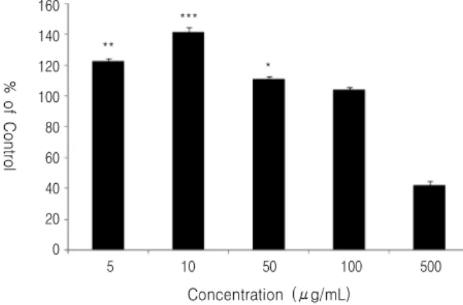
2. Effect of BK on splenocyte proliferation BK stimulated the proliferation of splenocytes.
Maximal splenocyte proliferation was observed at a concentration of BK 10 μg/mL (142.03±3.52%, p <
0.01, Fig. 2).
3. Effect of BK on mitogen- and
OVA-stimulated splenocyte proliferation in OVA-immunized mice
BK significantly enhanced splenocyte proliferation
(p<0.001) induced by the B cell mitogen LPS and by
the T cell mitogen Con A, compared with OVA
**
***
* 160
140 120 100 80 60 40 20 0
% of Control
5 10 50 100 500 Concentration (μg/mL)
Fig. 2. Effect of extract of BK on splenocyte proliferation. A spleen was removed from specific pathogen-free ICR mice to prepared single cell suspension. The splenocyte responses to BK were determined by ELISA. All values represent the mean ± SEM (n=3). Significant differences among the different concentrations in each extract were designated as ***p<0.001, **p<0.01 and *p<0.05.
400 350 300 250 200 150 100 50 0
% of Control
**
***
**
***
***
***
Control OVA/Alum BK-100 BK-300 BK-1000 OVA-10 LPS-2 Con A-4
Fig. 3. Effect of BK on OVA-, LPS- and Con A-stimulated splenocyte proliferation in vivo. C57BL/6 mice were immunized once on the intraperitoneal injection with OVA/alum (100/200 μg) at weekly intervals and oral administration with BK of 100, 300, 1000 mg/kg/day. Splenocytes were prepared 1 weeks after the last immunization and cultured with OVA (10 μg/mL), LPS (2 μg/mL) and Con A (4 μg/mL) for 48 hr. Splenocyte proliferation was measured by the CCK-8 method as described in the text, and shown as % of control. The values are presented as mean ± SEM (n=5). Significant differences with OVA/alum groups were designated as **p<0.01 and ***p<0.001.
immunized-, BK-untreated mice (Fig. 3). However, no significant difference was observed among mice given the three BK doses (100, 300, and 1000 mg/kg).
4. Effect of BK on IgM, IgG, and OVA-specific antibody levels in plasma
The level of total IgG, which was enhanced by
OVA/Alum, was not affected by BK (Fig. 4). In contrast, BK enhanced the plasma level of total IgM over that observed in OVA immunized mice (Fig.
4-A). In addition, BK enhanced plasma OVA- specific IgG1 but did not alter the OVA-specific IgG (Fig. 4-B).
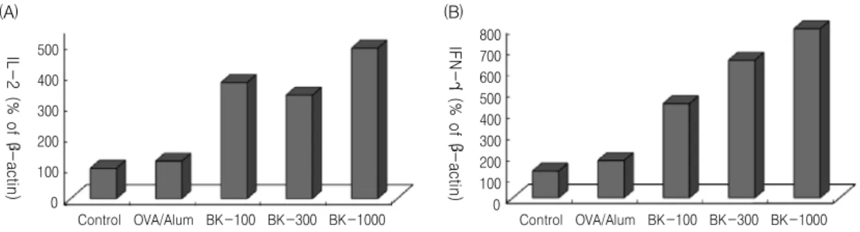
5. Effect of BK on cytokine mRNA levels in
splenocytes from OVA-immunized mice
350 300 250 200 150 100 50 0
Total IgG (g/mL)
800 700 600 500 400 300 200 100 0
Total IgM (g/mL)
IgG IgM
Control OVA/Alum BK-100 BK-300 BK-1000 (A)
30 25 20 15 10 5 0
OVA-specific IgG (g/mL)
300 250 200 150 100 50 0
OVA-specific IgG1 (g/mL)
Control OVA/Alum BK-100 BK-300 BK-1000
(B) IgG IgG1
Fig. 4. Effects of BK on and total IgM, IgG (A) and OVA-specific IgG, IgG1 (B) level in plasma. Total IgM, IgG and OVA-specific IgG, IgG1 level in plasma was assayed by ELISA. Each value represents a mean ± SEM (n=5).
No significant differences with OVA/alum groups were designated as p>0.05.
500 400 300 200 100 0
IL-2 (% of -actin)
Control OVA/Alum BK-100 BK-300 BK-1000 (A)
IFN-(% of -actin)
800 700 600 500 400 300 200 100 0
Control OVA/Alum BK-100 BK-300 BK-1000 (B)
Fig. 5. Effect of BK on the cytokines (A; IL-2, B; IFN-γ) mRNA gene expression in splenocytes from OVA-immunized mice.