신령버섯(Agaricus blazei Murill) 열수 추출물의 면역 활성에 미치는 영향
강인순1․김랑이1․김광섭1․김나리2․신중엽2․김채균1
1인하대학교 의과대학 약리학교실
2(주)건우에프피
Effects of Agaricus blazei Murill Water Extract on Immune Response in BALB/c Mice
In Soon Kang1, Rang Ie Kim1, Gwang Sub Kim1, Na Ri Kim2, Joong Yup Shin2, and Chaekyun Kim1
1Department of Pharmacology and Toxicology, Inha University School of Medicine
2Geonwoo Food & Pharm. Co., Ltd.
ABSTRACT The edible mushroom Agaricus blazei Murill is known to have many physiological functions, including antitumor, antiviral, and anti-inflammatory effects. Aqueous extracts were obtained by extracting A. blazei in water at 90°C for 15 h, followed by spray-drying with dextran at a 70:30 ratio. In this study, we examined the immuno- modulatory effect of A. blazei Murill water extract (ABM) in BALB/c mice. Mice were administered orally with 4, 20, and 100 mg/kg of ABM for 21 days. ABM-treated mice did not show significant differences in body and organ weights compare to saline-treated control mice. Splenocytes isolated from ABM-administered mice revealed similar levels of cellularity and proliferation compared to control mice, whereas they showed increased natural killer (NK) cell activity and decreased IL-4 and IL-12 production. Different from in vivo results, splenocytes isolated from normal mice showed increased proliferation and INF-γ production following ABM treatment in vitro. In addition, ABM treat- ment enhanced macrophage proliferation and nitric oxide (NO) production in a dose-dependent manner. However, ABM had no effect on LPS-induced NO production. These results suggest that A. blazei modulates immune function by increasing NK cell activity and macrophage function.
Key words: Agaricus blazei Murill, splenocytes, natural killer cell, macrophage, nitric oxide
Received 23 July 2015; Accepted 3 September 2015
Corresponding author: Chaekyun Kim, Department of Pharmacology
& Toxicology, Inha University School of Medicine, Incheon 22212, Korea
E-mail: [email protected], Phone: +82-32-860-9874
서 론
신령버섯(Agaricus blazei Murill)은 주름버섯과 주름버 섯속에 속하는 식용버섯으로 국내에서는 아가리쿠스버섯 또는 흰들버섯이라고 한다(1). 외형은 야생 양송이와 유사하 지만 양송이보다 향이 강하고 버섯대가 두껍고 길며 육질의 맛이 좋은 것이 특징이고, 항암 및 면역증강 활성물질을 함 유하고 있는 것으로 알려져 있다(2,3). 원산지는 브라질 산 악지대인 피에다데로(Piedade) 지역으로 고온다습한 기후 에서 야생한다. 1978년 일본의 Iwada Mushroom Institute 에서 인공재배를 보고한 이후 브라질과 일본에서 다량의 재 배가 이루어지고 있다(4). 국내에서는 자생하지 않고 소규모 의 재배가 이루어지고 있는 고가의 식용버섯이다.
일본의 학자들은 중남미 잉카지역 주민들의 암 관련 질환
환자가 적고 장수하는 사람이 많다는 점에 주목하여 그 원인 이 지역주민들이 식용하는 버섯에 있다는 점을 밝혀내 보고 하였다(5-7). 신령버섯의 유효성분으로는 다당체인 β-(1, 3)-glucan, α-(1,4)-glucan, α-(1,6)-glucan, β-(1,6)- glucan, glucomannan이 알려져 있다(8-11). 신령버섯에서 분리한 단백다당체는 탄수화물 38~45%와 단백질 40~45
%를 함유하고 있으며, 이들 단백질과 다당체의 복합체는 항 암 활성이 있다고 보고되었다(12-14). 주요 활성성분은 β- (1,6)-glycosyl 가지를 가진 β-(1,3)-glucan으로 복수암, 결장암, 난소암, 유방암, 폐암, 간암 등에 항암 효과가 있는 것으로 알려져 있다(4,8,12,15-17). 신령버섯은 항암 효과 외에도 항바이러스, 면역증강, 항염증, 항당뇨, 혈압 강하 효과와 콜레스테롤 저하 등의 생리작용이 있는 것으로 알려 져 있다(12,17-24).
자연살해세포(natural killer cell, NK cell)는 종양세포나 바이러스에 감염된 세포를 공격하여 용해와 사멸을 일으키 는 역할을 하며, 주로 바이러스 병원성이나 인터페론(inter- feron, IFN) 또는 interleukin(IL)-2, IL-12, IL-15 같은 사 이토카인에 의해 활성화된다. 선천면역을 담당하는 세포로
서 세포 용해 작용을 일으키는 NK 세포는 표적 세포에 직접 접촉하여 세포막을 파괴하는 perforin과 granzyme을 내어 표적 세포의 세포사멸을 유도하거나 표적 세포의 Fas/CD95 같은 death receptor가 NK 세포의 리간드에 결합하여 세포 사멸을 일으킨다. 신령버섯을 투여 받은 마우스의 NK 세포 의 활성이 증가되어 YAC-1 세포를 파괴시키는 것이 여러 연구에서 보고되었다(25-28).
본 연구에서는 신령버섯의 면역 활성에 미치는 효과를 BALB/c 마우스를 이용한 동물 실험과 대식세포인 RAW 264.7 세포를 이용하여 평가하였다.
재료 및 방법
신령버섯 농축분말 제조
본 실험에 이용된 신령버섯은 스톡미(Jeju, Korea)에서 구입하였으며, 덱스트린은 이씨케미컬(Seoul, Korea)에서 구 입하였다. 신령버섯을 후드믹서(MIX-30R, TongYang Mag- ic Corp., Seoul, Korea)로 분쇄하여 20 mesh를 통과한 분 말을 추출기에 넣고 원물 대비 15배의 물을 투입하여 90°C 에서 15시간 추출하였다. 추출액을 감압 여과한 후 감압농 축기로 농축하여 농축액을 얻었다. 이 농축액과 덱스트린을 고형분 비율 70:30으로 혼합하여 분무건조기로 분말을 제 조하였다. 분무건조의 조건은 8,000 rpm, 송풍온도 160°C, 배풍온도 90°C이며, 제조된 분말을 40 mesh로 선별하여 사용하였다.
신령버섯 농축분말의 β-glucan 함량은 건강기능식품공 전에 준하여 옵티팜(Osong, Korea)에서 분석하였으며 15.69 mg/g의 β-glucan을 함유하였다.
실험동물
수컷 BALB/c 마우스(6주령, 체중 19~23 g)를 오리엔트 바이오(Seoul, Korea)로부터 구입하여 일반 고형사료로 5 일간 순화, 사육하였다. 사육실의 온도는 22±2°C, 습도는 40~60%로 그리고 명암주기는 12시간으로 일정하게 유지 하였다. 동물실험은 인하대학교 의과대학 동물실험윤리위 원회의 승인(INHA 131217-255)을 받았으며, 그 규정을 준 수하여 실시하였다. 실험을 위하여 마우스는 6군으로 분류 하여 처치하였다. 시료는 day 1부터 day 21까지 생리식염 수(대조군, control group), 신령버섯 추출물 4 mg/kg (ABM(L)), 20 mg/kg(ABM(M)), 100 mg/kg(ABM(H))을 매일 1회 일정시간에 경구투여 하였다. 마지막 투여 다음날 에 마우스를 희생하여 체중 및 면역기관의 무게를 측정 후 비장을 적출하여 면역학적 분석에 사용하였다.
체중 및 면역장기 무게 측정
실험 시작일(day 1)과 종료일(day 22)에 실험동물의 체 중을 측정하였다. 또한 실험 종료일에 실험동물을 희생한 후 간, 흉선, 비장을 적출하여 그 무게를 측정하였다.
비장세포 준비
마우스 비장세포 분리는 Mishell과 Shiigi(29)의 방법에 의해 실행하였다. 실험동물에서 비장을 적출하여 40 μm nylon cell strainer(SPL, Pocheon, Korea)를 이용하여 파 쇄한 후 세포를 분리하였다. 세포를 300×g에서 5분간 원심 분리 한 후 red blood cell lysis buffer(Sigma-Aldrich Co., St, Louis, MO, USA)를 가하여 적혈구를 제거하였다.
비장세포를 배양액[RPMI 1640, 10% fetal bovine serum (FBS), 100 U/mL penicillin, 100 μg/mL streptomycin]에 현탁 후 실험에 사용하였다.
비장세포 및 대식세포의 증식능 측정
비장세포와 대식세포의 증식능은 3-(4,5-dimethylthia- zol-2-yl)-2,5-diphenyltetrazolium bromide(MTT) 방 법으로 측정하였다(30). 시료를 투여한 마우스에서 분리한 비장세포를 24-well plate에 5×10⁶ cells/well이 되도록 분주한 후, 1 μg/mL concanavalin A(ConA)를 넣고 5%
CO2가 포함된 37°C 배양기에서 48시간 배양하였다. 또한 in vitro에서 신령버섯 추출물의 비장세포 증식에 미치는 영 향을 측정하기 위하여 정상 마우스에서 분리한 5×10⁵ cells/
well 비장세포에 1 μg/mL ConA와 신령버섯을 각각 농도별 (0, 0.1, 1, 5, 10, 50, 100, 200, 500 μg/mL)로 처리하였다.
48시간 배양 후 250 μg/mL MTT 용액을 첨가하여 4시간 동안 배양 후 배양액을 제거하고 200 μL의 dimethyl sulf- oxide를 가하여 10분간 실온에서 반응 후 VERSAmax mi- croplate reader(Molecular Devices, Menlo Park, CA, USA)를 이용하여 570 nm에서 흡광도를 측정하였다. 대식 세포인 RAW 264.7 cell은 96-well plate에 5×10⁴ cells/
well이 되도록 분주하고 4시간 부착시킨 후 1 μg/mL LPS를 넣고 48시간 배양하여 MTT법으로 측정하였다.
사이토카인 분비능 측정
시료를 투여한 마우스에서 분리한 비장세포를 24-well plate에 5×10⁶ cells/well이 되도록 분주하고 1 μg/mL의 ConA를 첨가하여 48시간 동안 배양하였다(31,32). 배양액 에 유리된 IL-2, IL-4, IL-10, IL-12, IFN-γ, TNF-α의 양을 ELISA kit(R&D Systems, Minneapolis, MN, USA)을 이용하여 제조회사의 실험방법에 따라 실험하였다. 96- well plate에 capture antibody를 18시간 동안 부착시킨다.
Capture antibody가 부착된 well을 washing buffer(0.05
% Tween 20 in phosphate buffered saline, pH 7.2~7.4) 로 세척한 후 reagent diluent(0.1% bovine serum albu- min, 0.05% Tween 20 in Tris-buffered saline) 200 μL를 분주하여 실온에서 1시간 30분 동안 반응시킨다. 다시 washing buffer로 세척한 후 각 사이토카인 표준액 및 배양 액을 50 μL 분주하여 실온에서 2시간 동안 반응시킨다.
Washing buffer로 3회 세척한 후 detection antibody를 100 μL 첨가하여 실온에서 2시간 동안 더 반응시킨다. 다시
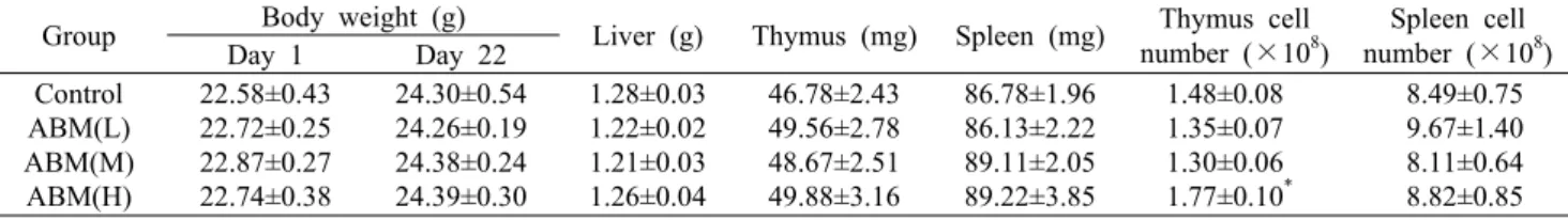
Table 1. Effect of Agaricus blazei Murill administration on the body and immune organ weights and cellularity of BALB/c mice, (n=9)
Group Body weight (g)
Liver (g) Thymus (mg) Spleen (mg) Thymus cell number (×108)
Spleen cell number (×108) Day 1 Day 22
Control ABM(L) ABM(M) ABM(H)
22.58±0.43 22.72±0.25 22.87±0.27 22.74±0.38
24.30±0.54 24.26±0.19 24.38±0.24 24.39±0.30
1.28±0.03 1.22±0.02 1.21±0.03 1.26±0.04
46.78±2.43 49.56±2.78 48.67±2.51 49.88±3.16
86.78±1.96 86.13±2.22 89.11±2.05 89.22±3.85
1.48±0.08 1.35±0.07 1.30±0.06 1.77±0.10*
8.49±0.75 9.67±1.40 8.11±0.64 8.82±0.85 Data are mean±SEM of 8∼9 mice, *P<0.05.
ABM(L): 4 mg/kg, ABM(M): 20 mg/kg, ABM(H): 100 mg/kg.
washing buffer로 세척한 후 horseradish peroxidase가 결합된 streptavidin을 100 μL 분주하여 20분간 암소에서 반응시킨다. 반응한 plate를 washing buffer로 세척한 후 tetramethylbenzidine 용액을 넣고 다시 20분간 암소에서 반응시킨다. 반응정지액인 2 N H2SO4를 50 μL 넣어 반응을 정지시킨 후 450 nm에서 흡광도를 측정하였다. 각 사이토 카인의 농도는 표준액을 사용하여 얻은 표준곡선에 따라 계 산하였다.
자연살해세포 활성 측정
자연살해세포의 세포독성은 NK 세포가 NK-sensitive 한 YAC-1 세포(NK-sensitive cell line)를 공격하여 파괴 된 YAC-1 세포로부터 유리된 lactate dehydrogenase를 측정하는 방법(LDH cytotoxic assay)을 이용하였다(31, 32). 96-well U-bottom culture plate에 1×10⁴ cells/100 μL의 YAC-1 세포(Korean Cell Line Bank, Seoul, Korea) 를 넣고 비장세포 : YAC-1 세포(effector-to-target) 비율 이 200:1, 100:1, 50:1이 되도록 비장세포를 넣은 후 37°C, 5% CO2 배양기에서 4시간 동안 배양하였다. 윈심분리 후 LDH가 유리된 상층액 100 μL를 채취하여 flat-bottom mi- croplate(Nunc, Roskilde, Denmark)에 옮겨 LDH assay kit(ABcam, Cambridge, UK)을 이용하여 실행하였다. 배양 액에 LDH working mixture를 100 μL 첨가하고 실온(15~
25°C)의 암소에서 30분간 반응 후, 반응정지 용액인 1 N HCl 50 μL를 첨가한 다음 490 nm에서 흡광도를 측정하였 다. NK 세포 활성도(%)는 [(test sample-low control)/
(high control-low control)]×100으로 계산하였다.
Nitric oxide(NO) 생성 측정
RAW 264.7 cell을 96-well plate에 5×10⁴ cells/well이 되도록 분주하여 4시간 부착시킨 후 1 μg/mL LPS와 시료를 농도별(0, 0.1, 1, 5, 10, 50, 100, 200, 500 μg/mL)로 처리 하여 48시간 배양하였다. 배양액 100 μL에 동량의 Griess reagent(0.1% N-(1-naphthyl)ethylenediamine, 1% sul- fanilamide, 5% phosphoric acid)를 혼합하여 실온에서 10 분간 반응시킨 후 550 nm에서 흡광도를 측정하였다. NO의 농도는 0~100 μM의 sodium nitrate를 사용하여 얻은 표준 곡선에 따라 계산하였다(30).
자료분석
본 실험 자료의 통계처리는 in vivo 실험은 Sigmaplot11 (Systat Software, San Jose, CA, USA)을 이용하여 one- way ANOVA로 분석 후 Dunn's method를 실시하였고, in vitro 실험은 Student's t-test를 실시하여 평균치(mean)와 표준오차(SEM) 또는 표준편차(SD)로 나타내었고, P 값이 0.05 미만일 때 통계적으로 유의성이 있다고 판정하였다.
결과 및 고찰
체중 및 면역장기의 무게
매일 1회씩 21일 동안 신령버섯을 경구투여 한 후 마우스 를 희생한 뒤 체중, 간 및 면역장기의 무게와 흉선 세포수와 비장 세포수를 측정하여 Table 1에 나타내었다. 실험이 진 행된 21일간 체중은 대조군이 7.6%, ABM(L)은 6.8%, ABM (M)은 6.6%, ABM(H)은 7.3%의 증가를 보였으며, 이는 마 우스가 성장함에 따른 자연적인 체중 증가로 신령버섯의 투 여가 대조군에 비하여 유의적인 체중 변화를 일으키지 않았 다. 대사 장기인 간의 무게는 변화가 없었으며 면역기관인 흉선과 비장의 무게와 세포수는 대조군과 신령버섯 투여군 사이에 대부분 차이를 보이지 않았으나, ABM(H)에서는 흉 선세포의 수가 유의하게 증가하였다.
마우스에 4~100 mg/kg의 ABM을 투여하였을 때 체중과 면역장기의 무게에는 영향을 미치지는 않았으나 100 mg/
kg을 투여한 경우에는 흉선세포의 수를 유의하게 증가시켰 다. 신령버섯을 경구로 투여한 후 마우스 면역기관 세포수 변화에 관한 연구는 많지 않으며, Liu 등(33)이 신령버섯 C3H/HeN 마우스에 열수 추출물을 14일간 투여하였을 때 비장 세포수에 유의할 만한 변화가 없음을 보고하였다.
신령버섯 투여가 비장세포 증식에 미치는 영향
신령버섯의 투여가 비장세포의 증식에 미치는 영향을 Fig.
1에 나타내었다. 비장세포에 ConA만을 처리한 실험군을 대 조군으로 하여 신령버섯 투여군의 비장세포 증식 정도를 백 분율로 나타내었다. ConA를 처리한 실험군은 처리하지 않 은 실험군에 비하여 약 5배 비장세포 증식을 증가시켰다.
그러나 각 농도의 신령버섯을 투여 받은 마우스 비장세포의 증식은 신령버섯을 투여 받지 않은 대조군과 차이가 없었다 (Fig. 1). 이 결과는 3~6 mg/kg의 신령버섯을 4~14일 동안
Fig. 1. Splenocyte proliferation of mice administered with Agaricus blazei Murill. Splenocytes were isolated from BALB/c mice administered with ABM for 21 days, then were stimulated with ConA for 48 h. The cell proliferation was measured with MTT assay. Data are mean±SEM of 8∼9 mice.
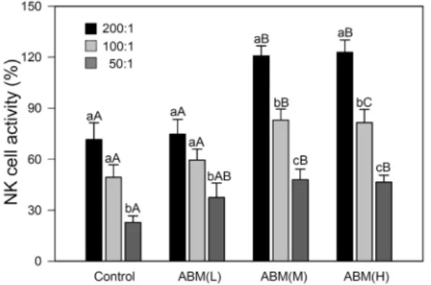
Fig. 2. Adminstration of Agaricus blazel Murill enhances NK cell activity. The splenocytes from BALB/c mice fed with ABM were incubated with different ratio of YAC-1 cells, and the re- leased LDH was determined by LDH assay. Data are mean±
SEM of 9 mice, and bar graphs with different small letters (a-c) are different at P<0.05 among the E:T ratios within a group, and bar graphs with different capital letters (A-C) are different at P<0.05 among the same E:T ratios in different groups.
투여 받은 마우스에서 비장세포 증식을 보고한 Tang 등(26) 의 결과와는 일치하지 않는다.
자연살해세포 활성에 미치는 영향
신령버섯의 투여가 NK 세포의 활성에 미치는 영향을 Fig.
2에 나타내었다. NK 세포의 세포 용해 활성(%)은 E:T ratio 가 200:1, 100:1, 50:1인 경우 대조군 71.5±9.9, 49.3±
7.5, 22.8±3.9%, ABM(L) 74.6±8.8, 59.5±6.5, 37.5±8.5
%, ABM(M) 120.7±6.0, 82.9±6.5, 48.0±6.2%, ABM(H) 122.8±7.2, 81.4±7.8, 46.5±4.1%였다. ABM(L)은 대조 군과 유사한 NK 세포 활성을 보였으며 ABM(M)과 ABM(H) 를 투여한 마우스에서 분리한 비장세포는 실험에 사용한 모 든 E:T 비율에서 생리식염수를 투여한 대조군보다 현저하 게 높은 세포 용해 활성을 나타내었다. 따라서 20 mg/kg 이상 의 신령버섯의 투여는 NK 세포 활성을 증가시켜 YAC-1
세포를 용해시킴을 알 수 있다. Takimoto 등(34)은 Meth A cell을 접종하여 종양을 유발한 BALB/c 마우스에 신령버 섯 열수 추출물을 투여하여 나타나는 항암작용이 NK 세포 의 활성화와 INF-γ의 증가에 기인한다고 보고하였다. 따라 서 암세포를 죽이는 대표적인 세포인 NK 세포의 활성 증가 는 암세포 또는 바이러스에 감염된 세포를 죽이는 데 중요한 역할을 하며, 여러 연구자에 의해 보고된 바 있는 신령버섯 의 항암 효과를 설명하는 주요 기전으로 생각된다.
신령버섯 섭취가 비장세포의 사이토카인 분비에 미치는 영향 신령버섯 경구투여가 비장세포의 사이토카인 생성에 미 치는 영향을 Fig. 3에 나타내었다. 신령버섯을 투여 받은 마 우스 비장세포의 사이토카인 생성을 측정한 결과 IL-12와 IL-4의 생성은 사용한 모든 용량(4~100 mg/kg)에서 억제 되었으며, IL-2, TNF-α와 IL-10은 100 mg/kg 투여군에서 억제되었다. IL-12는 대조군 84.2±5.2 pg/mL에 비해 ABM (H)에서 44.3±7.2 pg/mL로 52.6%, IL-4는 대조군 110.3
±2.6 pg/mL에 비해 ABM(H)에서 86.4±2.1 pg/mL로 78.3
%였다. 우리가 측정한 사이토카인 중 유일하게 IFN-γ는 대 조군 70.4±10.9 pg/mL에 비해 신령버섯 투여군에서는 ABM (M) 80.2±10.5(113.9%)로 4~20 mg/kg 투여군에서 증가 하는 경향이었으나 Student t-test와 one-way ANOVA 검 정에서 모두 유의성이 없었다. 이러한 결과는 신령버섯이 마 우스 비장세포의 IL-4의 생성을 억제하고 INF-γ의 생성을 증가시킨다는 보고(25,26,34,35)와 일치한다. 일반적으로 IL-2와 INF-γ는 NK cell의 활성을 증가시킨다고 알려진 사이토카인이므로 우리는 신령버섯이 이들의 생성을 증가 시킬 것을 기대하였으나, 본 연구에서는 IL-2의 생성은 억 제하고 INF-γ 생성은 증가시키는 경향을 보였다. 그러나 정상마우스의 비장세포에 ABM을 처리한 경우에는 INF-γ 의 생성을 현저하게 증가시켰다(Fig. 4). 신령버섯의 사이토 카인 생성에 미치는 영향은 연구자들 사이에서도 서로 상이 하다. Liu 등(33)은 신령버섯 추출물을 BALB/c 마우스에 7일간 투여하였을 때 TNF-α와 IL-6의 생성이 감소됨을 보 고한 반면, 비록 대부분 in vitro 연구이긴 하지만 여러 연구 자가 신령버섯이 TNF-α와 IL-6의 생성을 증가시킴을 보고 하였다(36-38). 또한 같은 연구자의 in vivo 실험과 in vitro 실험에서도 상이함이 나타났는데, Johnson 등(39)은 신령 버섯이 사람 혈액 중 IL-1β, TNF-α, IL-2, IL-16, IL-17의 생성을 억제한 반면, in vitro 실험에서는 이들 사이토카인의 생성을 증가시킴을 보고하였다.
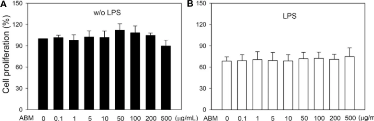
신령버섯이 in vitro에서 비장세포 증식과 사이토카인 분 비에 미치는 영향
신령버섯이 정상 마우스에서 분리한 비장세포의 증식에 미치는 영향을 알아보기 위해 정상 마우스에서 분리한 비장 세포에 1 μg/mL ConA와 신령버섯을 농도별(0~500 μg/
mL)로 처리하였다. 비장세포에 ConA만을 처리한 실험군을
A B C
D E F
Fig. 3. The cytokine production of splenocytes from Agaricus blazei Murill administered BALB/c mice. The splenocytes were activated with ConA and the production of cytokines were measured by ELISA. Data are mean±SEM of 8∼9 mice and significantly different at *P<0.05 compared to control.
A B
Fig. 4. The effect of Agaricus blazei Murill on the proliferation of splenocytes from normal BALB/c mice in vitro. Splenocytes were isolated from normal BALB/c mice and were treated with ConA and different doses of ABM for 48 h. (A) The cell proliferation was measured with MTT assay (n=9). (B) IFN-γ production was measured by ELISA (n=3). Data are mean±SD and significantly different at *P<0.05 compared to ConA.
100%로 하였을 때 각 실험군의 비장세포 증식 정도를 백분 율로 나타내었다. ConA를 처리한 군은 처리하지 않은 군에 비하여 4.5배의 세포증식을 보였고, 신령버섯 5, 10, 50, 100 μg/mL 처리군은 대조군에 비하여 112.6, 121.9, 126.3, 124.9% 비장세포 증식을 증가시켰다(Fig. 4A). 또한 신령 버섯이 정상마우스에서 분리한 비장세포의 사이토카인 생 성에 미치는 영향을 측정한 결과 신령버섯을 정상마우스에 서 분리한 비장세포에 직접 처리한 경우에는 INF-γ의 생성 을 현저하게 증가시켰다(Fig. 4B). 정상마우스에서 비장세 포를 분리한 후 신령버섯을 처리한 경우 농도 의존적으로 비장세포의 증식과 INF-γ 생성이 증가되는 결과(Fig. 4)는
신령버섯을 경구투여 한 마우스에서 분리한 비장세포의 증 식과 사이토카인 분비에 변화가 없는 결과(Fig. 1 & 3)와는 달랐다. 그 이유에 대해서는 더 많은 연구가 요구되며 동물 실험에서 투여 용량이나 투여 기간을 조절하여 연구하는 것 이 필요하다고 생각된다.
신령버섯의 대식세포의 증식에 미치는 영향
신령버섯이 면역과 염증반응에서 중요한 역할을 담당하 는 대식세포에 미치는 영향을 측정하기 위하여 신령버섯을 농도별(0~500 μg/mL)로 처리하였다. 신령버섯 50 μg/mL 에서 control에 비해 RAW 264.7 cell의 증식이 약 12% 증
A B
Fig. 5. Effect of Agaricus blazei Murill on RAW 264.7 cell proliferation. Cells were treated with different doses of ABM for 48 h in the absence or presence of LPS. The cell proliferation was measured with MTT assay. Data are mean±SD (n=3).
A B
Fig. 6. Effect of Agaricus blazei Murill on NO production in RAW 264.7 cells. Cells were treated with different doses of ABM for 48 h in the absence or presence of LPS. NO production was measured by Griess reaction. Data are mean±SD (n=3), and significantly different at *P<0.05 compared to control.
가하였으나 통계적으로 유의하지는 않았다(Fig. 5).
대식세포의 NO 생성에 미치는 영향
대식세포는 사이토카인, NO, prostaglandin을 생성하여 생체방어에 중요한 역할을 한다. NO는 혈압을 조절하고 면 역체계를 강화할 뿐만 아니라 암세포와 미생물을 죽이며 근 육활동을 조절하는 등 생체반응에 중요한 역할을 한다. LPS 자극에 의해 염증반응이 시작되면 iNOS가 발현되고 과량의 NO가 생성된다(30,39). 신령버섯을 농도별(0~500 μg/mL) 로 RAW 264.7 세포에 처리한 경우 200, 500 μg/mL에서 18.2±4.4, 32.2±7.1 μM의 NO가 생성되었으며 control의 7.7±4.6 μM에 비해 2.4배, 4.2배 증가되었다(Fig. 6). 그러 나 신령버섯은 LPS에 의해 유발된 과다한 NO 생성에는 영 향을 미치지 않았다. 따라서 신령버섯의 단독 처리는 농도 의존적으로 NO 생성을 증가시켜 면역력 증강에 기여하지 만, LPS에 의해 과량 생성되는 NO에는 영향을 미치지 않음 을 알 수 있다.
요 약
본 연구에서는 신령버섯의 면역기능 개선 효과를 마우스를 이용한 동물 모델에서 면역기관의 무게와 세포증식, 자연살
해세포 활성, 사이토카인 분비능을 측정하고 대식세포 증식 과 활성을 측정하여 평가하였다. BALB/c 마우스에 저(4 mg/kg), 중(20 mg/kg), 고(100 mg/kg) 농도의 신령버섯을 21일간 경구로 투여하였다. 마우스를 희생하여 체중 및 면 역장기 무게, 비장세포의 증식과 사이토카인 생성, 자연살해 세포의 활성을 측정하였다. 그 결과 신령버섯은 마우스의 체중, 간, 비장, 흉선의 무게에 영향을 주지 않았으며, 비장 세포의 증식에 유의한 효과가 없었다. 또한 비장세포의 IL- 4과 IL-12 생성을 억제하였으며, 마우스 자연살해세포의 활성을 현저하게 증가시켰다. 정상 마우스에서 분리한 비장 세포에 신령버섯을 처리한 in vitro 실험에서는 신령버섯 5~100 μg/mL에서 농도 의존적으로 비장세포의 증식과 IFN-γ 생성을 증가시켰다. 신령버섯은 대식세포인 RAW 264.7 세포의 증식을 100 μg/mL 농도까지 농도 의존적으 로 증가시켰으며, 대식세포에 의한 NO의 생성을 농도 의존 적으로 증가시켰다. 이상의 결과를 종합해 보면 신령버섯을 마우스에 3주간 투여하면 동물의 체중, 면역장기의 무게와 면역세포의 증식에는 영향을 미치지 않지만 자연살해세포 의 활성을 70% 가량 증가시키며 IL-4와 IL-12의 생성을 억제한다. 정상 마우스에서 분리한 비장세포에 신령버섯을 처리하면 세포증식과 IFN-γ 분비가 증가되고, 대식세포인 RAW 264.7 세포의 증식과 NO 생성이 증가된다. 따라서
신령버섯은 바이러스에 감염된 세포나 암세포를 죽이는 자 연살해세포와 대식세포의 활성을 증가시켜 면역반응 조절 에 중요한 역할을 할 것으로 기대된다.
감사의 글
본 연구는 산업통상자원부의 지역특화산업육성(R&D)사업 의 연구과제(R0003330)로 수행된 결과의 일부이며 이에 감 사드립니다.
REFERENCES
1. Ito H, Shimura K, Itoh H, Kawade M. 1997. Antitumor ef- fects of a new polysaccharide-protein complex (ATOM) prepared from Agaricus blazei (Iwade strain 101) "Him- ematsutake" and its mechanisms in tumor-bearing mice.
Anticancer Res 17: 277-284.
2. Choi WY, Park C, Lee JY, Kim GY, Park YM, Jeong YK, Lee WH, Choi YH. 2004. Anti-proliferative effects of water extract of Agaricus blazei Murill in human lung cancer cell line A549. J Korean Soc Food Sci Nutr 33: 1237-1245.
3. Sung JM, Yoo YB, Cha DY. 1998. Mushroom. Kyohaksa, Seoul, Korea. p 3-10.
4. Mizuno T, Hagiwara T, Nakamura T, Ito H, Shimura K, Sumiya T, Asakura A. 1990. Antitumor activity and some properties of water-soluble polysaccharides from "Himemat- sutake", the fruiting body of Agaricus blazei Murill. Agric Biol Chem 54: 2889-2896.
5. Ebina T, Fujimiya Y. 1998. Antitumor effect of a peptide- glucan preparation extracted from Agaricus blazei in a dou- ble-grafted tumor system in mice. Biotherapy 11: 259-265.
6. Mizuno TK. 1995. Bioactive biomolecules of mushrooms:
Food function and medicinal effect of mushroom fungi.
Food Rev Int 11: 5-21.
7. Mizuno TK. 1995. Kawariharatake, Agaricus blazei Murill:
medicinal and dietary effects. Food Rev Int 11: 167-172.
8. Kawagishi H, Inagaki R, Kanao T, Mizuno T, Shimura K, Ito H, Hagiwara T, Nakamura T. 1989. Fractionation and antitumor activity of the water-insoluble residue of Agaricus blazei fruiting bodies. Carbohydr Res 186: 267-273.
9. Mizuno M, Morimoto M, Minato K, Tsuchida H. 1998.
Polysaccharides from Agaricus blazei stimulate lymphocyte T-cell subsets in mice. Biosci Biotechnol Biochem 62: 434- 437.
10. Mizuno M, Minato K, Ito H, Kawade M, Terai H, Tsuchida H. 1999. Anti-tumor polysaccharide from the mycelium of liquid-cultured Agaricus blazei mill. Biochem Mol Biol Int 47: 707-714.
11. Ohno N, Furukawa M, Miura NN, Adachi Y, Motoi M, Yadomae T. 2001. Antitumor beta glucan from the cultured fruit body of Agaricus blazei. Biol Pharm Bull 24: 820-828.
12. Dong Q, Yao J, Yang XT, Fang JN. 2002. Structural charac- terization of water-soluble β-D-glucan from fruiting bodies of Agaricus blazei Murill. Carbohydr Res 337: 1417-1421.
13. Itoh H, Ito H, Amano H, Noda H. 1994. Inhibitory action of a (1→6)-β-D-glucan-protein complex (FIII-2-b) isolated from Agaricus blazei Murill ("Himematsutake") on Meth A fibrosarcoma-bearing mice and its antitumor mechanism.
Jpn J Pharmacol 66: 265-271.
14. Menoli RC, Mantovani MS, Ribeiro LR, Speit G, Jordão BQ. 2001. Antimutagenic effects of the mushroom Agaricus
blazei Murrill extracts on V79 cells. Mutat Res 496: 5-l3.
15. Mizuno T, Inagaki R, Kanao T, Hagiwara T. 1990. Antitumor activity and some properties of water-insoluble hetero-glu- cans from "Himematsutake" the fruiting body of Agaricus blazei Murill. Agric Biol Chem 5: 2897-2905.
16. Smith JE, Rowan NJ, Richard S. 2002. Medicinal mush- rooms: a rapidly developing area of biotechnology for can- cer therapy and other bioactivities. Biotechnol Lett 24: 1839- 1845.
17. Wang H, Fu Z, Han C. 2013. The medicinal values of culi- nary-medicinal royal sun mushroom (Agaricus blazei Mur- rill). Evidence-Based Complement Altern Med 2013: 842619- 842425.
18. Choi JM, Koo SJ. 2000. Effects of β-glucan from Agaricus blazei Murill on blood glucose and lipid composition in db/
db mice. Korean J Food Sci Technol 32: 1418-1425.
19. Kuo YC, Huang YL, Chen CC, Lin YS, Chuang KA, Tsai WJ. 2002. Cell cycle progression and cytokine gene ex- pression of human peripheral blood mononuclear cells mod- ulated by Agaricus blazei. J Lab Clin Med 140: 176-187.
20. Takaku T, Kimura Y, Okuda H. 2001. Isolation of an anti- tumor compound from Agaricus blazei Murill and its mech- anism of action. J Nutr 131: 1409-1413.
21. Sugjyama K, Saeki S, Tanaka A, Yoshida S, Sakamoto H, Ishiguro Y. 1992. Hypocholesterolemic activity of ningyo- take (Polyporus confluences) mushroom in rats. J Jpn Soc Nutr Food Sci 45: 265-270.
22. Yoshiaki F, Hidekazu K, Koichi O, Ryo S, Takusaburo E.
1998. Tumoricidal activity of high molecular weight poly- saccharides derived from Agaricus blazei via oral admin- istration in the mouse tumor model. Nippon Shokuhim Ka- gaku Kaishi 45: 246-252.
23. Bellini MF, Giacomini NL, Eira AF, Ribeiro LR, Mantovani MS. 2003. Anticlastogenic effect of aqueous extracts of Agaricus blazei on CHO-k1 cells, studying different devel- opmental phases of the mushroom. Toxicol In Vitro 17:
465-469.
24. Martins de Oliveira J, Jordão BQ, Ribeiro LR, Ferreira da Eira A, Mantovani MS. 2002. Anti-genotoxic effect of aque- ous extracts of sun mushroom (Agaricus blazei Murill line- age 99/26) in mammalian cells in vitro. Food Chem Toxicol 40: 1775-1780.
25. Ni WY, Wu MF, Liao NC, Yeh MY, Lu HF, Hsueh SC, Liu JY, Huang YP, Chang CH, Chung JG. 2013. Extract of medicinal mushroom Agaricus blazei Murill enhances the non-specific and adaptive immune activities in BALB/c mice. In Vivo 27: 779-786.
26. Tang NY, Yang JS, Lin JP, Hsia TC, Fan MJ, Lin JJ, Weng SW, Ma YS, Lu HF, Shen JJ, Lin JG, Chung JG. 2009.
Effects of Agaricus blazei Murill extract on immune re- sponses in normal BALB/c mice. In Vivo 23: 761-766.
27. Niu YC, Liu JC, Zhao XM, Su FQ, Cui HX. 2009. Immu- nostimulatory activities of a low molecular weight anti- tumoral polysaccharide isolated from Agaricus blazei Murill (LMPAB) in Sarcoma 180 ascitic tumor-bearing mice.
Pharmazie 64: 472-476.
28. Yuminamochi E, Koike T, Takeda K, Horiuchi I, Okumura K. 2007. Interleukin-12- and interferon-gamma-mediated natural killer cell activation by Agaricus blazei Murill. Im- munology 121: 197-206.
29. Mishell BB, Shiigi SM. 1980. Selected methods in cellular immunology. 1st ed. WH Freeman and Co., San Francisco, CA, USA. p 4-27.
30. Kim JW, Kim C. 2005. Inhibition of LPS-induced NO pro-
duction by taurine chloramine in macrophages is mediated though Ras-ERK-NF-κB. Biochem Pharmacol 70: 1352-1360.
31. Kim H, Lee T, Kwon Y, Son M, Kim C. 2012. Immunomod- ulatory activities of ethanol extract of Cordyceps militalis in immunocompromised mice. J Korean Soc Food Sci Nutr 41: 494-500.
32. Kang IS, Kim H, Lee TH, Kwon YS, Son M, Kim C. 2014.
Effect of Cordyceps militaris on immune activity. Yakhak Hoeji 58: 81-90.
33. Liu Y, Fukuwatari Y, Okumura K, Takeda K, Ishibashi K, Furukawa M, Ohno N, Mori K, Gao M, Motoi M. 2008.
Immunomodulating activity of Agaricus brasiliensis KA21 in mice and in human volunteers. Evid Based Complement Alternat Med 5: 205-219.
34. Takimoto H, Wakita D, Kawaguchi K, Kumazawa Y. 2004.
Potentiation of cytotoxic activity in naive and tumor-bearing mice by oral administration of hot-water extracts from Agar- icus brazei fruiting bodies. Biol Pharm Bull 27: 404-406.
35. Lin JG, Fan MJ, Tang NY, Yang JS, Hsia TC, Lin JJ, Lai KC, Wu RS, Ma CY, Wood WG, Chung JG. 2012. An ex- tract of Agaricus blazei Murill administered orally promotes immune responses in murine leukemia BALB/c mice in
vivo. Integr Cancer Ther 11: 29-36.
36. Sorimachi K, Akimoto K, Ikehara Y, Inafuku K, Okubo A, Yamazaki S. 2001. Secretion of TNF-α, IL-8 and nitric ox- ide by macrophages activated with Agaricus blazei Murill fractions in vitro. Cell Struct Funct 26: 103-108.
37. Bernardshaw S, Hetland G, Ellertsen LK, Tryggestad AM, Johnson E. 2005. An extract of the medicinal mushroom Agaricus blazei Murill differentially stimulates production of pro-inflammatory cytokines in human monocytes and hu- man vein endothelial cells in vitro. Inflammation 29: 147- 153.
38. Johnson E, Førland DT, Saetre L, Bernardshaw SV, Lyberg T, Hetland G. 2009. Effect of an extract based on the medic- inal mushroom Agaricus blazei murill on release of cyto- kines, chemokines and leukocyte growth factors in human blood ex vivo and in vivo. Scand J Immunol 69: 242-250.
39. Higuchi M, Higashi N, Taki H, Osawa T. 1990. Cytolytic mechanisms of activated macrophages. Tumor necrosis fac- tor and L-arginine-dependent mechanisms act synergistically as the major cytolytic mechanisms of activated macrophages.
J Immunol 144: 1425-1431.