Lutein Modulates Th2 Immune Response in Ovalbumin-Induced Airway Inflammation
Jun Young Song
1, Chang-Min Lee
2and Min Ki Lee
1*
1
Department of Internal Medicine, Pusan National University School of Medicine, Busan 626-870, Korea
2
Section of Pulmonary and Critical Care Medicine, Department of Internal Medicine, Yale University School of Medicine, New Haven, Connecticut 06520-8057, USA
Received January 10, 2012 /Revised March 16, 2012 /Accepted March 16, 2012
The general term flavonoids is often used to categorize a family of natural compounds that are highly abundant in all higher plants, and which in recent years have attracted scientific interest as therapeutics. Lutein is a xanthophyll and one of 600 known naturally occurring carotenoids. It is found in green vegetables such as spinach and kale, and has been demonstrated to exert anti-in- flammatory activities. However, its anti-allergic effect in the Th1/Th2 immune response is poorly understood. In this study, we attempt to determine whether lutein regulates inflammatory mediators in an ovalbumin (OVA)-induced murine asthma model. To address this, mice were sensitized and challenged with OVA, and then treated with lutein before the last OVA challenge. Administration of lutein significantly suppressed the OVA-induced airway hyper-responsiveness. It also resulted in a significant alleviation of the infiltration of inflammatory cells into the bronchoalveolar lavage.
Additionally, lutein attenuated the increased expression of Th2 responses in OVA-challenged mice.
These results demonstrate that lutein is a potent inhibitor that reduces Th2 immune responses.
Furthermore, they show that the immunopharmacological function is mediated by a pathway that in- volves and is regulated by Th2 immune response.
Key words : Asthma, lutein, GATA-3, Th1/Th2 balance, STAT-6
*Corresponding author
*Tel:+82-51-240-7216, Fax:+82-51-254-3127
*E-mail : [email protected]
Introduction
Asthma is a chronic inflammatory lung disease that is characterized by airway hyperresponsiveness (AHR) to al- lergens, airway edema and increased mucus secretion [4,9].
Inflammatory lung disease usually results from the infiltra- tion of eosinophils, neutrophils, macrophages and lympho- cytes into the bronchial lumen and lung tissues [20,21].
Recruitment of these inflammatory cells from blood to the site of inflammation is regarded as a critical event in the development and persistence of airway inflammation [8,22].
In particular, it has been reported that eosinophil infiltration into the asthmatic lung leads to degranulation and release of eosinophil peroxidase (EPO), resulting in airway epi- thelial damage and the development of AHR [24,26].
Previous studies have shown that eosinophils infiltrating in- to the lung preferentially stimulate T-helper type 2 (Th2) cell responses by presenting antigens [1,15,16]. Therefore, Th2 cells are dominant in the airways [2] and Th2 cytokines, such as IL-4, IL-5 and IL-13, play a pivotal role in the pathophysi-
ology of asthma [10,17,18].
The balance between Th1 and Th2 cells is tuned by inter-
actions between transcription factors. One of the major tran-
scription factors regulating the expression of Th2 cytokine
is STAT6 [5,7,25]. STATs have been shown to be important
in the regulation of cytokines and growth factor-inducible
transcription factors in immune response [19,23]. Recent
studies using STAT6-deficient mice the demonstrated that
phosphorylation and nuclear translocation of STAT6 are crit-
ical for the development of Th2 cell differentiation and air-
way responses. The significant role of STAT6 in airway in-
flammation was further supported by findings in asthmatic
patients who showed increased levels of STAT6 expression
in their lungs [14]. IL-4, a prototype Th2 cytokine, enhances
Th2 cell development through STAT6, which activates
GATA3 genes [6]. GATA3, a downstream transcriptional fac-
tor of STAT6, plays a key role in Th2 cell development by
promoting Th2 cytokine expression by binding to a variety
of regulatory regions of Th2 cytokines [7]. On the other
hand, IL-12 drives Th1 cell differentiation through the acti-
vation of STAT4 and T-box expressed in T cells (T-bet), Th1
transcription factors, which up-regulates IFN-γ and
down-regulates IL-4 and IL-5 production [12]. Lutein plays
a role in various pharmacological responses, including an- ti-inflammatory, anticarcinogenic and free radical-scaveng- ing activities, in a variety of in vitro systems [11]. In this study, we attempted to characterize the effects of lutein in a murine model of asthma and to determine whether lutein treatment would inhibit asthmatic syndrome and suppress OVA-induced gene expressions of STAT-6 and GATA-3.
Materials and methods Animals and experimental protocol
Female BALB/c mice, 6 weeks of age and free of mur- ine-specific pathogens, were obtained from Charles River Laboratories (Yokohama, Japan). All experimental animals used in this study were maintained under a protocol ap- proved by the Institutional Animal Care and Use Committee of Pusan National University Medical School. Mice were im- munized intraperitoneally (i.p.) with 15 μg of OVA (Sigma-Aldrich, St. Louis, MO, USA) emulsified in 1 mg of aluminum hydroxide (Pierce Chemical Co., Rockford, IL, USA) on days 0 and 14.
Mice were challenged via the airway with OVA (3% OVA) for 30 minutes every day from day 20 to day 22. BAL fluid was obtained at 24 hours after the last challenge. For the extraction of lavage, mice (10 mice in each group) were kil- led with an overdose of ether. The chest cavity was exposed for expansion, after which the trachea was carefully in- tubated and a catheter was secured with ligatures.
Administration of lutein
We injected 200 μl of either 1 or 10 mg/kg lutein (Sigma-Aldrich) i.p. into each mouse every day from day 16 to day 19.
Total cell counts
The total number of cells was counted with a hemocytometer. Smears of BAL cells prepared with a cyto- spin II (Shandon, Runcorn, UK) were stained with Diff-Quik solution (Dade Diagnostics of P.R. Inc, Aguada, PR) for differential cell counting. Two independent, blind- ed investigators counted the cells using a microscope.
Approximately 200 cells were counted in each of the 4 ran- dom locations.
Histopathology
The mice were sacrificed and their lungs were removed
48 hours after the last challenge. Prior to removal, the lungs and trachea were intratracheally filled with 4% paraf- ormaldehyde, a fixative, using a ligature around the trachea.
The specimens were dehydrated and embedded in paraffin.
For histological examination, 5-μm sections of fixed em- bedded tissues were cut on a Leica model rotary microtome (Leica, Nussloch, Germany), placed on glass slides, depar- affinized and sequentially stained with hematoxylin 2 and eosin-Y (Richard-Allan Scientific, Kalamazoo, MI, USA).
Measurement of Th1/Th2 cytokines and IgE levels
The levels of IL-4, IL-5, IL-13 and IFN-γ were measured in the supernatant of BAL fluid by enzyme immunoassays performed according to the manufacturer’s protocol (IL-4, IL-5, IL-13 and IFN-γ; R&D Systems, Inc, Minneapolis, MN, USA). The levels of serum IgE were measured using an ELISA kit according to the manufacturer’s protocol (R&D Systems, Inc).
Western blot analysis
The lung tissues were homogenized, washed with PBS and incubated in lysis buffer containing a protease in- hibitor cocktail (Sigma-Aldrich) in order to obtain lung protein extracts. The samples were loaded on 10%
SDS-PAGE gels and were separated at 120 V for 90 mi- nutes and transferred electronically to polyvinylidene fluo- ride (PVDF) membranes. The PVDF membranes were then blocked with 5% non-fat milk in a washing buffer (50 mM Tris-HCl, pH 8.0; 150 mM NaCl; 0.1% Tween 20) and in- cubated with the indicated antibodies in the buffer (50 mM Tris-HCl, pH 8.0; 150 mM NaCl; 0.1% Tween 20; 1%
nonfat milk) for 1 hour at room temperature. The mem- branes were subsequently washed and incubated with the appropriate secondary antibodies conjugated with horse- radish peroxidase (Amersham Pharmacia Biotech, Uppsala, Sweden) for 1 hour at room temperature. Protein bands were visualized using an enhanced chemiluminescence system (Amersham Pharmacia Biotech).
Determination of airway responsiveness to methacholine
Airway responsiveness was measured with the mice in an unrestrained conscious state, 24 hours after the last challenge. Mice were placed in a barometric plethysmo- graphic chamber (All Medicus Co, Seoul, Korea) and base- line readings were taken for 3 minutes and averaged.
Aerosolized methacholine was nebulized in increasing con-
centrations (from 2.5 to 50 mg/ml) through an inlet of the main chamber for 3 minutes. Readings were taken for 3 mi- nutes after each nebulization and averaged. Enhanced pause (Penh), calculated as (expiratory time/relaxation time-1) × (peak expiratory flow/peak inspiratory flow) according to the manufacturer’s protocol, is a dimensionless value that represents a function of the proportion of maximal ex- piratory to maximal inspiratory box pressure signals and a function of the timing of expiration. Here, Penh was used as a measure of airway responsiveness to methacholine.
Results are expressed as the percentage increase in Penh fol- lowing challenge with each concentration of methacholine, where the baseline Penh (after saline challenge) is expressed as 100%. Penh values were averaged for 3 minutes after each nebulization and evaluated.
Densitometric analysis and statistical analysis
Experiments were repeated at least 3 times with consistent results. Unless stated otherwise, data are expressed as the mean ± S.E.M. ANOVA was used to compare the ex- perimental and control groups, while comparisons between individual groups were performed using Tukey’s multiple comparison test. A P value of less than 0.05 was considered statistically significant.
Results
Lutein reduces the number of inflammatory cells in BAL fluid
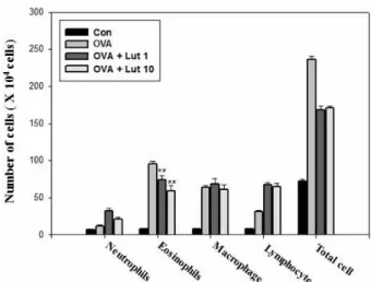
Inflammatory lung disease including asthma usually re- sults from the infiltration of eosinophils, neutrophils, macro- phages and lymphocytes into the bronchial lumen and lung tissues. The total numbers of cells, eosinophils, lymphocytes and macrophages in BAL fluid were more significantly in- creased 24 hours after OVA inhalation than after saline inhalation. The increased number of eosinophils was sig- nificantly reduced by the administration of lutein (Fig. 1).
Lutein reduces the levels of Th2 cytokines in the lung tissues of mice sensitized to and challenged with OVA
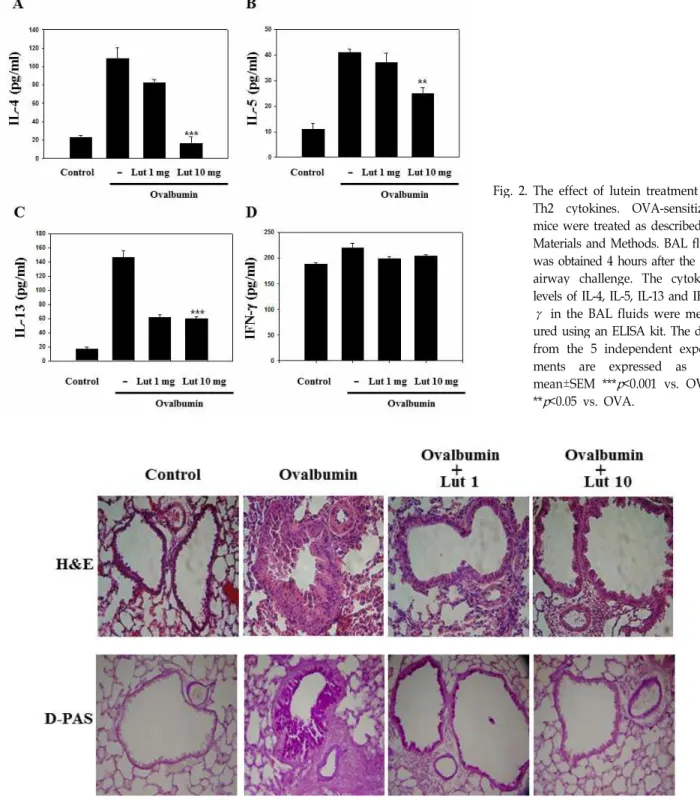
The Th2-type cytokines IL-4, IL-5, and IL-13, produced by activated CD4+ T cell play an important role in the pathogenesis of asthma by controlling the key process of immunoglobulin (IgE) production, growth of mast cells and the differentiation and activation of mast cells and
Fig. 1. The effect of lutein on total and differential cellular com- ponents in the BAL fluid of OVA-sensitized and OVA-challenged mice. Mice were treated with PBS (CON), OVA plus lutein 1 mg/kg/day (OVA + Lut 1 mg), OVA plus lutein 10 mg/kg/day (OVA + Lut 10 mg) and OVA (OVA), respectively. The BAL fluid cells were collected 1 day after OVA challenge, and different cell types were enumerated. This experiment used 5 mice (n=5) in each group. Statistical significance: **
p
<0.05 when compared with mice treated with OVA.eosinophils. BAL fluid was obtained 4 hours after the last airway challenge. The levels of IL-4, IL-5 and IL-13 in the BAL fluid were more significantly increased by airway chal- lenge with OVA than in the control state. The administration of lutein reduced the increased levels of IL-4, IL-5, IL-13 (Fig.
2). The levels of Th2 cytokines, including IL-4, IL-5 and IL-13, were higher in OVA-sensitized and OVA-challenged mice compared to saline-sensitized and saline-challenged mice; however, the levels of IFN-γ, among the Th1 cyto- kines, was not changed. These results indicate that lutein functions as an attenuator of Th2 cytokines rather than Th1 cytokines in airways challenged with OVA.
Lutein ameliorates pathological changes in OVA- induced asthma
Histological analyses of OVA-exposed mice revealed the typical pathologic features of asthma: numerous in- flammatory cells, including eosinophils, remarkably in- filtrated around the bronchioles as compared to the control mice. Mice treated with lutein showed significantly reduced infiltration of inflammatory cells in the lung tissues. Severe lung inflammation was observed in lung tissue 24 hours af- ter OVA inhalation compared to that after saline inhalation.
OVA-induced lung inflammation was significantly reduced
Fig. 2. The effect of lutein treatment on Th2 cytokines. OVA-sensitized mice were treated as described in Materials and Methods. BAL fluid was obtained 4 hours after the last airway challenge. The cytokine levels of IL-4, IL-5, IL-13 and IFN- γ in the BAL fluids were meas- ured using an ELISA kit. The data from the 5 independent experi- ments are expressed as the mean±SEM ***
p
<0.001 vs. OVA,**
p
<0.05 vs. OVA.Fig. 3. Lutein inhibits lung inflammation and inflammatory cell infiltration. Mice were sensitized and challenged as described in Materials and Methods. Sections were obtained from the lungs of the mice which received the control treatment (CON), OVA plus lutein at 1 mg/kg/day (OVA + Lut 1 mg), OVA plus lutein at 10 mg/kg/day (OVA + Lut 10 mg) and OVA (OVA). The lungs were removed 2 days after the last airway challenge. Sections were stained with hematoxylin and eosin (×200) and PAS staining.
by the administration of lutein (Fig. 3). The above-mentioned pathophysiological phenomena were dramatically decreased
by the administration of lutein, and these results suggest
that lutein inhibits OVA-induced inflammation in the lungs.
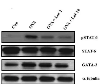
Fig. 4. The effect of lutein on expressions of STAT-6 and GATA-3 in the lung tissues of OVA-sensitized and OVA-challenged mice. The lung tissues were obtained 24 hours after the last challenge in the mice that re- ceived the control treatment (CON), OVA plus lutein at 1 mg/kg/day (OVA + Lut 1 mg), OVA plus lutein at 10 mg/kg/day (OVA + Lut 10 mg) and OVA (OVA).
The data from the 5 independent experiments are ex- pressed as the mean±SEM.
Lutein decreases STAT6 and GATA3 expression in the lung tissues of mice sensitized to and challenged with OVA
Western blot analysis revealed that the expressions of STAT6 and GATA3 proteins in lung tissues was more sig- nificantly increased 24 hours after OVA inhalation than after saline inhalation (Fig. 4). The increased protein expressions of STAT6 and GATA3 were reduced when lutein was ad- ministered compared to control protein expression.
Lutein decreases airway hyperresponsiveness
Airway responsiveness was assessed as the percentage in- crease in the basis of Penh in response to increasing doses of methacholine. In OVA-sensitized and OVA-challenged mice, the dose-response curve of percentage Penh was shift- ed to the left compared to that of the control mice. In addi- tion, methacholine administration (2.5 mg/ml to 50 mg/ml) significantly increased the percentage Penh in OVA-sensi- tized and OVA-challenged mice compared to the controls.
OVA-sensitized and OVA-challenged mice treated with lu- tein showed a dose-response curve of percentage Penh that was shifted to the right compared to that in untreated mice, and this shift was dose-dependent (Fig. 5). These data in- dicate that lutein treatment reduces OVA-induced airway hyperresponsiveness.
Fig. 5. The effect of lutein on airway responsiveness in OVA-sensitized and OVA-challenged mice. Airway re- sponsiveness was measured 24 hours after the last chal- lenge in the mice which received the control treatment (CON), OVA plus lutein at 1 mg/kg/day (OVA + Lut 1 mg), OVA plus lutein at 10 mg/kg/day (OVA + Lut 10 mg) and OVA (OVA). Airway responsiveness to aer- osolized methacholine was measured in conscious un- restrained mice. The mice were placed in the main chamber, nebulized with PBS, and then administered increasing doses of methacholine (2.5 to 50 mg/ml) for 3 minutes for each nebulization. Readings of breathing parameters were taken for 3 minutes after each nebu- lization while Penh values were determined. The data from the 5 independent experiments are expressed as the mean±SEM. Statistical significance: ***
p
<0.001.Lutein decreases IgE levels in serum
Because Th2 cytokines promote airway inflammation in asthma through increased IgE levels, we investigated the expression of IgE associated with Th2 response in airway inflammation. We measured how lutein modu- lates serum IgE levels in OVA-challenged mice. As pre- dicted, we observed that IgE expression was remarkably increased by OVA challenge. However, administration of lutein significantly reduced the level of IgE in serum (Fig. 6). These data indicate that lutein modulates IgE levels associated with the Th2 response in an OVA-in- duced asthma model.
Lutein inhibits ROS generation in bronchoalveolar lavage fluid
We indirectly measured tissue injury status via ROS levels
in BALF. The ROS levels were higher in OVA-sensitized and
OVA-challenged mice. However, administration of lutein
significantly reduced the ROS level in BALF (Fig. 7). This
result indicates that lutein impaired tissue injury status
through diminishment of ROS generation.
Fig. 6. The effect of lutein on IgE levels in the serum of OVA-sensitized and OVA-challenged mice. To measure IgE in serum, blood was collected by cardiac punctures.
IgE levels were measured 4 hours after the last chal- lenge in the mice which received the control treatment (CON), OVA plus lutein at 1 mg/kg/day (OVA + Lut 1 mg), OVA plus lutein at 10 mg/kg/day (OVA + Lut 10 mg) and OVA (OVA). ELISA was used for the analy- ses in 3 independent experiments.
Fig. 7. The effect of lutein on ROS levels in the BALF of OVA-sensitized and OVA-challenged mice. ROS levels were measured in the mice which received the control treatment (CON), OVA plus lutein at 1 mg/kg/day (OVA + Lut 1 mg), OVA plus lutein at 10 mg/kg/day (OVA + Lut 10 mg) and OVA (OVA). ELISA was per- formed for the analyses in 5 independent experiments.
***
p
<0.001 vs. OVA, **p
<0.05 vs. OVA.Discussion
This study is the first to provide experimental evidence
demonstrating that lutein inhibits OVA-induced airway in- flammation in a murine model of asthma. OVA-induced asthma has been recognized as a disease that results from chronic airway inflammation characteristically associated with the infiltration of lymphocytes, eosinophils and neu- trophils into the bronchial lumen [1]. Lutein markedly in- hibited asthmatic reactions, such as leukocytic recruitment into the airway and lung inflammation.
It has been well recognized that in chronic asthma, Th2 lymphocytes infiltrate into the lungs and produce in- flammatory cytokines, including IL-4, IL-5 and IL-13 [27].
These cytokines may induce the expression of inflammatory molecules in both the endothelial cells of bronchial circu- lation and the epithelial cells in the airway and may also promote recruitment of lymphocytes and eosinophils [13].
IL-4, induces isotype switching in B cells from IgG to IgE production [3]. Based on animal studies, immunological processes involved in airway inflammation of asthma are characterized by the proliferation and activation of Th2 CD4
+T-cells. Ultimately, mentioned cytokines lead to the de- granulation of effector/proinflammatory cells with the re- lease of mediators and oxidants, which causes the injury and inflammation noted in asthma. In the present study, the ad- ministration of lutein reduced the increased levels of Th2 cytokines, including IL-4, IL-5 and IL-13 in OVA-sensitized and OVA-challenged mice.
In addition, ROS such as superoxide, hydrogen peroxide, and possibly hydroxyl radicals lead to inflammatory changes in the asthmatic airway. High levels of ROS are produced in the lungs of asthmatic patients by activated inflammatory cells, i.e., eosinophils, alveolar macrophages, and neutrophils. According to the results of the present study, lutein significantly reduced the ROS level in BALF and this result indicates that lutein impaired tissue injury status through diminishment of ROS generation.
Lutein reduced expressions of STAT-6 and GATA-3 in OVA-sensitized and OVA-challenged mice (Fig. 4). This sug- gested that lutein treatment is a novel, selective way to si- multaneously suppress Th2 immune responses in asthmatic reactions in vivo. We also examined Th1/Th2 cytokine pro- duction in BAL fluid cells and found that lutein reduced the increased levels of IL-4, a Th2 cytokine, in OVA-sensi- tized and OVA-challenged mice.
In summary, our results strongly suggest that lutein may
reduce allergic airway inflammation through an alteration
of the Th1/Th2 balance by suppression of STAT6 and
GATA3. It is believed that administration of lutein might be a new therapeutic approach to allergic airway diseases.
References
1. Bousquet, J., Chanez, P., Lacoste, J. Y., Barneon, G., Ghavanian, N., Enander, I., Venge, P., Ahlstedt, S., Simony-Lafontaine, J., Godard, P. and Michel, F. B. 1990.
Eosinophilic inflammation in asthma.
N. Engl. J. Med.
323, 1033-1039.2. Busse, W. W., Calhoun, W. F. and Sedgwick, J. D. 1993.
Mechanism of airway inflammation in asthma.
Am. Rev.
Respir. Dis.
147, 20-24.3. Chen, C. C., Chow, M. P., Huang, W. C., Lin, Y. C. and Chang, Y. J. 2004. Flavonoids inhibit tumor necrosis fac- tor-alpha-induced up-regulation of intercellular adhesion molecule-1 (ICAM-1) in respiratory epithelial cells through activator protein-1 and nuclear factor-kappaB: structure-ac- tivity relationships.
Mol. Pharmacol.
66, 683-693.4. Corrigan, C. J. and Kay, A. B. 1992. T cells and eosinophils in the pathogenesis of asthma.
Immunol. Today
13, 501-507.5. Darcan-Nicolaisen, Y., Meinicke, H., Fels, G., Hegend, O., Haberland, A., Kuhl, A., Loddenkemper, C., Witzenrath, M., Kube, S., Henke, W. and Hamelmann, E. 2009. Small inter- fering RNA against transcription factor STAT6 inhibits aller- gic airway inflammation and hyperreactivity in mice.
J.
Immunol.
182, 7501-7508.6. Das, J., Chen, C. H., Yang, L., Cohn, L., Ray, P. and Ray, A. 2001. A critical role for NF kappa B in GATA3 expression and TH2 differentiation in allergic airway inflammation.
Nat. Immunol.
2, 45-50.7. Hirota, J. A., Ask, K., Fritz, D., Ellis, R., Wattie, J., Richards, C. D., Labiris, R., Kolb, M. and Inman, M. D. 2009. Role of STAT6 and SMAD2 in a model of chronic allergen ex- posure: a mouse strain comparison study.
Clin. Exp. Allergy
39, 147-158.8. Iwamoto, I., Nakajima, H., Endo, H. and Yoshida, S. 1993.
Interferon gamma regulates antigen-induced eosinophil re- cruitment into the mouse airways by inhibiting the infiltra- tion of CD4+ T cells
. J. Exp. Med.
177, 573-576.9. Karp, M. and Oker-Blom, C. 1999. A streptavidin-luciferase fusion protein: comparisons and applications.
Biomol. Eng.
16, 101-104.
10. Kay, A. B. 1991. Asthma and inflammation.
J. Allergy Clin.
Immunol.
87, 893-910.11. Kim, J. S., Ahn, J., Lee, S. J., Moon, B., Ha, T. Y. and Kim, S. 2011. Phytochemicals and Antioxidant Activity of Fruits and Leaves of Paprika (Capsicum Annuum L., var. Special) Cultivated in Korea.
J. Food Sci.
76, C193-198.12. Lee, C. C., Huang, H. Y. and Chiang, B. L. 2008.
Lentiviral-mediated GATA-3 RNAi decreases allergic air- way inflammation and hyperresponsiveness.
Mol. Ther.
16, 60-65.13. Lee, C. M., Chang, J. H., Moon, D. O., Choi, Y. H., Choi, I. W., Park, Y. M. and Kim, G. Y. 2008. Lycopene suppresses
ovalbumin-induced airway inflammation in a murine model of asthma.
Biochem. Biophys. Res. Commun.
374, 248-252.14. Lee, J. S., Lee, C. M., Jeong, Y. I., Jung, I. D., Kim, B. H., Seong, E. Y., Kim, J. I., Choi, I. W., Chung, H. Y. and Park, Y. M. 2007. D-pinitol regulates Th1/Th2 balance via sup- pressing Th2 immune response in ovalbumin-induced asthma.
FEBS. Lett.
581, 57-64.15. Mapp, C. E. 1989. Assessment of occupational asthma.
Med.
Lav.
80, 275-280.16. Mapp, C. E., Boschetto, P., Dal Vecchio, L., Maestrelli, P.
and Fabbri, L. M. 1988. Occupational asthma due to isocyanates.
Eur. Respir. J.
1, 273-279.17. Murphy, G. and Docherty, A. J. 1992. The matrix metal- loproteinases and their inhibitors.
Am. J. Respir. Cell Mol.
Biol.
7, 120-125.18. Okada, S., Kita, H., George, T. J., Gleich, G. J. and Leiferman, K. M. 1997. Migration of eosinophils through basement membrane components
in vitro
: role of matrix metal- loproteinase-9.Am. J. Respir. Cell Mol. Biol.
17, 519-528.19. Pernis, A. B. and Rothman, P. B. 2002. JAK-STAT signaling in asthma.
J. Clin. Invest.
109, 1279-1283.20. Punnonen, J., Aversa, G., Cocks, B. G. and de Vries, J. E.
1994. Role of interleukin-4 and interleukin-13 in synthesis of IgE and expression of CD23 by human B cells.
Allergy
49, 576-586.21. Renz, H., Bradley, K., Saloga, J., Loader, J., Larsen, G. L.
and Gelfand, E. W. 1993. T cells expressing specific V beta elements regulate immunoglobulin E production and air- ways responsiveness
in vivo
.J. Exp. Med.
177, 1175-1180.22. Saito, H., Hatake, K., Dvorak, A. M., Leiferman, K. M., Donnenberg, A. D., Arai, N., Ishizaka, K. and Ishizaka, T.
1988. Selective differentiation and proliferation of hema- topoietic cells induced by recombinant human interleukins.
Proc. Natl. Acad. Sci. USA
85, 2288-2292.23. Simeone-Penney, M. C., Severgnini, M., Tu, P., Homer, R.
J., Mariani, T. J., Cohn, L. and Simon, A. R. 2007. Airway epithelial STAT3 is required for allergic inflammation in a murine model of asthma.
J. Immunol.
178, 6191-6199.24. Sur, S., Lam, J., Bouchard, P., Sigounas, A., Holbert, D. and Metzger, W. J. 1996. Immunomodulatory effects of IL-12 on allergic lung inflammation depend on timing of doses.
J.
Immunol.
157, 4173-4180.25. Tamachi, T., Takatori, H., Fujiwara, M., Hirose, K., Maezawa, Y., Kagami, S., Suto, A., Watanabe, N., Iwamoto, I. and Nakajima, H. 2009. STAT6 inhibits T-bet-independent Th1 cell differentiation.
Biochem. Biophys. Res. Commun.
382, 751-755.26. Tanaka, H., Komai, M., Nagao, K., Ishizaki, M., Kajiwara, D., Takatsu, K., Delespesse, G. and Nagai, H. 2004. Role of interleukin-5 and eosinophils in allergen-induced airway remodeling in mice.
Am. J. Respir. Cell Mol. Biol.
31, 62-68.27. Yoon, M. S., Lee, J. S., Choi, B. M., Jeong, Y. I., Lee, C. M., Park, J. H., Moon, Y., Sung, S. C., Lee, S. K., Chang, Y. H., Chung, H. Y. and Park, Y. M. 2006. Apigenin inhibits im- munostimulatory function of dendritic cells: Implication of immunotherapeutic adjuvant.