Korean J Gastroenterol Vol. 73 No. 6, 370-372 https://doi.org/10.4166/kjg.2019.73.6.370 pISSN 1598-9992 eISSN 2233-6869
IMAGE OF THE MONTH
Korean J Gastroenterol, Vol. 73 No. 6, June 2019 www.kjg.or.kr
내시경적 절제술을 시행한 목이 있는 폴립 형태의 대장 혈관확장증
이상훈, 박성철, 이성준, 남승주, 이승구
1강원대학교 의학전문대학원 내과학교실, 해부병리과학교실1
Endoscopic Resection of Colonic Vascular Ectasia Mimicking as a Pedunculated Polypoid Lesion
Sang Hoon Lee, Sung Chul Park, Sung Joon Lee, Seung-Joo Nam and Seung Koo Lee1
Departments of Internal Medicine and Anatomic Pathology1, Kangwon National University School of Medicine, Chuncheon, Korea
CC This is an open access article distributed under the terms of the Creative Commons Attribution Non-Commercial License (http://creativecommons.org/licenses/
by-nc/4.0) which permits unrestricted non-commercial use, distribution, and reproduction in any medium, provided the original work is properly cited.
Copyright © 2019. Korean Society of Gastroenterology.
교신저자: 박성철, 24289, 춘천시 백령로 156, 강원대학교 의학전문대학원 강원대학교병원 내과
Correspondence to: Sung Chul Park, Department of Internal Medicine, Kangwon National University Hospital, Kangwon National University School of Medicine, 156 Baengnyeong-ro, Chuncheon 24289, Korea. Tel: +82-33-258-2405, Fax: +82-33-258-7146, E-mail: [email protected], ORCID: https://orcid.org/0000-0003-3215-6838 Financial support: None. Conflict of interest: None.
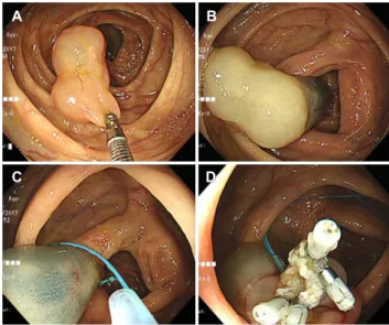
Fig. 1. Colonoscopic findings. (A) A 3 cm-sized, bluish-red polypoid mass was noted at the ileocecal valve. (B) The epinephrine solution diluted with normal saline was administered to the lesion. (C) A detachable snare was placed at the base of the stalk and a standard electrical snare was then used for polypectomy. (D) Hemoclips were used to prevent bleeding from the polypectomy site.
증례: 63세 여자 환자가 1년 전부터 발생한 간헐적인 복부 팽만감으로 내원하였다. 환자는 수년 전부터 고지혈증으로 약 을 복용하는 중이었으며 수술력은 없었고 음주 및 흡연은 하 지 않았다. 환자는 복통이나 흑색변, 혈변 등 특별히 호소하는 증상은 없었다. 내원 당시 신체 활력징후에서 혈압 110/70 mmHg, 맥박 69회/분, 호흡수 20회/분이었으며 복부에 만져 지는 종괴나 압통, 반발통은 없었다. 말초혈액 검사에서 백혈 구 4,100/μL, 혈색소 14.3 g/dL, 헤마토크리트 42.2%, 혈소판 172,000/μL였으며 생화학 검사에서 총 단백 7.8 g/dL, 알부 민 4.6 g/dL, 총 빌리루빈 0.9 mg/dL, 아스파르테이트아미노 전달효소(AST) 31 U/L, 알라닌아미노전달효소(ALT) 28 U/L, 혈액요소질소 18.6 mg/dL, 크레아티닌 0.8 mg/dL로 특이 소 견은 없었다. 환자는 내원 3년 전에 외부에서 시행한 대장 내 시경 검사에서는 특이 소견이 없었다고 하였다.
위 내시경에서 전정부에 미란성 위염이 있었으며 대장 내 시경에서 회맹판에 3 cm 크기의 푸르스름한 색상을 띤 목이 있는 폴립 형태의 병변이 관찰되었다(Fig. 1A). 복부 전산화 단층촬영에서는 근위부 상행 결장에 3 cm 크기를 가진 난원 형(ovoid)의 균질한 저음영 종괴가 관찰되었다(Fig. 2). 폴립 형 병변에서 시행한 조직 검사상 비특이적 소견을 보였으며
A B
C DD
Lee SH, et al. Endoscopic Resection of Colonic Polypoid Vascular Ectasia
371
Vol. 73 No. 6, June 2019
Fig. 3. Histopathological findings. (A) The specimen showed submucosal cystic dilation of the venous vessel associated with proliferation of the reactive focal muscle bundle (black arrow) (H&E, ×100). (B) It showed CD31-positive endothelial cells (black arrows) in the dilated blood vessels (CD31, ×400). (C) It showed D2-40-negative endothelial cells (black arrows) in the dilated blood vessels (D2-40, ×200).
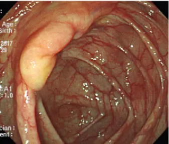
Fig. 4. Colonoscopic finding at 4 months after endoscopic polypectomy. A scar was noted at the ileocecal valve.
Fig. 2. Abdominopelvic computed tomography finding. Transverse plane (A) and coronal plane (B). An ovoid and low-density mass measuring about 3.0 cm in size (arrows) was noted in the proximal ascending colon.
낭종형 병변(cystic lesion)과도 감별이 필요하여 환자에게 양 성 병변의 가능성에 대하여 설명하였으나 환자가 병변을 제거 하기 원하여 병변에 대하여 내시경적 절제술을 시행하기로 하 였다. 출혈을 예방하기 위하여 병변의 목 부위에 생리식염수 로 1:10,000으로 희석한 에피네프린 용액을 약 5 mL 정도 주입하고 박리성 올가미를 이용하여 병변의 기저 부위를 결찰 하였다(Fig. 1B, C). 이후 올가미를 사용하여 병변을 제거하였 고 클립으로 절제 부위를 봉합하였다(Fig. 1D). 시술 후 환자 는 별다른 합병증 없이 퇴원하였다.
조직병리학적 소견에서 대장 점막하층의 혈관이 비정상적 으로 확장되어 있고 구불구불한 양상을 보였다(Fig. 3A). 또 한, 확장된 혈관에서 혈관 내피세포의 표지자인 CD31 양성 소견과 혈관 주위에 평활근 증식 소견을 보였으며, 림프관종 (lymphangioma)에서 보이는 림프관 내피세포 표지자인 D2-40 염색에는 음성 소견을 보여 혈관확장증(vascular ec- tasia)을 시사하였다(Fig. 3B, C). 4개월 후 시행한 추적 대장 내시경에서 회맹판의 시술 부위에는 절제술 후 반흔 소견을 A B
A B C
372
이상훈 등. 내시경적 절제술을 한 폴립양 대장 혈관확장증The Korean Journal of Gastroenterology
보였다(Fig. 4).
진단: 폴립 형태의 대장 혈관확장증
대장 혈관확장증은 혈관이형성증(angiodysplasia)이라고 도 하며 퇴행성 변화로 인하여 점막하층과 점막에 비정상적으 로 확장된 혈관을 수반한 상태로, 60세 이상의 연령에서 하부 위장관 출혈의 흔한 원인으로 보고되고 있다.1혈관확장증은 주로 맹장과 상행결장에서 호발하고 약 10%가 소장 말단부에 서 발견된다.2 내시경 소견은 일반적으로 편평하거나 약간 융 기된 붉은 병변으로 나타나며, 본 증례와 같은 목이 있는 폴립 형태는 국내외에 보고된 적이 있으나 극히 드물다.3,4
혈관확장증과 감별해야 할 질환은 혈관종, 동정맥기형 (arteriovenous malformation), 동맥경화성 동맥류, 림프관종 등이다. 혈관확장증은 동정맥기형으로도 알려져 있었으나 병 리학적 소견상 서로 다른 질환으로 보고되고 있다.5즉, 혈관 확장증은 점막층과 점막하층 내에 확장된 정맥, 세정맥, 모세 혈관으로 구성되며 고유근층은 침범하지 않는다. 이에 비하여 동정맥기형은 동맥과 정맥이 모세혈관을 통하지 않고 바로 연 결되는 병변으로 동맥과 세동맥을 포함하며 고유근층을 포함 한 장벽 전층을 침범한다.6따라서, 동정맥기형에서는 동맥의 특성인 내탄력층(internal elastic layer)이 있어 Verhoeff’s 탄력섬유(elastic fiber) 염색으로 염색이 되지만 혈관확장증 은 동맥 혈관이 보이지 않아 동정맥기형과 구별될 수 있다.
림프관종의 경우 내시경적으로 반투명한 백색의 상피하 종양 양상으로 보이며 절제 시 절단면에서 낭종액의 유출이 있고 병리 소견상 림프관 내피세포 표지자인 D2-40에 양성을 보인 다. 본 증례의 경우 병변의 모양이 이전에 보고된 폴립 형태의 혈관확장증 증례들과 비교하여 림프관종에 더 가까운 양상을 보였으나 면역조직화학 염색을 통하여 감별할 수 있었다.
혈관확장증의 치료는 만성적인 빈혈, 제한적인 출혈, 중증 의 만성 질환이 있는 경우에 시행하며 치료 방법은 병변의 크기와 형태에 따라 결정된다.7치료로는 대개 아르곤 플라즈 마 응고술, 열탐침, 응고소작술, 레이저 광응고술(laser pho- tocoagulation), 클립 등이 시행되며 폴립형 병변의 경우 폴
립절제술 또는 내시경 점막절제술을 적용할 수 있고 본 증례 와 같이 병변의 목의 두께가 5 mm 이상으로 두꺼운 경우에 는 박리성 올가미로 목의 기저부를 결찰한 다음 절제할 수 있다.8 병변의 크기가 크거나 대량 출혈이 동반되는 경우에는 수술적 절제가 고려된다.9 본 증례와 같이 혈관확장증은 특별 한 증상 없이 목이 있는 폴립 형태의 단일 병변으로 나타날 수 있으며 크기가 큰 경우 박리성 올가미를 이용하여 내시경 적 절제술로 안전하게 제거할 수 있다.
REFERENCES
1. Tada Y, Okamura S, Okita Y, et al. Vascular ectasia of the colon treated by argon plasma coagulation: report of a case. Dig Endosc 2001;13:37-40.
2. Koziara FJ, Brodmerkel GJ, Boylan JJ, Ciambotti GF, Agrawal RM.
Bleeding from polypoid colonic arteriovenous malformations.
Am J Gastroenterol 1996;91:584-586.
3. Yu BH, Shin SJ, Lee KW, et al. A large polypoid vascular ectasia removed by using a polypectomy with a detachable snare in an asymptomatic patient. Ann Coloproctol 2013;29:31-33.
4. Lin IT, Chang WH, Shih SC, et al. Successful endoscopic poly- pectomy for colonic vascular ectasia presenting as peduncu- lated polypoid lesion. Endoscopy 2007;39 Suppl 1:E253-E254.
5. Boley SJ, Sammartano R, Adams A, DiBiase A, Kleinhaus S, Sprayregen S. On the nature and etiology of vascular ectasias of the colon. Degenerative lesions of aging. Gastroenterology 1977;72(4 Pt 1):650-660.
6. Kim JW, Oh HC, Kim MK, Kim JG. Polypoid vascular and lymphatic malformation of the duodenum: a case report. J Gastrointestin Liver Dis 2010;19:85-88.
7. Roberts PL, Schoetz DJ Jr, Coller JA. Vascular ectasia. Diagnosis and treatment by colonoscopy. Am Surg 1988;54:56-59.
8. Nasseri-Moghaddam S, Mohamadnejad M, Malekzadeh R, Tavangar SM. Images of interest. Gastrointestinal: polypoid arte- riovenous malformation of the colon. J Gastroenterol Hepatol 2004;19:1419.
9. Gong EJ, Kim DH, Jung HY, et al. An arteriovenous malformation in the jejunum mimicking a gastrointestinal stromal tumor.
Korean J Gastroenterol 2014;63:42-46.