Korean J Gastroenterol Vol. 61 No. 5, 294-296 http://dx.doi.org/10.4166/kjg.2013.61.5.294 pISSN 1598-9992 eISSN 2233-6869
IMAGE OF THE MONTH
Korean J Gastroenterol, Vol. 61 No. 5, May 2013 www.kjg.or.kr
내부에 낭성 부분을 보인 상피하종양 형태의 십이지장 Brunner선 선종
최동욱, 박성철, 장세중, 이성준
강원대학교 의학전문대학원 내과학교실
Duodenal Brunner’s Gland Adenoma Presenting as Subepithelial Tumor with Internal Cystic Portion
Dong Wook Choi, Sung Chul Park, Sei Joong Chang and Sung Joon Lee
Department of Internal Medicine, Kangwon National University School of Medicine, Chuncheon, Korea
CC This is an open access article distributed under the terms of the Creative Commons Attribution Non-Commercial License (http://creativecommons.org/licenses/
by-nc/3.0) which permits unrestricted non-commercial use, distribution, and reproduction in any medium, provided the original work is properly cited.
교신저자: 박성철, 200-722, 춘천시 백령로 156, 강원대학교 의학전문대학원 강원대학교병원 내과학교실
Correspondence to: Sung Chul Park, Department of Internal Medicine, Kangwon National University Hospital, Kangwon National University School of Medicine, 156 Baengnyeong-ro, Chuncheon 200-722, Korea. Tel: +82-33-258-2405, Fax: +82-33-258-7146, E-mail: [email protected]
Financial support: None. Conflict of interest: None.
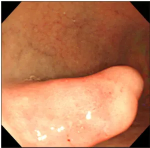
Fig. 1. Endoscopic finding. An elongated polypoid lesion with hyperemia and erosion was seen at the anterior wall of the duodenal bulb.
증례: 63세 여자 환자가 개인병원에서 시행한 상부위장관 내시경검사에서 십이지장에 용종형 병변이 발견되어 정밀 검 사를 위해 내원하였다. 환자는 수 년 전부터 고혈압 진단 후 투약 중이었으며, 복식 전 자궁절제술, 우측 갑상샘절제술을 시행받은 기왕력이 있었다. 음주 및 흡연은 하지 않았다. 환자 는 복통 등 특별히 호소하는 증상은 없었다. 내원 당시 신체활 력징후에서 혈압 130/80 mmHg, 맥박 70회/분, 호흡수 16회/
분이었으며 신체 검진에서 복부에 만져지는 종괴나 압통, 반 발통은 없었다. 말초 혈액검사에서 백혈구 5,200/mm3, 혈색 소 12.8 g/dL, 혈소판 244,000/mm3이었으며 혈청 생화학검 사에서 총 단백 6.7 g/dL, 알부민 3.6 g/dL, 총 빌리루빈 0.6 mg/dL, AST 24U/L, ALT 24U/L, 혈액 요소 질소 15.4 mg/dL, 크레아티닌 0.5 mg/dL로 특이소견은 없었다. 환자는 내원 4년 전에 본원에서 시행한 상부위장관 내시경검사에서 도 십이지장 구부에 용종형 병변이 있어 조직검사를 시행한 결과 만성 염증 소견을 보인 적이 있었다.
본원에서 시행한 상부위장관 내시경에서 십이지장 구부 전 벽에 20 mm 정도 크기의 기다란 모양의 종괴가 발견되었다 (Fig. 1). 종괴의 표면에는 발적을 동반한 미란이 관찰되었으 며 생검 겸자로 눌렀을 때에 매우 단단하지는 않았다. 4년 전 검사 당시와 비교하여 크기나 모양의 변화 여부는 뚜렷하지
않았다. 통상적인 소편 생검(pinch biopsy)으로 조직검사를 시행하였고 병리학적 결과는 만성 염증 소견 외에 특이소견은 관찰되지 않았다. 내시경초음파검사에서 내부에 낭성 부분을
Choi DW, et al. Duodenal Brunner’s Gland Adenoma as Subepithelial Tumor with Internal Cystic Portion
295
Vol. 61 No. 5, May 2013 Fig. 2. Endoscopic ultrasonography finding. In the submucosal layer,
about 2 cm sized elongated hyperechoic mass with internal cystic portion was noted.
Fig. 3. Computed tomography finding. In the coronal view, about 2 cm sized well-defined ovoid lesion of highly enhancement was seen at the duodenal bulb (arrow).
Fig. 4. Microscopic findings. (A) The resected specimen had internal cystic portion with thin fibrous wall (H&E, ×10). (B) It showed proliferation of Brunner’s gland covered with normal duodenal mucosa (H&E, ×40).
포함한 고 에코성의 병변이 제3층을 중심으로 보였으며(Fig. 2), 복부 CT에서는 십이지장 구부에 약 20 mm 크기의 조영 증강 이 잘되는 난원형 병변이 관찰되었다(Fig. 3). 크기가 20 mm 이상으로 상피하종양의 형태이며 일반적인 조직검사에서 확 진이 되지 않아서 십이지장 절개술 및 종양절제술을 시행하였 다. 절제된 표본의 육안적 크기는 32×17×15 mm였으며, 현 미경 소견에서 종양 내부에 낭성 변화가 관찰되어(Fig. 4A), 세포 기저에 핵이 놓여진 호산성의 깨끗한 세포질을 갖는 전 형적인 Brunner선 세포의 과다증식을 보이는 Brunner선 선 종으로 진단되었다(Fig. 4B). 환자는 수술 이후에 합병증 없이 퇴원하였다.
진단: 내부에 낭성 부분을 보인 상피하종양 형태의 십이지 장 Brunner선 선종
Brunner선은 십이지장의 점막하층에 주로 위치하며, 관상 선 구조로 알칼리성의 점액을 분비한다.1 이러한 점액은 위산 으로부터 십이지장을 보호하며 십이지장 내 알칼리성 환경을 조성하여 장내 소화효소의 활성화에 기여한다. Brunner선의 과다 증식에 의한 Brunner선 선종은 발병기전이 정확히 알려 져 있지 않으나, 앞서 언급한 위산의 중화와 연관되어 위산 노출이 자극이 되어 발생하는 것으로 추정하고 있다.2 또한 Helicobacter pylori 감염도 병태생리에 영향을 줄 것으로 여 겨진다.3
296
최동욱 등. 내부에 낭성 부분을 보인 상피하종양 형태의 십이지장 Brunner선 선종The Korean Journal of Gastroenterology
Levine 등4이 Brunner선 선종 27예를 분석한 결과에서 Brunner선 선종은 십이지장 구부에서 가장 호발하고 대개 유 경성 형태를 가지며 40-50대에 잘 생기고 남녀 성별은 차이가 없다고 하였다. Kang 등,5 Lee 등6의 국내 연구에서도 비슷한 결과를 제시하여 우리나라에서도 외국과 유사한 양상인 것으 로 보인다. 임상 증상은 대개 무증상이지만 증상을 보이는 경 우 복통, 복부 불편감, 오심, 포만감 등의 비특이적인 증상을 나타낼 수 있다.1 그러나 일부에서는 종양 표면에서의 궤양의 출혈로 인한 상부위장관 출혈 및 이로 인한 철결핍성 빈혈, 장폐색, 바터 팽대부 압박에 의한 황달 등 다양한 증상으로 나타나기도 한다.7
진단은 대개 상부위장관 조영술, 상부위장관 내시경검사 등에서 우연히 발견되어 확인되며, 복부 CT, 복부 초음파 등 여러 진단 방법들이 있으나 내시경 겸자를 이용한 조직검사로 확진할 수 있다. 그러나 Brunner선 선종은 정상 점막으로 덮 여 있으므로 점막이 두껍고 선종이 깊은 층에 위치하는 경우 나 조직검사가 까다로운 부위에서는 충분한 깊이에서 조직을 얻지 못하여 진단을 하지 못하는 경우가 있다.8 Brunner선 선 종의 진단에 내시경초음파도 도움이 되는데 점막 및 점막하층 에서 기원하며 경계가 불분명하고 종양 내 확장된 Brunner선 으로 인하여 단방성 또는 다방성의 낭종 형태로 보이거나 고 형 형태의 종양으로 보이는 등 다양한 반향성을 보이는 것이 특징이다.1 일반적으로 크기가 작고 증상이 없다면 별다른 치 료는 필요 없다고 알려져 있으나 드물게 악성화에 대한 보고 가 있어 치료에 대하여 논란의 여지가 있다.9,10 국내의 한 연 구에서는 종양의 정확한 진단 및 치료를 위해 가능하면 제거 할 것을 권고하였으며 우선적으로 내시경 치료를 고려하고 출 혈, 장폐색과 같은 합병증이 동반된 경우, 내시경 치료에 제한 이 있는 경우, 악성화 가능성이 높은 경우에는 수술 치료를 권고하였다.5 이번 증례의 경우 증상은 없으나 크기가 2 cm 이상으로 크고 조직검사에서 악성 여부에 대한 확인이 되지
않았으며 상피하종양의 형태를 띤 무경성이어서 십이지장 절 개술 및 종양절제술을 시행하여 Brunner선 선종을 확진하였 다. 이를 통해 점막하층 내에 고형이나 낭성 형태를 보이는 Brunner선 선종의 내시경초음파 소견의 특징을 확인할 수 있 었다.
REFERENCES
1. Patel ND, Levy AD, Mehrotra AK, Sobin LH. Brunner's gland hy- perplasia and hamartoma: imaging features with clinicopatho- logic correlation. AJR Am J Roentgenol 2006;187:715-722.
2. Peetz ME, Moseley HS. Brunner's gland hyperplasia. Am Surg 1989;55:474-477.
3. Kovacević I, Ljubicić N, Cupić H, et al. Helicobacter pylori in- fection in patients with Brunner's gland adenoma. Acta Med Croatica 2001;55:157-160.
4. Levine JA, Burgart LJ, Batts KP, Wang KK. Brunner's gland ha- martomas: clinical presentation and pathological features of 27 cases. Am J Gastroenterol 1995;90:290-294.
5. Kang JH, Lim YJ, Hahn SJ, Choi JS, Koh MS, Lee JH. Clinical char- acteristics of large brunner's gland tumors in Korea. Korean J Gastrointest Endosc 2010;40:297-302.
6. Lee SE, Hwang DW, Lim CS, et al. The clinical features and the clinical outcome of duodenal brunner's gland adenoma. Korean J Hepatobiliary Pancreat Surg 2008;12:284-286.
7. Gupta V, Gupta P, Jain A. Giant Brunner's gland adenoma of the duodenal bulb presenting with ampullary and duodenal ob- struction mimicking pancreatic malignancy. JOP 2011;12:413- 419.
8. Gao YP, Zhu JS, Zheng WJ. Brunner's gland adenoma of duode- num: a case report and literature review. World J Gastroenterol 2004;10:2616-2617.
9. Akino K, Kondo Y, Ueno A, et al. Carcinoma of duodenum arising from Brunner's gland. J Gastroenterol 2002;37:293-296.
10. Brookes MJ, Manjunatha S, Allen CA, Cox M. Malignant potential in a Brunner's gland hamartoma. Postgrad Med J 2003;79:416- 417.