Korean J Gastroenterol Vol. 65 No. 4, 258-260 http://dx.doi.org/10.4166/kjg.2015.65.4.258 pISSN 1598-9992 eISSN 2233-6869
IMAGE OF THE MONTH
Korean J Gastroenterol, Vol. 65 No. 4, April 2015 www.kjg.or.kr
방사선 치료 30년 후 진단된 직장의 만성 방사선결장염 및 직장골반루
김효선, 백광호
한림대학교 의과대학 춘천성심병원 내과학교실
Chronic Radiation Colitis with Rectopelvic Fistula Diagnosed 30 Years after Radiation Therapy
Hyo Sun Kim and Gwang Ho Baik
Department of Internal Medicine, Hallym University Chuncheon Sacred Heart Hospital, Hallym University College of Medicine, Chuncheon, Korea
CC This is an open access article distributed under the terms of the Creative Commons Attribution Non-Commercial License (http://creativecommons.org/licenses/
by-nc/3.0) which permits unrestricted non-commercial use, distribution, and reproduction in any medium, provided the original work is properly cited.
Copyright © 2015. Korean Society of Gastroenterology.
교신저자: 백광호, 200-704, 춘천시 삭주로 77, 한림대학교춘천성심병원 내과
Correspondence to: Gwang Ho Baik, Department of Internal Medicine, Hallym University Chuncheon Sacred Heart Hospital, 77 Sakju-ro, Chuncheon 200-704, Korea.
Tel: +82-33-240-5810, Fax: +82-33-255-4291, E-mail: [email protected] Financial support: None. Conflict of interest: None.
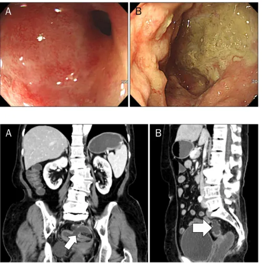
증례: 72세 여자 환자로 잦은 대변 배출 및 간헐적인 복통 을 주소로 개인의원에서 시행한 대장내시경검사 결과 직장S 자결장경계부에서 종괴가 발견되어 전원되었다. 환자는 자궁 경부암으로 30년 전 방사선치료를 받았던 과거력이 있었고 기저질환으로는 15년 전 진단받은 당뇨가 있었으며 흡연력과 음주력은 없는 상태였다. 입원 당시 활력징후는 혈압 120/60 mmHg, 맥박수 60회/분, 호흡수 20회/분, 체온 36.4oC로 측 정되었고, 신체검사에서 복부의 압통 등의 이상소견은 없었 다. 혈액검사에서 백혈구 7,960/mm3, 혈색소 13.4 g/dL, 혈 소판 481,000/mm3였고, 총 단백질은 5.9 g/dL, 알부민은 3.5 g/dL였다. 환자는 이전에 대장내시경검사를 받은 적이 없는 환자였고, 입원 당일 시행한 대장내시경검사에서는 항문피부 선에서 15 cm부터 20 cm까지 내강의 협착이 관찰되었고 주 변 점막의 부종과 확장된 모세혈관 및 궤양들이 관찰되었다 (Fig. 1A). 위내시경으로 교체한 후에 협착 부위를 통과한 후 정상 장점막이 관찰되지 않는 공간이 확인되었고 골반강으로 추정하였다. 골반강 추정 병변에서는 궤양을 동반한 불규칙한 점막과 주변의 삼출물이 관찰되었다(Fig. 1B). 이 부위에서 조 직검사를 시행하였고 병리검사 결과 육아조직과 궤양 소견을 보였으나 악성을 시사하는 소견은 보이지 않았다. 복부전산화
단층촬영에서도 직장S자결장에 장벽의 비후와 13 mm 크기 의 결손된 부위가 관찰되었으며(Fig. 2A) 주변에 액체저류가 관찰되었다(Fig. 2B). 직장S자결장 절제술(Hartmann’s oper- ation)을 시행하였고 병리검사 결과 직장의 천공과 농양 소견 을 보였으나 악성을 시사하는 소견은 없었다.
진단: 직장골반루를 동반한 직장의 만성 방사선결장염 최근 방사선 치료는 전체 암 환자의 70% 정도에서 시행되 고 있을 정도로 광범위하게 시행되고 있는 추세이지만, 복부 의 방사선 치료로 인한 위장관의 손상은 환자의 경제적인 부 담을 가중시키고 삶의 질을 떨어뜨릴 뿐만 아니라 치료 자체 에 있어서도 충분한 방사선 조사량을 투여하지 못하는 한계가 있다.1 방사선에 의한 조직 손상의 기전은 산화반응, 염증, 세 포자멸사, 그리고 유전자의 변형 등 복합적인 원인들이 작용 한다.2 복부의 방사선 조사는 장벽 전체에 걸쳐 위와 같은 손 상을 주게 되며 발생 시기에 따라 급성형과 만성형으로 나눌 수 있다. 급성형 방사선결장염은 수시간에서 수일 내에 발생 하여 수주 이내에 호전되며, 만성형은 2개월부터 길게는 10-30년 후까지도 보고되고 있다.3,4 동반될 수 있는 만성 합 병증들 중에는 장출혈, 장루, 그리고 장의 폐쇄 등이 있으며,5
Kim HS and Baik GH. Radiation Colitis 30 Years after Radiation Therapy
259
Vol. 65 No. 4, April 2015
Fig. 1. Endoscopic findings. (A) Diffuse edematous mucosa with capillary dilatation and luminal narrowing is seen on rectosigmoid juction. (B) Irregular pelvic wall with ulcers and pus-like material are observed when the scope is passed into the recto- pelvic fistula.
Fig. 2. Abdomen-pelvis computed tomography. (A) Diffuse wall thicken- ing is noted on rectosigmoid junction with 13-mm colonic wall defect (arrow). (B) Complicated fluid collec- tion (5.5 cm, arrow) is present around the pericolic area.
만성 방사선결장염의 빈도는 5-15% 정도로 발생할 수 있다는 보고가 있었으나6 정확한 발생률에 대한 대단위 연구는 아직 이루어지지 않은 상태이다. 만성 방사선결장염의 예방 방법에 대해 몇몇 연구들이 있는데, 방사선 조사 전 부데소나이드를 경구 투여하는 방법이 방사선 결장염의 예방에 효과적이라는 연구가 있으나 아직은 동물실험까지만 연구가 진행되어 있는 상태이고,7 골반의 방사선 치료 전 글루타민을 경구 투여하는 무작위 대조군 연구가 있으나 만성 방사선결장염에 예방효과 는 없다는 결과를 보였다.2
이 증례의 환자의 경우 30년이라는 긴 무증상 기간이 있었 고 검사 결과 만성 방사선 결장염의 전형적인 소견들에 부합 하는 장벽의 섬유화와 비후, 그로 인한 장의 협착, 모세혈관의 확장, 그리고 장루의 형성 등이 관찰되었다. 단, 이 환자의 경 우 직장암이 의심되어 전원되었고 골반의 방사선 치료와 연관 된 직장암도 여러 증례 보고들이 있었기 때문에8 입원 당시 직장암 가능성을 배제할 수는 없었다. 그러나 병리결과 악성 을 시사하는 소견은 없었기 때문에 결국 만성 방사선결장염으 로 진단을 하게 되었고, 장루가 동반되어 있었기 때문에 수술 을 통한 치료를 시행하였다.
이 증례는 오랜 시간이 경과된 치료받지 않은 만성 방사선 결장염이 장의 협착 및 누공까지 진행하는 자연경과를 볼 수 있었던 드문 증례이다. 또한 복부의 방사선 치료를 받은 환자 에게 암의 재발에 대한 추적관찰뿐만 아니라 방사선 치료 후 의 합병증에 대한 추적관찰의 중요성, 30년이라는 오랜 기간 이 지난 후에도 방사선결장염이 발생하여 진행할 수 있다는 가능성, 그리고 향후 만성결장염 예방을 위한 방법에 대한 추 가연구 필요성에 대해 생각해 볼 수 있는 흥미로운 증례로 생각되어 이 달의 영상으로 소개하고자 한다.
REFERENCES
1. Shadad AK, Sullivan FJ, Martin JD, Egan LJ. Gastrointestinal radi- ation injury: prevention and treatment. World J Gastroenterol 2013;19:199-208.
2. Vidal-Casariego A, Calleja-Fernández A, Cano-Rodríguez I, Cordido F, Ballesteros-Pomar MD. Effects of oral glutamine dur- ing abdominal radiotherapy on chronic radiation enteritis: a randomized controlled trial. Nutrition 2015;31:200-204.
3. Harb A, Abou Fadel C, Sharara A. Radiation enteritis. Curr Gastroenterol Rep 2014;16:383.
260
김효선, 백광호. 방사선 치료 30년 후 진단된 직장의 방사선결장염The Korean Journal of Gastroenterology 4. Sher ME, Bauer J. Radiation-induced enteropathy. Am J Gastro-
enterol 1990;85:121-128.
5. Coia LR, Myerson RJ, Tepper JE. Late effects of radiation therapy on the gastrointestinal tract. Int J Radiat Oncol Biol Phys 1995;31:1213-1236.
6. Abayomi J, Kirwan J, Hackett A, Bagnall G. A study to investigate women's experiences of radiation enteritis following radio-
therapy for cervical cancer. J Hum Nutr Diet 2005;18:353-363.
7. Odabasi M, Gokdemir S, Muftuoglu T, Aktekin A, Saglam A, Aker F. Prophylactic and therapeutic effects of oral budesonide for acute radiation-induced enteritis and colitis in rats. Int J Clin Exp Med 2014;7:940-946.
8. Tamai O, Nozato E, Miyazato H, et al. Radiation-associated rectal cancer: report of four cases. Dig Surg 1999;16:238-243.