Yeungnam Univ J Med 2017;34(2):279-284
18F-FDG PET/CT를 통해 진단된 주폐동맥 협착 소견의 폐동맥 육종
이훈희1, 박한빛1, 조윤경1, 안정민4, 이상민5, 이재승2,3, 김대희3,4
울산대학교 의과대학 서울아산병원 1내과, 2호흡기내과, 3폐 고혈압 정맥혈전 센터, 4심장내과, 5영상의학과
Pulmonary artery sarcoma manifesting as a main pulmonary artery stenosis diagnosed by 18F-FDG PET/CT
Hoonhee Lee1, Han-bit Park1, Yun Kyung Cho1, Jung-Min Ahn4, Sang Min Lee5, Jae Seung Lee2,3, Dae-Hee Kim3,4
Departments of
1Internal Medicine,
2Pulmonary and Critical Care Medicine,
3Pulmonary Hypertension and Venous Thrombosis Center,
4
Cardiology,
5Radiology, University of Ulsan College of Medicine, Asan Medical Center, Seoul, Korea
Pulmonary artery sarcoma (PAS) is a rare and fatal disease that often mimics chronic thromboembolic pul- monary hypertension (CTEPH); therefore, diagnosis of PAS is often delayed. Herein, a healthy 74-year-old man was presented with a 4-month history of dyspnea. Chest computed tomography showed wall thickening and stenosis in the main pulmonary artery as well as in both proximal pulmonary arteries. In order to diffe- rentiate between unusual CTEPH, vasculitis, and PAS, we performed right heart catheterization and pulmo- nary angiography. The mean pulmonary arterial pressure was 21 mmHg, and there was severe pulmonary artery stenosis. Thrombi on the pulmonary arterial wall lesions were observed in intravascular ultrasound and optical coherence tomography. Furthermore, the patient had a history of deep vein thrombosis. There- fore, we diagnosed unusual CTEPH. After 6 months of rivaroxaban anticoagulation therapy, a chest X-ray re- vealed a left lower lobe lung mass, and a positron emission tomography later showed hypermetabolic lesions in the main pulmonary artery wall, in both pulmonary arteries walls, in the lung parenchyma, and in the bones.
A biopsy of the right proximal humerus lesion revealed undifferentiated intimal sarcoma. Pulmonary sarcoma is rare, but should be considered when differentially diagnosing main pulmonary artery wall thickening and stenosis. A positron emission tomography may aid in this diagnosis.
Keywords: Pulmonary artery; Stenosis; Sarcoma; Positron emission tomography
Copyright ©2017 Yeungnam University College of Medicine
This is an Open Access article distributed under the terms of the Creative Commons Attribution Non-Commercial License (http://creative- commons.org/licenses/by-nc/4.0/) which permits unrestricted non-commercial use, distribution, and reproduction in any medium, provided the original work is properly cited.
Received: July 8, 2016, Revised: September 28, 2016 Accepted: October 4, 2016
Corresponding Author: Jae Seung Lee, Departments of Pulmonary and Critical Care Medicine, Pulmonary Hypertension and Venous Thrombosis Center, Asan Medical Center, University of Ulsan College of Medicine, 88 Olympic-ro 43-gil, Songpa-gu, Seoul 05505, Korea Tel: +82-2-3010-3994, Fax: +82-2-2045-4039
E-mail: [email protected]
서 론
원발성 폐동맥 육종(pulmonary artery sarcoma)은 드물지 만 예후가 불량한 질환으로, 완전 절제가 가능할 경우 평균 생존기간은 36개월이지만 절제가 불가능한 경우 11개월로 보고되었다[1]. 폐색전증과 임상양상이 유사하여 항응고제 에 호전이 없어 뒤늦게 진단이 되는 사례가 많으며, 이외에도
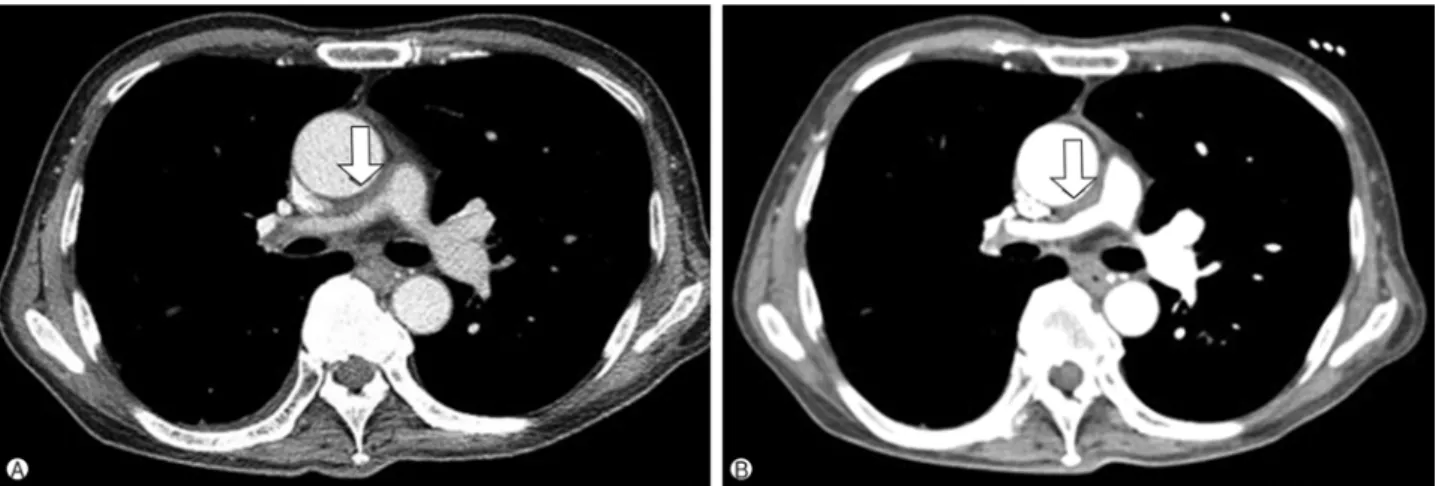
Fig. 1. Chest computed tomography showing (A) wall thickening and stenosis in the main pulmonary artery and in both proximal pulmonary arteries (right>left) and (B) no interval change after 4 months (arrow).
폐동맥 협착에 의한 폐동맥 고혈압을 일으키는 폐동맥 혈관 염, 섬유화 종격염 등과 같은 다양한 질환에서 감별해야 할 질환이다[2]. 국내에도 증례 형식으로 몇 차례 보고된 적 있 으나 대부분 폐색전증으로 오인된 폐동맥 육종 증례였다.
그러나 현재까지 국내에서 주폐동맥 협착으로 발현했던 원 발성 폐동맥 육종 사례는 보고된 적 없다. 이에 저자들은 주폐 동맥 협착에서부터 진단된 폐동맥 육종 1예를 통해 그 질병 의 발현 양상 및 진단에 도움이 되었던 검사들에 대해 알리고 자 한다.
증 례
환 자: 남자, 74세
주 소: 내원 4개월 전부터 발생한 호흡곤란
현병력: 내원 12년 전 왼쪽 다리의 심부정맥혈전증으로 치료 받은 병력이 있던 자로 4개월 전에 호흡곤란이 발생해 서 본원 외래로 내원하였다. 당시 흉부 조영증강 컴퓨터단층 촬영(computed tomography, CT)을 시행했고, 폐 실질의 이상 소견은 없었지만 폐 구간(pulmonary trunk)인 주폐동맥에서 부터 양측 근위부 폐동맥의 두께가 증가되어 있었고, 이로 인한 협착이 우측에서 더 심하게 확인되었다. 흉부경유심초 음파에서는 삼첨판역류 최대속도 3.2 m/s, 우심실 수축기 압 력 50 mmHg로 경증 폐동맥 고혈압 외 다른 소견은 정상이 었다. 지속되는 호흡곤란 및 폐동맥 협착에 대한 원인을 평가 하기 위해서 입원하였다.
과거력 및 사회력: 46년 전 결핵신장염으로 우측 신장절제 술을 받았고, 20년 전 진단받은 고혈압으로 항고혈압제를 복용 중이었다. 매주 소주 1잔씩 50년 동안 음주하고 있었으
며, 하루 0.5갑씩 50년 동안 흡연하였고, 10년 전부터 금연 중이었다.
가족력: 특이사항 없었다.
신체 검사: 혈압 116/79 mmHg, 맥박수 85회/분, 호흡수 22회/분, 체온 36.6℃였고, 맥박산소측정기로 측정한 산소포 화도는 92%였다. 다른 신체검사상 특이 소견은 없었다.
혈액검사 소견: 혈액요소질소 28mg/dL, 혈액크레아티닌 1.41 mg/dL, 사구체여과율(glomerular filtration rate) 49 mL/min/
1.73m2의 경도 신기능 저하가 관찰되었다. 동맥혈가스분석 에서는 pH 7.407, PaCO2 30.2 mmHg, PaO2 77.4 mmHg, HCO3- 19.2 mEq/L, SaO2 95.6%로 측정되었다. 호흡성알 칼리증 소견과 함께 폐포동맥간산소분압차(alveolar-arterial oxygen gradient)는 35 mmHg로 증가되어 있었다. 기타 다른 이상소견은 관찰되지 않았다.
흉부영상 소견: 흉부 조영증강 CT를 다시 시행하여 4개월 전 검사 결과와 비교하였다. 양측 폐동맥의 두꺼워진 혈관벽 및 협착 병변은 큰 변화 없었다(Fig. 1).
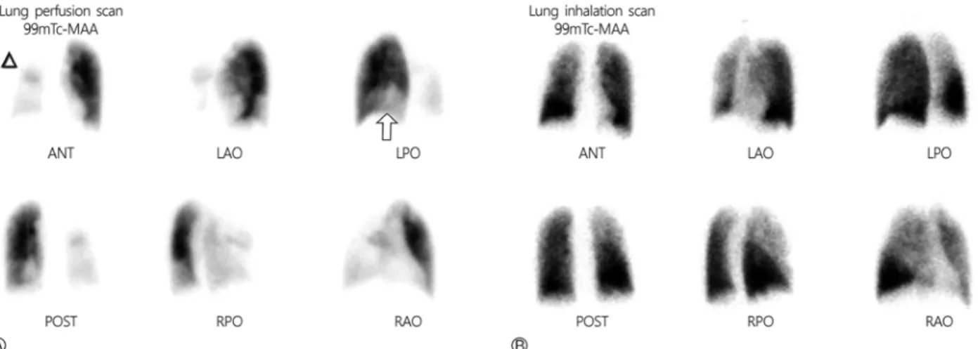
폐환기-관류 스캔(ventilation-perfusion scan): 우상엽 꼭대 기 뒤구역(apicoposterior segment), 우중엽 내측구역 및 가측 구역(medial, lateral segments)에서 큰 관류결손(large perfu- sion defect)이 관찰되었다. 이 외 우상엽 및 좌하엽의 전방구역 (anterior segment), 우하엽, 좌하엽의 바닥구역(basal segment) 에서 관류감소가 관찰되었다. 환기 스캔에서는 양측 폐에서 환기 결손이 관찰되지 않았다(Fig. 2).
치료 및 경과: 주폐동맥의 혈관벽 두께 증가 및 협착 병변 에 대한 감별진단으로 폐동맥 혈관염, 비전형적(unusual) CTEPH, 폐동맥 육종과 같은 악성질환을 고려했다. 베흐체 트병, 다카야스병과 같은 혈관염을 배제하기 위해서 항핵항
Fig. 2. (A) Lung perfusion scan showing severe perfusion defects in the right upper lobe (apical and posterior segments), and in the right middle lobe (medial and lateral segments), as well as a large perfusion decrement in the right upper lobe anterior segment, and in the right lower lobe (arrow head) and in the left lower lobe basal segments (arrow). (B) Technetium-99m sodium pertechnegas lung inhalation scan showing no ventilation defect.
Fig. 3. (A) Pulmonary angiography showing 90% diffuse stenosis in the proximal right pulmonary artery (arrow) and 20% stenosis in the left pulmonary artery (arrow head). (B) Intravascular ultrasound and (C) optical coherence tomography showing a well-organized thrombus in the wall of the proximal right pulmonary artery (arrow).
체검사(antinuclear antibody), 항중성구세포질항체검사(anti- neutrophil cytoplasmic antibody), 적혈구침강속도(erythro- cyte sedimentation rate), C-반응단백질(C-reactive protein) 검사를 시행했으나, 검사 결과는 정상 범위에 있었고, 혈관염 을 시사하는 기타 다른 임상 소견은 없었다. 폐동맥 협착 병변에 대한 조직 검사는 폐동맥 파열과 같은 시술 관련 합병 증의 위험이 높아 시행하지 않았고, 대안으로 우측 심장도관 삽입 및 폐동맥 혈관조영술을 시행하기로 하였다. 또한 폐동 맥의 혈관벽 두께 증가 소견에 대해서 혈관 내 초음파(intra- vascular ultrasound) 및 광간섭단층촬영(optical coherence tomography)을 시행했다.
폐동맥 혈관 조영술에서 우측 근위부 폐동맥의 90% 협착 과 함께 우상엽 폐동맥 분지, 우하엽 폐동맥 분지의 완전 폐쇄 및 좌측 폐동맥의 20% 협착이 관찰되었으며, 심장도관삽입
검사에서 평균 폐동맥 압력은 21 mmHg로 측정되었다. 우측 근위부 폐동맥 협착에 대한 혈관 내 초음파 및 광간섭단층촬 영 검사를 시행했는데, 표면이 비교적 규칙적이고 잘 기질화 된 혈전(well organized thrombus)이 관찰되었다(Fig. 3).
환자의 심부정맥혈전증 병력 및 혈관 내 초음파와 광간섭 단층촬영 검사를 통해 확인된 혈전, 그리고 평균 폐동맥 압력 이 21 mmHg로 측정되어 정상 범위 이상인 점을 모두 함께 종합하여 비전형적(unusual) CTEHP으로 진단했다. 항응고 제 rivaroxaban을 투약하며 경과관찰을 계획하고 퇴원하였 다. 퇴원 후 3개월 째 특별한 변화 없었으나, 6개월 째 흉부X 선에서 좌하엽에 3 cm 크기의 종괴가 새롭게 발견되어 재입 원했다.
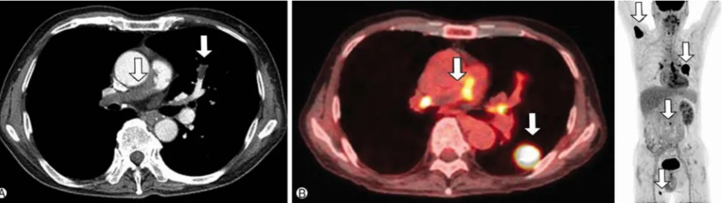
이어 시행한 흉부 조영증강 CT에서 좌하엽으로 3 cm 크기 의 원형 종괴가 보였고, 우측 폐동맥의 협착이 더 악화되었으
Fig. 4. (A) Chest computed tomography showing aggravated wall thickening with stenosis in the main pulmonary artery and newly developed filling defect in the anterior segmental pulmonary artery of the left upper lobe (arrows), compared with the previous image, taken 6 months ago. (B) Whole body positron-emission tomography/computed tomography showing hypermetabolic lesions (arrows) in the left lower lobe mass (maximum standardized uptake values [maxSUV]: 30.8), bilateral pulmonary arteries (maxSUV: 9.6), right humeral head (maxSUV: 22.2), L3 body (maxSUV: 8.0), and right vastus medialis (maxSUV: 14.0), suggesting malignancy.
Fig. 5. (A) Biopsy of the right humeral head showing marked nuclear pleomorphism and spindle-like cell morphology, indicating an undifferentiated spindle-cell malignancy, most likely a metastatic intimal sarcoma (H&E stain). (B) The immunohistochemical stainings showing all negative results (immunohistochemical stain).
며, 좌상엽 전방 폐동맥의 충만 결손이 새롭게 확인되었다.
좌하엽 폐 종괴는 악성 종양 가능성이 높아 양전자방출컴퓨터 단층촬영(18F-fluorodeoxyglucose positron emission tomo- graphy/computed tomography, 18F-FDG PET/CT)을 시행했 다. 우심실 출구부터 양측 폐동맥에 이르기까지 FDG 섭취율 이 높은 혈관 내 병변과 함께 좌하엽, 좌상엽으로 FDG 섭취 율이 높은 종괴가 확인되었다. 이외에도 우측 상완골 근위부, 제 3허리 척추체, 우측 내측광근(vastus medialis)에서 전이가 의심되는 병변이 관찰되었다(Fig. 4).
흉부경유심초음파에서 삼첨판역류 최대속도 3.9 m/s, 우심 실 수축기 압력 70 mmHg로 폐동맥 고혈압이 악화되었다.
우측 상완골 근위부 병변에서 조직검사를 시행했고, 미분 화 내막육종(undifferentiated intimal sarcoma)이 확인되었다
(Fig. 5). 중증 폐동맥 고혈압에 대한 완화 치료를 위해 폐동 맥 폐쇄 병변에 완화 방사선 치료를 먼저 시행하기로 하였고, 이후 항암치료를 계획하였다. 진단 1주일 뒤에 폐동맥에 대한 완화 방사선 치료를 시행했고, 골절 위험이 높은 우측 상완골 근위부 병변에 대해서도 방사선 치료를 함께 시행했다. 방사선 치료 종료 2주일 뒤부터 전신 항암치료를 시행했다. 2개월 간 3주기의 항암치료 이후 종양의 크기가 감소되었지만 환자 의 활동도(performance status)가 악화되어 항암치료를 중단 하고 현재 경과 관찰 중이다.
고 찰
주폐동맥 협착은 선천성 질환과 후천성 질환으로 크게 나
뉘며, 후천성 질환의 원인 질환에는 섬유화 종격염(fibrosing mediastinitis), 결합조직병(connective tissue disease), 만성 폐혈 전색전증 및 드물게 폐동맥 육종과 같은 악성 질환이 있다.
이번 증례에서 섬유화 종격염은 흉부 조영증강 CT에서 종격 의 이상 병변이 관찰되지 않았기 때문에 배제할 수 있었다[3].
혈관염, 사르코이드증과 같은 결합조직병에 대해서는 이와 관련된 다른 임상 소견이 없었고, 혈액 검사에서 항핵항체검 사, 항중성구세포질항체, 적혈구침강속도, C-반응단백질 검 사가 음성으로 나왔기에 결합조직병의 가능성은 낮았다. 악성 질환을 배제하기 위한 폐동맥 조직검사는 합병증 위험이 높 았기에 폐동맥의 혈관벽 두께 증가 및 협착 병변에 대한 평가 를 위해 혈관 내 초음파 및 광간섭단층촬영을 시행했다. 이 두 검사 방법은 만성 폐혈전색전증에서 혈관 내 혈전을 가시화 하여 진단을 내리는데 도움이 된다고 알려져 있다[4,5]. 이를 통해서 확인된 혈관 내 혈전 및 심부정맥혈전증의 과거력으 로 인하여 CTEPH으로 오인하여 진단이 어려웠던 증례이다.
6개월 뒤 전이를 동반한 질환으로 진행했고, 18F-FDG PET/
CT에서 FDG 섭취율이 높은 병변들이 관찰되었는데, 이러한 소견은 폐동맥 육종을 포함한 악성 질환을 시사하는 소견이다.
원발성 폐동맥 육종은 드물지만 평균 생존기간이 6개월에 서 2년 정도로 예후가 매우 불량한 질환이다. 수술을 통한 종양의 완전 적출이 어려울 경우 질환의 예후는 더욱 불량하 다[1]. 이번 증례의 경우 첫 입원 당시에 원격 전이가 없는 상태에서 진단이 되어 종양의 수술적 절제가 가능했다면 환 자의 예후가 더 개선되었을 것이다. 임상양상은 폐동맥 폐쇄 로 인해 폐동맥 고혈압이 발생하게 되는데 대부분의 환자들 이 호흡곤란을 호소한다. 이와 비슷한 임상양상을 보이는 폐혈전색전증, 폐동맥 혈관염, 섬유화 종격염, 악성 종양 등 을 감별하여야 한다.
폐혈전색적증에 비해 폐동맥 육종을 시사하는 임상소견은 발열, 체중 감소 등의 전신 증상, 항응고제에 반응이 없는 경우, 그리고 주폐동맥 혹은 양측 폐동맥을 동시에 침범하는 경우 등이 있다[6]. 흉부 조영증강 CT에서 주폐동맥 혹은 근 위부 폐동맥 전체에서 저음영 충만 결손과 함께 분절 폐동맥 으로 충만 결손이 진행하는 소견, 폐동맥 팽창 소견, 그리고 혈관벽 밖으로 진행된 소견은 폐동맥 육종에서 더 흔한 소견 이다[7]. 그리고 폐동맥 육종에서 조영 이후 Hounsfield units 과 요오드 농도가 더 높은 점도 진단에 도움이 된다[8]. CT 이외 폐동맥 육종 진단에 도움이 되는 검사 소견은 다음과 같다. 가돌리늄 조영증강 자기공명영상에서는 단순 혈전에 비해 폐동맥 육종에서 조영증강이 더 잘된다[1]. 혈관 내 초
음파를 시행할 경우 폐동맥 육종과 같은 혈관 내 종양에서 불규칙한 표면, 끈 모양의 돌출 소견, 모자이크 패턴을 보이 는 에코발생덩이(echogenic mass) 소견을 보일 수 있다[4].
본 증례의 경우 혈관 내 초음파에서 위와 같은 소견들이 없이 잘 기질화된 에코발생덩이(well orgniazed echogenic mass) 소견이 확인되어 혈전으로 판단 후 CTEPH으로 진단되었다.
광간섭단층촬영은 높은 해상력의 영상을 통해 폐혈관을 가 시화할 수 있는데, 이를 병리 소견과 비교, 분석하여 폐고혈 압의 감별 진단 및 폐암 진단에 있어서의 역할에 대해 연구가 이루어지고 있다[9-12]. 하지만 폐동맥 육종에서의 광간섭단 층촬영 소견에 대해서는 아직까지 보고된 자료가 없다. 18F- FDG PET/CT에서는 단순 혈전에 비해 폐동맥 육종에서 FDG 최대 standardized uptake value가 유의하게 높다[13,14]. 이 러한 검사 소견은 이번 증례에서 폐동맥 육종의 진단을 내리 는 데에 보조적인 역할을 한다.
주폐동맥의 혈관벽 두께 증가 및 협착 병변이 보일 때 원발 성 폐동맥 육종은 드물지만 반드시 감별이 필요한 중요한 질환이다. 그리고 감별 진단 과정에서 18F-FDG PET/CT는 폐동맥 육종과 같은 악성 종양 진단에 있어서 유용한 검사이다.
CONFLICT OF INTEREST
No potential conflict of interest relevant to this article was reported.
ORCID
Hoonhee Lee, https://orcid.org/0000-0003-2926-2197
REFERENCES