290 Received:April 27, 2020, Revised:May 22, 2020, Accepted:June 4, 2020 Corresponding to:Jung Hee Koh http://orcid.org/0000-0002-6617-1449
Division of Rheumatology, Department of Internal Medicine, Bucheon St. Mary’s Hospital, College of Medicine, The Catholic University of Korea, 327 Sosa-ro, Wonmi-gu, Bucheon 14647, Korea. E-mail:[email protected]
Copyright ⓒ 2020 by The Korean College of Rheumatology. All rights reserved.
This is an Open Access article, which permits unrestricted non-commerical use, distribution, and reproduction in any medium, provided the original work is properly cited.
Clinical Image
pISSN: 2093-940X, eISSN: 2233-4718
Journal of Rheumatic Diseases Vol. 27, No. 4, October, 2020 https://doi.org/10.4078/jrd.2020.27.4.290
Pneumatosis Intestinalis and Pneumomediastinum in Patients With Granulomatosis With Polyangiitis
Aran Kim, M.D.1, Jung Hee Koh, M.D., Ph.D.2
1Division of Rheumatology, Department of Internal Medicine, Pusan National University Hospital, Busan, 2Division of Rheumatology, Department of Internal Medicine, Bucheon St. Mary’s Hospital, College of Medicine, The Catholic University of Korea, Seoul, Korea
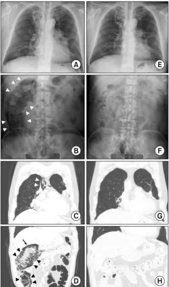
A 66-year-old male patient presented with dyspnea and blood tinged sputum for 2 weeks. He had a 4-month his- tory of granulomatosis with polyangiitis, with protei- nase-3 antineutrophil cytoplasmic antibodies (ANCA), involving the lungs, left orbit, and nasopharynx. He had received 3 courses of monthly intravenous cyclo- phosphamide (cumulative dose: 2.25 g). Prednisolone 25 mg per day (cumulative dose: 680 mg) and trimetho- prim/sulfamethoxazole at a prophylactic dose had been prescribed. Plain chest and abdomen radiographs and a chest computed tomography (CT) scan revealed in- creased size of the cavitary lung mass, nodular con- solidation, and new onset of pneumomediastinum and pneumoperitoneum (Figure 1A∼C). Subsequent ab- dominal CT images with the lung window setting showed a large amount of intramural air on the wall of the ascend- ing colon, and pneumatosis intestinalis (PI) was diag- nosed (Figure 1D). The patient did not complain of ab- dominal pain. Intravenous antibiotics along with fasting, fluid supplementation, and oxygen therapy was main- tained for 3 days. However, the amount of intramural air on the ascending colon and pneumomediastinum did not change in follow-up plain radiographs. Meanwhile, the presence of blood tinged sputum continued, and the pro- teinase-3 ANCA titer (cut-off 5 U/ml; Orgentec, Mainz, Germany) was elevated compared to a previous study (from 19.0 U/ml to 61.3 U/ml). All blood and sputum culture sets were reported as “no growth.” The pre- dnisolone dose was increased to 1 mg/kg, leading to grad- ual improvement of intramural air on the ascending colon and blood tinged sputum. The patient was able to tolerate
a regular diet and was discharged in stable condition. An additional set of plain radiographs of the chest and abdo- men and CT scan obtained 4 weeks later revealed that pneumomediastinum and intestinal gas accumulation had completely resolved (Figure 1E∼H).
PI is defined as the presence of gas within the wall of the gastrointestinal tract [1]. It has been reported in various pulmonary, intestinal bowel, infectious, and autoimmune diseases [2]. In addition, medication-associated or post- operative etiologies have been described [2]. Treatment with glucocorticoid could be associated with develop- ment of PI in patients with ANCA-associated vasculitis [3]. In this case, PI appeared simultaneously with he- moptysis, which is considered an aggravation of gran- ulomatosis with polyangiitis, over the course of pre- dnisolone tapering. The rapid radiographic improvement after increasing the dose of prednisolone, without evi- dence of infection in this patient, supports the hypothesis that PI was associated with exacerbation of vasculitis. A vasculitis-based pathogenesis within the intestinal terri- tory has been proposed as a cause for development of PI among patients with systemic lupus erythematosus and systemic necrotizing vasculitis [4,5]. While many pa- tients with autoimmune disease who develop PI respond to conservative therapy and have a benign course, it is prudent for rheumatologists caring for such patients to consider PI in the differential diagnosis. The treatment decision of PI should be based on the patient’s clinical fea- tures and underlying etiologies since treatment of the un- derlying condition often improves PI, while glucocorti- coids can often exacerbate PI.
Pneumatosis Intestinalis in GPA
www.jrd.or.kr 291
Figure 1. Plain radiographs and computed tomography (CT) scans at the time of pneumatosis intestinalis and follow-up af- ter 4 weeks from the onset. Chest radiograph on admission (A).
Supine abdominal radiograph showing linear pneumatosis in- testinalis of the colon (arrowheads) (B). The coronal CT scan (C) of the chest shows pneumomediastinum (arrowheads). The coronal abdominal CT images using the lung window setting (D) show free air (long arrows) and colon pneumatosis in- testinalis (arrowheads). Follow-up plain radiographs (E and F) and CT scan using the lung window setting (G and H) show the complete resolution of pneumomediastinum and pneuma- tosis intestinalis.
CONFLICT OF INTEREST
No potential conflict of interest relevant to this article was reported.
AUTHOR CONTRIBUTIONS
J.H.K. had full access to all data in the study and takes re- sponsibility for the integrity of the data and accuracy of the data analysis. J.H.K. and A.K. contributed to the study concept, design, data acquisition, analysis and interpretation.
J.H.K. and A.K. draft the manuscript and contributed to the critical revision. All authors read and approved the fi- nal manuscript.
REFERENCES
1. Ho LM, Paulson EK, Thompson WM. Pneumatosis in- testinalis in the adult: benign to life-threatening causes.
AJR Am J Roentgenol 2007;188:1604-13.
2. Khalil PN, Huber-Wagner S, Ladurner R, Kleespies A, Siebeck M, Mutschler W, et al. Natural history, clinical pat- tern, and surgical considerations of pneumatosis intestinalis.
Eur J Med Res 2009;14:231-9.
3. Nakagawa S, Akimoto T, Takeda S, Okada M, Miki A, Yamamoto H, et al. Antineutrophil cytoplasmic antibody- associated glomerulonephritis complicated by pneumatosis intestinalis. Clin Med Insights Case Rep 2015;8:65-70.
4. Mizoguchi F, Nanki T, Miyasaka N. Pneumatosis cystoides intestinalis following lupus enteritis and peritonitis. Intern Med 2008;47:1267-71.
5. Pagnoux C, Mahr A, Cohen P, Guillevin L. Presentation and outcome of gastrointestinal involvement in systemic ne- crotizing vasculitides: analysis of 62 patients with poly- arteritis nodosa, microscopic polyangiitis, Wegener gran- ulomatosis, Churg-Strauss syndrome, or rheumatoid ar- thritis-associated vasculitis. Medicine (Baltimore) 2005;84:
115‐28.