207
Effect of adrenalectomy on gene expression of adrenoceptor subtypes in the hypothalamic paraventricular nucleus
Kyung-Yoon Kam1, Seung Yub Shin, Seong Kyu Han2, Long Hua Li, Wonee Chong, Dae Hyun Baek, So Yeong Lee, and Pan Dong Ryu*
Department of Pharmacology, College of Veterinary Medicine and School of Agricultural Biotechnology, Seoul National University, Seoul 151-742, Korea
OAccepted: May 20, 2004)
Abstract : It is well known that the hypothalamic-pituitary-adrenocortical (HPA) axis is under the negative feedback control of adrenal corticosteroids. Previous studies have suggested that glucocorticoids can regulate neuroendocrine cells in the paraventricular nucleus (PVN) by modulating catecholaminergic transmission, a major excitatory modulator of the HPA axis at the hypothalamic level. But, the effects of corticosteroids on the expression of adrenoceptor subtypes are not fully understood. In this work, we examined mRNA levels of six adrenoceptor subtypes (α1A, α1B, α2A, α2B, β1 and β2) in the PVN of normal and adrenalectomized (ADX) rats. Total RNA (2.5 µg) was extracted from PVN micropunches of brain slices (500 µm) and analyzed by reverse transcription-polymerase chain reaction (RT-PCR). The levels of corticotropin-releasing hormone (CRH) mRNA were increased in the ADX rats relative to normal rats, indicating that the PVN had been liberated from the negative feedback of corticosteroids. Among the six adrenoceptor subtypes examined, mRNA levels for α1B- and β1-adrenoceptors were increased, but the level for β2-adrenoceptors was decreased in the ADX rats. The mRNA levels for the other three subtypes and for the general and neuronal specific housekeeping genes, glyceroaldehyde-3-phosphate dehydrogenase (GAPDH) and N-enolase, respectively, were not changed in the ADX rats. In conclusion, the results indicate that adrenal steroids selectively regulate the gene expression of adrenoceptor subtypes in the PVN.
Key words : corticosteroid, RT-PCR, micropunch, corticotropin-releasing hormone
Introduction
The hypothalamic-pituitary-adrenocortical (HPA) axis is critical to an animal’s response to stress. Parvocellular secretory neurons of the hypothalamic paraventricular nucleus (PVN) in the HPA axis release corticotropin- releasing hormone (CRH) into the portal circulation and thereby induce the release of adrenocorticotropin from corticotrophs in the anterior pituitary. Adrenocorticotropin subsequently stimulates the release of glucocorticoids from the adrenal cortex.
Among the neuronal systems involved in the central control of the HPA axis [16], the catecholaminergic
system appears to play a major role [12, 23, 30, 39]
in mediating various stress responses. At receptor level, adrenergic α1, α2 [4] and α1B receptors [8] have been identified in CRH neurons of the PVN. The presence of β receptor in the PVN has also been demonstrated by radio-ligand binding assay [24]. However, the effects of specific subtypes of adrenoceptors and their modulations by glucocorticoids are not fully understood yet.
The activity of PVN neurons in the HPA axis is under the close control of corticosterones from the adrenal gland. For example, CRH-containing neurons in the PVN are negatively regulated by microiontophoresed
*Corresponding author: Pan Dong Ryu
Department of Pharmacology, College of Veterinary Medicine, Seoul National University, Seoul 151-742, Korea [Tel: +82-2-880-1254, Fax: +82-2-879-0378, E-mail: [email protected]]
Current address :1Division of Endocrinology, Diabetes and Hypertension, Brigham and Women’s Hospital and Harvard Medical School, 221 Longwood Avenue, Boston, MA, 02115, USA
2Center for Neuroendocrinology and Department of Physiology, School of Medical Sciences, The University of Otago, Dunedin, New Zealand
glucocorticoids [34], whereas the number of CRH- immunoreactive neurons in the PVN is increased by adrenalectomy, as are the CRH mRNA levels and CRH synthesis or release rates [35]. It is known that the function and gene expression of different subtypes of α- and β-adrenoceptors are affected by either adrenalectomy or chronic glucocorticoid administration in cultured hypothalamic slices [10] and non-neuronal cells [14, 32].
Therefore, the modulation of gene expression of adrenoceptor subtypes in the PVN by corticosteroids and consequent shifts in adrenoceptor density in this region may be an important mechanism in the regulation of HPA activity. However, the effects of corticosteroids on the gene expression of adrenoceptor subtypes are not well understood at the PVN level yet. The aim of the present study is to examine whether the removal of corticosteroids by adrenalectomy affects the gene expression in the PVN of the following six adrenoceptor subtypes: α1A, α1B,α2A,α2B,β1 and β2.
Materials and Methods
Animals
Animal experiments were carried out according to the protocol for the care and use of animals approved by the Laboratory Animal Care Advisory Committee of Seoul National University. Male Sprague-Dawley rats (4 weeks old) were housed under the condition of constant temperature and humidity, on a 12 hr light/
dark cycle (lights on at 08:00 h), with ad libitum access to food and water. Animals were adrenalectomized (ADX) bilaterally under light ether anesthesia, and housed two or three per cage, with drinking water replaced with a solution containing 0.9% saline and 5% dextrose. Animals were sacrificed by decapitation 7 days after surgery [8]. Brains were rapidly removed, frozen in isopentane cooled to -40 to -50oC and stored at -70oC. Trunk blood was collected and serum separated by centrifugation (3,000 g, 10 min). The sera were stored at -70oC until corticosterone radioimmunoassay.
Corticosterone radioimmunoassay
The serum corticosterone was measured by double antibody radioimmunoassay using a rat corticosterone kit (ICN Pharmaceuticals, Inc. USA) according to the manufacturer’s manual. Assay sensitivity was 5 ng/ml.
All concentrations were measured in duplicate.
Brain section and micropunch
The brain micropunch method followed the protocols of Palkovits et al. [27]. Briefly, frozen brains were sliced into 500 µm thick coronal sections in a cryostat (Leica, USA) at -10oC. The PVN micropunches were obtained bilaterally by using a blunted needle (19G, i.d.
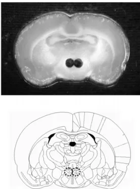
1.1 mm). Samples were collected in polypropylene tubes and stored at -70oC. Fig. 1 shows a representative thawed section immediately after the removal of micropunches.
Total RNA extraction and reverse transcription- polymerase chain reaction (RT-PCR)
Total RNA was extracted by RNA extraction kit (iNtron Co. Korea) according to the manufacturer’s manual. Briefly, a 500 µl of RNA extraction buffer was added to the tube containing micropunch samples and
Fig. 1. Photograph (upper panel) and brain map (lower panel) showing microdissection of the paraventricular nucleus.
Brains were removed immediately following decapitation and frozen in isopentane. Sections (500 µm) were then cut on a cryostat and mounted onto glass slides. Punching was done on a frozen stage with 19G blunted needles. The circles (upper panel) represent punches microdissected out from rat brain sections and the dotted circles (lower panel) indicate the paraventricular nucleus area with the corresponding diameter.
the mixture was sonicated. Subsequently, 100 µl of chloroform was added, allowed to sit for 15 min on ice, and then spun at 14,000 g for 10 min to recover only the aqueous phase. Then, 0.2 µg of glycogen and 250 µl of isopropanol were added and the recovered RNA pellets following centrifugation at 14,000 g were washed twice with 70% ethanol and reconstituted in 10 µl RNase-free water. The RNA content was measured with an ultraviolet spectrophotometer. Only those RNA samples with a 260/280 ratio above 1.6 were used and 2.5 µg of total RNA was used in the RT reaction. First strand cDNA was synthesized for 1 h at 37oC in a total reaction volume of 25 µl containing 4 µM random hexamer primers (Takara, Japan), 10 mM dithiothreitol, 0.2 mM each of four deoxyribonucleotide triphosphates (dNTP; Takara), 15 U of ribonuclease inhibitor (Takara) and 100 U of reverse transcriptase (MMLV-RT; Gibco- BRL, USA). The cDNA was kept at -20oC until PCR amplification. Following RT reaction, the cDNA template was PCR amplified using the corresponding primer set for glyceroaldehyde-3-phosphate dehydrogenase (GAPDH), N-enolase, CRH and six adrenoceptor
subtypes (Table 1). GAPDH and N-enolase were tested as invariant controls because corticosterone and adrenalectomy have been confirmed not to affect the mRNA level of GAPDH [37], and its enzyme activity [19] or N-enolase level [33].
The PCR was performed as a hot start in a final volume of 20 µl containing 1X PCR buffer, 1.5 mM MgCl2, 0.2 mM each of dNTPs, 100 µM of each primer, 1 U of taq polymerase (Elpis Biotech, Korea) and either 1 µl (for GAPDH and N-enolase) or 1.5 µl (for CRH and adrenoceptor subtypes) of RT reaction in a PTC- 100TM thermal controller (MJ Research Inc. USA) under the following cycling protocol: after 5 min at 94oC, 37
~43 cycles (each 2 min cycle consisting of: 94oC, 30 s;
60oC, 30 s; 72oC, 1 min) of PCR were performed with specific primers. After 22 (for GAPDH and N-enolase), 25 (for CRH and α1A- and α1B-adrenoceptors), 28 (for α2A- , α2B- and β2-adrenoceptors) or 31 (for β1- adrencoceptor) cycles, and at every three additional cycles, 3 µl of the reaction product was aliquoted and stored until the reaction was over. For the six adrenoceptor subtypes, samples were collected during the
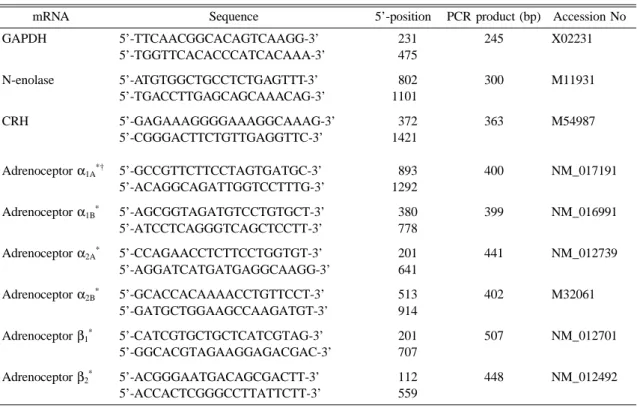
Table 1. PCR primer sets
mRNA Sequence 5’-position PCR product (bp) Accession No
GAPDH
N-enolase
CRH
Adrenoceptor α1A*†
Adrenoceptor α1B*
Adrenoceptor α2A*
Adrenoceptor α2B*
Adrenoceptor β1*
Adrenoceptor β2*
5’-TTCAACGGCACAGTCAAGG-3’
5’-TGGTTCACACCCATCACAAA-3’
5’-ATGTGGCTGCCTCTGAGTTT-3’
5’-TGACCTTGAGCAGCAAACAG-3’
5’-GAGAAAGGGGAAAGGCAAAG-3’
5’-CGGGACTTCTGTTGAGGTTC-3’
5’-GCCGTTCTTCCTAGTGATGC-3’
5’-ACAGGCAGATTGGTCCTTTG-3’
5’-AGCGGTAGATGTCCTGTGCT-3’
5’-ATCCTCAGGGTCAGCTCCTT-3’
5’-CCAGAACCTCTTCCTGGTGT-3’
5’-AGGATCATGATGAGGCAAGG-3’
5’-GCACCACAAAACCTGTTCCT-3’
5’-GATGCTGGAAGCCAAGATGT-3’
5’-CATCGTGCTGCTCATCGTAG-3’
5’-GGCACGTAGAAGGAGACGAC-3’
5’-ACGGGAATGACAGCGACTT-3’
5’-ACCACTCGGGCCTTATTCTT-3’
231 475 802 1101 372 1421
893 1292 380 778 201 641 513 914 201 707 112 559
245
300
363
400
399
441
402
507
448
X02231
M11931
M54987
NM_017191
NM_016991
NM_012739
M32061
NM_012701
NM_012492
*Nomenclature as agreed by the NC-IUPHAR Subcommittee on Adrenoceptors.
†The clone originally called the α1C-adrenoceptor corresponds to the pharmacologically defined α1A-adrenoceptor (see [1]).
exponential amplification period based on preliminary data for each gene. The PCR products for all examined genes were produced from the same RT reaction product of the same RNA sample, although PCR reactions were separately performed for each gene.
Data analysis and statistics
The PCR products were resolved on 2.0% agarose gel by electrophoresis and the gel was stained by immersing it in electrophoresis buffer containing ethidium bromide (0.5 µg/ml ). The band images were digitally captured with a CCD camera (Bio-Rad, USA). The band density of each PCR product was quantified with image analysis software (Kodak, USA). All RT-PCR data were normalized into arbitrary units by the densitometric value of 500 bp band of molecular ladder marker (Elpis- Biotech Co.) loaded by the same amount in inter- electrophoresis.
Differences between the control and ADX groups were examined for statistical significance using two- way ANOVA followed by student’s t-test for comparison of data at the same cycle number. The level of statistical significance was set at a p-value of less than 0.05.
Results
Quantitative comparison of different amounts of mRNAs by PCR profile as a function of cycle number Reverse transcriptase reaction was performed at various starting amounts of total RNA (1, 2.5 and 5 µg), and then the same volume of each RT product was used for subsequent PCR for GAPDH, a general housekeeping gene, and N-enolase, a neuronal marker, to provide internal controls (Fig. 2). For GAPDH, a detectable band by image analysis software began to appear after 28-, 25- and 22-cycles for 1, 2.5 and 5 µg of total RNA, respectively. The plots showed a rather linear increase until 40-cycles except that the plot for 5 µg showed saturation at 37-cycles which reflected the differences in amount of total RNA in the samples (Fig. 2A and 2B). The plots for N-enolase also showed quantitative differences between the three samples (Fig. 2C).
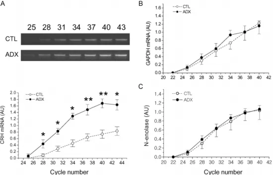
Effect of adrenalectomy on gene expression of CRH, GAPDH and N-enolase in the micropunched PVN
The relative band density for CRH mRNA levels was
significantly increased in the ADX group from cycle 28 to cycle 43 of PCR while the relative densitometric values for mRNA levels of both GAPDH and N- Fig. 2. Amplification of GAPDH and N-enolase mRNAs by RT-PCR according to cycle number with different starting amounts (1, 2.5 and 5 µg) of total RNA. Panel A illustrates PCR products (245 bp) for GAPDH on 2% agarose gel. The numbers over the photograph represent PCR cycle number. Panel B shows the quantitative plot of the bands depicted in panel A. The densitometric values of each band were normalized into arbitrary units by the density of 500 bp fragment (white arrowhead) of the size ladder marker loaded equally at every electrophoresis. Panel C shows the quantitative plot of the PCR product bands for N-enolase (photograph not shown). Abbreviation: GAPDH, glyceraldehyde -3-phosphate dehydrogenase; N-enolase, neuronal enolase;
AU, arbitrary unit.
enolase, the general and neuronal specific house keeping genes, did not differ between the control and ADX groups at any point throughout the entire cycle (Fig.
3). The serum corticosterone level of ADX rats 7 days after bilateral adrenalectomy (10.3 ± 0.5 ng/ml, mean ± SEM, n =5) was significantly lower than that of the control rats (259.5±23.3 ng/ml, n=5; p<0.001), indicating that the amount of endogenous corticosterone was almost depleted.
Gene expression of six adrenoceptor subtypes in the PVN of ADX rats
The effects of adrenalectomy on the gene expression of six adrenoceptor subtypes in the PVN are illustrated in Fig. 4. Although preliminary data were used to allow sample collection during expected exponential am- plification periods, the relationship of relative densitometric value to cycle number for some adrenoceptors exhibited a somewhat linear rather than exponential pattern. Two- way ANOVA indicated that adrenalectomy produced
significantly increased mRNA levels of α1B- (p<0.001) and β1-adrenceptors (p < 0.05), but decreased the level of β2- adrenoceptors (p < 0.001). By student’s t-test, performed as post-hoc analysis between samples obtained from the corresponding cycle, the relative densitometric values of α1B-adrenoceptor mRNA levels in the ADX group showed a significant increase at cycles 37 and 40, with respective levels of significance of 0.015 and 0.014. For the β1- adrenoceptor subtype, significant difference was shown only at cycle 43 (p=0.034) although the values for the ADX group were higher than those for the control group throughout all cycle numbers examined. For β2- adrenoceptors, the densitometric values for the ADX group were lower than the control group throughout all cycle numbers, to a significant extent at cycles 40 (p=0.024) and 43 (p=0.040). The mRNA levels for the other three adrenoceptor subtypes in the ADX rats were not significantly different from those in the control rats.
Fig. 3. Effect of adrenalectomy on mRNA levels of CRH (A), GAPDH (B) and N-enolase (C) in the PVN micropunches.
The inset (A) illustrates the bands of PCR products for CRH at cycles 25, 28, 31, 34, 37, 40 and 43. The PCR products were analyzed as described in Fig. 2. Open circles indicate control and solid circles indicate ADX group data. Each data point represents mean±SE (n=5). CRH, corticotropin-releasing hormone. *, p < 0.05; **, p < 0.01 compared with corresponding control value.
Discussion
Among the six adrenoceptor subtypes examined in this study, mRNA level of α1B and β1-adrenoceptors was increased, whereas that of β2-adrenoceptors was decreased in the ADX rats. The increase of α1B- adrenoceptor mRNA observed in the micropunches of PVN after removal of the adrenal gland is consistent with the results of previous studies using in situ hybridization [8] and semi-quantitative RT-PCR analysis [10]. Stimulation of α1-adrenoceptors within the central nervous system is known to be predominantly excitatory,
via decreasing potassium conductance and slow depolarization [2, 25, 28]. Since α1B subtype mRNA has been detected in the parvocellular region of the PVN [7, 26, 29, 40] and expression of α1A mRNA has been shown to be restricted primarily to the lateral magnocellular region [9], it is likely that the α1B- adrenoceptor subtype plays a major role in HPA axis stimulation by noradrenaline. The dual in situ hybridization study [8] demonstrated that α1B-adrenoceptor mRNA is expressed within CRH cells of the PVN and that the cells expressing this receptor are active after stress, due to the co-localization of c-fos with α1B-adrenoceptor Fig. 4. Effect of adrenalectomy on mRNA levels in the PVN micropunches of six adrenoceptor subtypes. The PCR products were quantitated as described in Fig. 2. Open circles indicate control and solid circles indicate ADX group data. Each data point represents mean ±SE (n = 5). Two way ANOVA indicated significant changes in mRNA levels of α1B- ( p < 0.001), β1-adrenceptors (p < 0.05) and β2-adrenoceptors (p < 0.001). *, p < 0.05 compared with corresponding control value. AU, arbitrary unit.
mRNA in cells of the PVN after the restraint, swimming, or immune stress. These reports and the present findings indicate that α1B-adrenoceptors may play important roles in HPA axis regulation by acting directly on the cell membranes of CRH containing neurons [8] and indirectly on the presynaptic GABAergic [15] and glutamatergic neurons [5].
The results that α2-adrenoceptor mRNA levels were not affected by adrenalectomy were rather unexpected.
Our results are not consistent with those studies which showed a decrease in mRNA expression in organotypic slices of anterior hypothalamus cultured in corticosteroid- free medium [10] and a decrease in the number of binding sites on the PVN of ADX rats [17]. These discrepancies could be due to the differences in experimental pre- parations; i.e. the use of whole animals vs. cultured slices.
It could also be have resulted from translational regulation or posttranslational modification by cor- ticosteroids as previously noted in the rat skeletal muscle [3], where the biogenesis of acetylcholinesterase is down-regulated, although its mRNA level remains normal, by dexamethasone treatment for 7 days.
As for β-adrenoceptors, they have been known to mediate the noradrenaline-induced hyperpolarization in type II PVN neurons [5], the stress-induced increase in corticosterone concentration in the PVN [31] and the isoproterenol-induced increase in corticosterone [6]. In addition, it is known that corticosterone pretreatment attenuates the isoproterenol-induced desensitization of β-adrenoceptors [36]. The results of the present study and the previous cited works suggest that catecholaminergic transmission mediated by β-adrenoceptors in the PVN also participates in HPA axis regulation. However, to our knowledge, this is the first study to report the changes in mRNA levels of β-adrenoceptors in the paraventricular nucleus of ADX rats. Further study is required to clarify the roles of the two β-adrenoceptors in relation to HPA activity regulation.
In this study, α1B-adrenoceptor mRNA levels were increased by adrenalectomy, while β2- adrenoceptor mRNA levels were decreased. It has been known that some physiological or pathological conditions are associated with inverse changes in α- and β-receptor- mediated responses in various tissues [22], including hepatic cells [21]. The results of the present study are also consistent with previous findings that the gene for α1B-adrenoceptors contains a glucocorticoid response element (GRE) within its promoter region [13] and that
the β2-adrenoceptor also contains putative GRE [18].
The changes in β1-adrenoceptor mRNA levels were found to be opposite those of β2-adrenoceptors. Furthermore, dexamethasone did not change the content of total β- adrenoceptors in rat C6 glial cells, but rather shifted the β1:β2 ratio from 80:20 to 50:50 due to its down- regulation of β1-receptor transcription [20]. The phy- siological meaning of these inverse relations in gene expression of α1B - and β2-adrenoceptors in the PVN remains to be elucidated.
Concerning the steroid modulation of adrenergic activity in the PVN, it was demonstrated that the lesions of the ventral noradrenergic ascending bundle (VNAB) inhibit the adrenocorticotrophic hormone (ACTH) and corticosterone surges induced by ether stress in the rats with intact adrenals. On the contrary, adrenalectomized and VNAB-lesioned rats showed a greatly amplified ACTH stress response relative to the controls; an adrenalectomy amplification effect was reversed completely by oral corticosterone administration [11]. In organotypic cultures of hypothalamic slices, norepinephrine acted as a potent secretagogue for CRH release in the presence of low levels of glucocorticoids. In the absence of glucocorticoids, however, the basal CRH release was high and norepinephrine strongly inhibited CRH secretion [38]. In this viewpoint, changes in gene expression of adrenergic receptor subtypes is likely to be an important mechanism responsible for the functional and steroid- dependent plasticity of adrenergic activity in the PVN, especially on CRH secreting neurons. Further study is needed to identify which one of the two adrenal corticosteroids is responsible for the observed changes in mRNA levels of adrenoceptor subtypes in the PVN of ADX rats, since adrenalectomy depletes endogenous glucocorticoids as well as mineralocorticoids.
In conclusion, the removal of corticosteroids by adrenalectomy produced an increase in the gene expression of α1B-, β1-adrenoceptors and a decrease in the gene expression of β2-adrenoceptor subtypes in the PVN, suggesting that consequent alterations in density of these adrenoceptors in the region, especially on CRH- secreting neurons, may be an important mechanism by which HPA activity is modulated.
Acknowledgements
We are grateful to Professors Kyungjin Kim, Seoul National University College of Natural Sciences and
Kyungza Ryu, Yonsei University College of Medicine for their kind help and advice for this study. This study was supported by the Brain Science Project of the Korea Ministry of Science and Technology, and the Research Institute of Veterinary Science, Seoul National University.
References
1. Alexander, S. P. H., Mathie, A. and Peters, J. A.
TiPS, Nomenclature supplement, pp. 15-18. Elsevier, Amsterdam, 2001.
2. Bergles, D. E., Doze, V. A., Madison, D. V. and Smith, S. J. Excitatory actions of norepinephrine on multiple classes of hippocampal CA1 interneurons. J.
Neurosci. 1996, 16, 572-585.
3. Brank, M., Zajc-Kreft, K., Kreft, S., Komel, R. and Grubic, Z. Biogenesis of acetylcholinesterase is impaired, although its mRNA level remains normal, in the glucocorticoid-treated rat skeletal muscle. Eur. J.
Biochem. 1998, 251, 374-381.
4. Cummings, S. and Seybold, V. Relationship of alpha 1- and alpha 2-adrenergic-binding sites to regions of the paraventricular nucleus of the hypothalamus containing corticotropin-releasing factor and vasopressin neurons.
Neuroendocrinology. 1988, 47, 523-532.
5. Daftary, S. S., Boudaba, C. and Tasker, J. G.
Noradrenergic regulation of parvocellular neurons in the rat hypothalamic paraventricular nucleus. Neuroscience.
2000, 96, 743-751.
6. Daniels, W. M., Jaffer, A., Russell, V. A. and Taljaard, J. J. Alpha 2- and beta-adrenergic stimulation of corticosterone secretion in rats. Neurochem. Res.
1993, 18, 159-164.
7. Day, H. E., Campeau, S., Watson, S. J. Jr and Akil, H. Distribution of alpha 1a-, alpha 1b- and alpha 1d- adrenergic receptor mRNA in the rat brain and spinal cord. J. Chem. Neuroanat. 1997, 13, 115-139.
8. Day, H. E. W., Campeau, S., Watson, S. J. Jr and Akil, H. Expression of a1b adrenoceptor mRNA in corticotropin-releasing hormone-containing cells of the rat hypothalamus and its regulation by corticosterone.
J. Neurosci. 1999, 19, 10098-10106.
9. Domyancic, A. V. and Morilak, D. A. Distribution of alpha 1A adrenergic receptor mRNA in the rat brain visualized by in situ hybridization. J. Comp. Neurol.
1997, 386, 358-378.
10. Feuvrier, E., Aubert, M., Malaval, F., Szafarczyk, A.
and Gaillet, S. Opposite regulation by glucocorticoids of the alpha1B- and alpha2A-adrenoreceptor mRNA levels in rat cultured anterior hypothalamic slices.
Neurosci. Lett. 1999, 271, 121-125.
11. Gaillet, S., Alonso, G., Le Borgne, R., Barbanel, G., Malaval, F., Assenmacher, I. and Szafarczyk, A.
Effects of discrete lesions in the ventral noradrenergic ascending bundle on the corticotropic stress response depend on the site of the lesion and on the plasma levels of adrenal steroids, Neuroendocrinology. 1993, 58, 408-419.
12. Gaillet, S., Lachuer, J., Malaval, F., Assenmacher, I.
and Szafarczyk, A. The involvement of noradrenergic ascending pathways in the stress-induced activation of ACTH and corticosterone secretions is dependent on the nature of stressors. Exp. Brain Res. 1991, 87, 173- 180.
13. Gao, B. and Kunos, G. Isolation and characterization of the gene encoding the rat alpha-1B adrenergic receptor. Gene, 1993, 131, 243-247.
14. Hadcock, J. R. and Malbon, C. C. Regulation of beta- adrenergic receptors by “permissive” hormones:
glucocorticoids increase steady-state levels of receptor mRNA. Proc. Natl. Acad. Sci. U S A. 1988, 85, 8415- 8419.
15. Han, S. K., Chong, W., Li, L. H., Lee, I. S., Murase, K. and Ryu, P. D. Noradrenaline excites and inhibits GABAergic transmission in parvocellular neurons of rat hypothalamic paraventricular nucleus. J. Neurophysiol.
2002, 87, 2287-2296.
16. Herman, J. P. and Cullinan, W. E. Neurocircuitry of stress: central control of the hypothalamo-pituitary- adrenocortical axis. Trends Neurosci. 1997, 20, 78-84.
17. Jhanwar-Uniyal, M. and Leibowitz, S. F. Impact of circulating corticosterone on alpha 1- and alpha 2- noradrenergic receptors in discrete brain areas. Brain Res. 1986, 368, 404-408.
18. Jiang, L. and Kunos, G. Sequence of the 5’ regulatory domain of the gene encoding the rat beta-2-adrenergic receptor. Gene, 1995, 163, 331-332.
19. Joe, I. and Ramirez, V. D. Binding of estrogen and progesterone-BSA conjugates to glyceraldehyde-3- phosphate dehydrogenase (GAPDH) and the effects of the free steroids on GAPDH enzyme activity:
physiological implications. Steroids, 2001, 66, 529-538.
20. Kiely, J., Hadcock, J. R., Bahouth, S. W. and
Malbon, C. C. Glucocorticoids down-regulate beta 1- adrenergic-receptor expression by suppressing transcription of the receptor gene. Biochem. J. 1994, 302, 397-403.
21. Kunos, G., Ishac, E. J., Gao, B. and Jiang, L. Inverse regulation of hepatic alpha1B- and beta2- adrenergic receptors. Cellular mechanisms and physiological implications. Ann. N. Y. Acad. Sci. 1995, 757, 261-271.
22. Kunos, G., Kunos, I., Hirata, F. and Ishac, E. J.
Adrenergic receptors: possible mechanism of inverse regulation of alpha and beta receptors. J. Allergy Clin.
Immunol. 1985, 76, 346-351.
23. Li, H. Y., Ericsson, A. and Sawchenko, P. E. Distinct mechanisms underlie activation of hypothalamic neurosecretory neurons and their medullary catecholaminergic afferents in categorically different stress paradigms. Proc. Natl. Acad. Sci. U S A. 1996, 93, 2359-2364.
24. Little, K. Y., Duncan, G. E., Breese, G. R. and Stumpf, W. E. Beta-adrenergic receptor binding in human and rat hypothalamus. Biol. Psychiatry, 1992, 32, 512-522.
25. McCormick, D. A. and Prince, D. A. Noradrenergic modulation of firing pattern in guinea pig and cat thalamic neurons, in vitro. J. Neurophysiol. 1988, 59, 978-996.
26. McCune, S. K., Voigt, M. M. and Hill, J. M.
Expression of multiple alpha adrenergic receptor subtype messenger RNAs in the adult rat brain.
Neuroscience, 1993, 57, 143-151.
27. Palkovits, M. and Brownstein, M. J. Microdissection of brain areas by the punch technique; In Brain Microdissection Technique, Cuello, A.C. (eds), pp. 1- 36, Wiley, New York. 1983.
28. Pan, Z. Z., Grudt, T. J. and Williams, J. T. Alpha 1-adrenoceptors in rat dorsal raphe neurons: regulation of two potassium conductances. J. Physiol. 1994, 478 Pt 3, 437-447.
29. Pieribone, V. A., Nicholas, A. P., Dagerlind, A. and Hokfelt, T. Distribution of alpha 1 adrenoceptors in rat brain revealed by in situ hybridization experiments utilizing subtype-specific probes. J. Neurosci. 1994, 14, 4252-4268.
30. Plotsky, P. M., Cunningham, E. T. Jr. and Widmaier, E. P. Catecholaminergic modulation of corticotropin- releasing factor and adrenocorticotropin secretion.
Endocr. Rev. 1989, 10, 437-458.
31. Richardson-Morton, K. D., Van de Kar, L. D., Brownfield, M. S., Lorens, S. A., Napier, T. C. and Urban, J. H. Stress-induced renin and corticosterone secretion is mediated by catecholaminergic nerve terminals in the hypothalamic paraventricular nucleus.
Neuroendocrinology, 1990, 51, 320-327.
32. Sakaue, M. and Hoffman, B. B. Glucocorticoids induce transcription and expression of the alpha1B adrenergic receptor gene in DTT1 MF-2 smooth muscle cells. J. Clin. Invest. 1991, 88, 385-389.
33. Sanne, J. L., Scarna, H., Grange, E. and Bobillier, P. Restraint stress and adrenalectomy do not affect the level of rat cerebral enolase. Neurosci. Lett. 1990, 119, 94-96.
34. Saphier, D. and Feldman, S. Iontophoretic application of glucocorticoids inhibits identified neurones in the rat paraventricular nucleus. Brain Res. 1988, 453, 183-190.
35. Sawchenko, P. E. Evidence for a local site of action for glucocorticoids in inhibiting CRF and vasopressin expression in the paraventricular nucleus. Brain Res.
1987, 403, 213-223.
36. Stone, E. A., Egawa, M. and Colbjornsen, C. M.
Catecholamine-induced desensitization of brain beta adrenoceptors in vivo and reversal by corticosterone.
Life Sci. 1989, 44, 209-213.
37. Subramaniam, M., Colvard, D., Keeting, P. E., Rasmussen, K., Riggs, B. L. and Spelsberg, T. C.
Glucocorticoid regulation of alkaline phosphatase, osteocalcin, and proto-oncogenes in normal human osteoblast-like cells. J. Cell. Biochem. 1992, 50, 411- 424.
38. Szafarczyk, A., Feuvrier, E., Siaud, P., Rondouin, G., Lacoste, M., Gaillet, S., Malaval, F. and Assenmacher, I. Removal of adrenal steroids from the medium reverses the stimulating effect of catecholamines on corticotropin-releasing hormone neurons in organotypic cultures. Neuroendocrinology, 1995, 61, 517-524.
39. Szafarczyk, A., Malaval, F., Laurent, A., Gibaud, R.
and Assenmacher, I. Further evidence for a central stimulatory action of catecholamines on adrenocorticotropin release in the rat. Endocrinology, 1987, 121, 883-892.
40. Williams, A. M. and Morilak, D. A. Alpha 1B adrenoceptors in rat paraventricular nucleus overlap with, but do not mediate, the induction of c-Fos expression by osmotic or restraint stress. Neuroscience, 1997, 76, 901-913.