․교신저자: 서운교 경기도 성남시 분당구 수내3동 87-2 동국대학교 분당한방병원 2내과
TEL: 031-710-3751 FAX: 031-710-3780 E-mail: [email protected]
健肺湯이 호흡기 뮤신의 생성 및 유전자 발현에 미치는 영향
정병진, 김 호, 서운교 동국대학교 한의과대학 내과학교실
Effect ofGeonpye-tang(GPT) on Production and Gene Expression of Respiratory Mucin
Byeong-jin Jung, Ho Kim, Un-kyo Seo
Dept. of Oriental Internal Medicine, College of Oriental Medicine, Dong-Guk University ABSTRACT
Objectives :In this study, the author tried to investigate whetherGeonpye-tang(GPT) significantly affects PMA-, EGF- or TNF-alpha-induced MUC5AC mucin production and gene expression from human airway epithelial cells.
Materials and Methods :Effects of the agent on PMA-, EGF- or TNF-alpha-induced MUC5AC mucin production and gene expression from human airway epithelial cells (NCI-H292) were investigated. Confluent NCI-H292 cells were pretreated for 30 min in the presence of GPT and treated with PMA (10ng/ml) or EGF (25ng/ml) or TNF-alpha (0.2nM), to assess both effect of the agent on PMA- or EGF- or TNF-alpha-induced MUC5AC mucin production by enzyme-linked immunosorbent assay (ELISA) and gene expression by reverse transcription-polymerase chain reaction (RT-PCR). Possible cytotoxicity of the agent was assessed by examining the rate of survival and proliferation of NCI-H292 cells after treatment with the agent over 72 hrs (SRB assay).
Results : (1) GPT significantly inhibited PMA-induced and EGF-induced MUC5AC mucin production from NCI-H292 cells. However, GPT did not affect TNF-alpha-induced MUC5AC mucin production. (2) GPT significantly inhibited the expression levels of PMA-, EGF- or TNF-alpha-induced MUC5AC genes in NCI-H292 cells (3) GPT did not show significant cytotoxicity to NCI-H292 cells.
Conclusion :This result suggests that GPT can affect the production and gene expression of respiratory mucin observed in diverse respiratory diseases accompanied by mucus hypersecretion. This can explain the traditional use of GPT in oriental medicine. Effects of GPT with their components should be further investigated using animal experimental models that reflect pathophysiology of airway diseases through future studies.
Key words : Airway mucin,Geonpye-tang, RTSE, NCI-H292
Ⅰ. 緖 論
인체의 호흡기에 존재하는 점액(mucus)은 섬모 세포와의 협동적 작용을 통해, 인체에 불필요하거 나 혹은 유해한 물질의 제거에 있어서 중요한 역
할을 한다1. 기도 점액의 인체 방어 기능은 주로 점 액의 구성요소인 mucin의 점탄성(viscoelasticity)에 기인하는데, 이러한 mucin의 양과 질의 이상은, 기 도 생리의 이상 뿐 아니라 인체의 방어작용에 영 향을 주어 병리 현상을 유발할 수 있다. 즉, 천식, 만성 기관지염, 폐기종, 기관지 확장증, 낭포성 섬 유증 등의 기도 질환에서 관찰되는 객담 혹은 점 액의 과다분비는 이러한 질환군의 예후를 악화시 키는 주된 요인으로 알려져 있다2,3,4. 서양의학계에
서도 기도점액(객담) 과다분비를 조절하기 위해 여러 약물을 사용하고 있으나, 그 약효나 부작용 면에서 문제가 있어서 효율적인 약물요법을 시행 하기가 쉽지 않은 것으로 알려져 있다5,6. 현재, 기 존의 서양의학적 약물 외에 많은 신약후보물질이 연구되어 학계에 보고되고 있으나, 아직까지 실용 화되어 임상에서 응용되는 약물은 없다7,8.
健肺湯(Geonpye-tang, GPT)은 蔘蘇飮과 平胃散 加味方의 合方으로 解表, 理氣化痰, 補氣하는 효능 이 있어 氣虛한 사람의 外感風寒, 內有痰飮으로 인 한 咳嗽多痰, 胸膈痞滿, 食無味 등의 증상을 치료 하는 효과가 있다.
蔘蘇飮은 和劑局方에 최초로 인용되고 있으며, 辛溫解表, 理氣化痰하는 효능으로 虛弱者의 外感風 寒 內有痰飮을 치료하는 처방으로9 기존의 실험 연 구는 주로 蔘蘇飮의 항알레르기 효과에 대한 연구
10,11,12,13이 이루어졌다. 또한 호흡기 점액의 주요 구
성요소인 mucin 분비에 대한 연구로는 加味治哮散 및 加味理中湯14, 加味六君子湯15, 加味腎氣湯 및 加 味淸肺湯16, 紫莞治哮散 및 瓜蔞枳實湯17, 加味鎭咳 湯18, 杏蘇湯 및 取淵湯19, 大靑龍湯 및 <石室秘籙>
急治法方20, 射干麻黃湯 및 <石室秘籙> 逆醫法方21, 淸燥救肺湯과 理陰煎22, 小靑龍湯加石膏 및 <石室 秘籙> 小治法方23 등이 보고되어 있다. 그러나 현재 까지 健肺湯이 호흡기 mucin의 생성 및 유전자 발 현에 미치는 영향은 보고되어 있지 않다. 따라서 본 연구에서는 임상에서 호흡기 질환 치료에 사용 되고 있는 健肺湯이 호흡기 mucin의 생성 및 mucin 유전자의 발현에 어떠한 영향을 미치는 지
를 검증해 보고자 하였다.
이에 본 논문에서는 인간의 호흡기 상피세포인 NCI-H292 세포에 종양괴사인자(TNF-alpha), 상 피세포 성장인자(EGF), phorbol ester(PMA)로 각 각 호흡기 mucin의 생성(production) 및 유전자 발 현을 자극한 상태에서, 주된 호흡기 mucin인 MUC5AC의 생성 및 그 유전자 발현에 대한 健肺 湯의 영향을 측정하였다. 또한 健肺湯 투여에 의해 NCI-H292 세포에 독성이 유발될 수 있는 지를 알 아보기 위해 세포의 생존 및 증식에 미치는 약물 의 영향을 검증하였다. 이상의 실험연구를 통하여, 임상에서 호흡기 점액 과다분비 관련 질환의 치료 를 목표로 사용되고 있는 健肺湯의 약리작용에 대 한 기초과학적 정보를 얻고자 하였다.
Ⅱ. 實 驗
1. 재 료 1) 藥 材
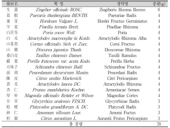
健肺湯의 구성 약물은 동국대학교 부속 한방병 원 약제실에서 공급받아 조제하였으며 한 첩의 분 량은 Table 1과 같다. 한 첩 분량에 800 - 1,000ml 의 탈이온 2차 증류수를 가하고 100℃로 가온된 상 태에서 3시간 동안 전탕하여, 최종 80ml의 탕액을 수거하였다. 각 탕액을 실온 정도로 방냉한 후, 멸 균 청정 후드 내에서 0.22μm filter를 이용, 가압 여 과하고 멸균용기에 저장하여, 4℃ 냉장고에 보관하 였다(Table 1).
藥材名 학 명 생약명 중량(g) 生 薑 Zingiber officinale ROSC. Zingiberis Rizoma Recens 6
葛 根 Pueraria thunbergiana BENTH. Puerariae Radix 4
麥 芽 Hordeum Vulgare L. Hordei Fructus Germiniatus 4
半 夏 Pinellia ternata Breit. Pinelliae Rhizoma 4
白茯苓 Poria cocos Wolf. Poria 4
白 朮 Atractylodes macrocepha-la Koidz. Atractylodis Rhizoma Alba 4 山茱萸 Cornus officinalis Sieb. et Zucc. Corni Fructus 4
山 藥 Discorea japonica Thunb. Dioscoreae Rhizoma 4
桑白皮 Taxillus chinensis Danser. Taxilli Ramulus 4
蘇 葉 Perilla frutescens var. acuta Kudo. Perilla Herba 4 五味子 Schizandra chinensis Baill. Schizandrae Fructus 4
前 胡 Peucedanum decursivum Maxim. Peucedani Radix 4
陳 皮 Citrus unshiu Markovich. Citri Pericarpium 4
蒼 朮 Atractylodes lancea DC. Atractylodis Rhizoma 4
杏 仁 Prunus mandshurica Koehne. Armeniacae Semen 4
厚 朴 Magnolia officinalis Rehder et Wilson Magnoliae Cortes 4 甘 草 Glycyrrhiza uralensis FISCH. Glycyrrhizae Radix 3 桔 梗 Platycodon grandiflorum A. DC. Platycodi Radix 3
砂 仁 Amomum villosum Lour. Amomi Fuctus 3
枳 殼 Citrus aurantium L. Aurantii Frutus Pericarpium 3
총 중량 78
Table 1. The Composition & Dosage of Geonpye-tang(GPT)
2) 배양세포
인간 호흡기 상피세포주인 NCI-H292 세포는 American Type Culture Collection 사 (Manassas, VA, U.S.A.)에서 구입하였다.
3) 시 약
Protease inhibitor cocktail은 Roche사 (Indianapolis, IN, U.S.A.)에서, diethylpyrocarbonate (DEPC), 3,3',5,5'-tetramethyl-benzidine peroxide solution (TMB), sulforhodamine B (SRB), trypsin-EDTA, Trizma base, trichloroacetic acid (TCA), NP-40, EDTA, EGTA, Phorbol 12-Myristate 13-Acetate (PMA), HEPES, dimethyl sulfoxide (DMSO), Tween 20, bovine serum albumin (BSA), sodium dodecyl sulfate (SDS), tumor necrosis factor-alpha (TNF-alpha), epidermal growth factor (EGF) 등
은 Sigma사(St. Louis, Mo., U.S.A.)에서, penicillin-G, streptomycin, fetal bovine serum (FBS), RPMI 1640은 GIBCO-BRL사 (Grand Island, New York, U.S.A.)에서, mouse anti-MUC5AC clone 45M1 및 HRP-Goat Anti-Mouse IgG Conjugate는 NeoMarkers 사(Freemont, CA, U.S.A.)에서, Easy-Blue RNA extraction kit는 INTRON biotechnology사 (Kyung-gi, Korea)에서, Accuprep RT premix kit와 Accuprep PCR premix kit는 Bioneer사 (Daejeon, Korea)에 서, 기타 제반 시약들은 일급시약 등급 이상의 것 들을 구입하여 사용하였으며, 실험에 사용된 물은 탈이온 2차 증류수이었다24,25.
2. 방 법
1) NCI-H292 세포 배양
세포는 습도가 충분히 유지되고 95% 공기, 5%
CO2를 함유하는 37℃ 조건의 배양기 내에서 HEPES (25mM), penicillin G (100 U/ml), streptomycin (100μg/ml ), FBS (10%, V/V)등이 첨가된 RPMI 1640 배양액을 이용하여 배양되었으며, 1주에 2회 빈도로 subculture하였다. MUC5AC mucin 생성에 대한 검액의 작용을 검증하기 위하여, 24 well culture plate를 기준으로 well 당 2.0 x 10⁴cells/well 의 밀도로 세포를 도포하고 배양하였다. 세포가 다 자라면 FBS의 농도를 0.2%로 감소시킨 배양액을 주고 24 시간 동안 배양하고, 이후 serum을 첨가하 지 않은 배양액(serum-free medium)으로 세포를 세척하였다. 이렇게 준비된 세포에 검액을 각각 5 - 20μl씩을 함유하는 배양액 200μl를 well(24 well plate 기준)마다 가하고, 30분이 지난 시점에 PMA 10ng/ml, 또는 EGF 25ng/ml, 또는 TNF-alpha 0.2nM을 각 well마다 투여한 후 37℃에서 24시간동 안 배양하였다.
2) MUC5AC mucin 생성량 측정(ELISA) 24 시간의 배양이 종료된 시점에 세포 용해용 완충액 (20mM Tris, 0.5% NP-40, 250mM NaCl, 3mM EDTA, 3mM EGTA, protease inhibitor cocktail)을 가하여 세포 내에 존재하는 MUC5AC 를 추출한 후 이후의 실험에 사용하였다. 즉, 수거 된 세포 용해 추출액 (cell lysate)을 PBS로 1/10배 희석하고 희석된 각 sample을 ELISA 전용의 96-well plate에 각각 100㎕씩 분포시킨 후, 42℃에 서 완전히 건조될 때까지 incubation하였다. 그 후 PBS-Tween 20 (0.05%, PBS-T) 용액 200㎕/well 을 이용, 각 well 당 3회씩 세척하였다. 세척 후 PBS-T에 용해된 2% BSA 용액 200㎕를 각 well당 가하고, 다시 1시간 동안 incubation하였다. 1시간 후, PBS-T 200㎕로 3회 세척하고, MUC5AC에 대 한 monoclonal antibody인 mouse anti-MUC5AC clone 45M1을, 2% BSA에 1: 200의 비율로 희석한 후에, 각 well당 100㎕씩 첨가하고, 1시간 동안 incubation하였다. 1시간 후 PBS-T로 3회 세척하고,
2차 항체인 Horse radish peroxidase (HRP)-Goat Anti-Mouse IgG Conjugate를 2% BSA에 1: 3,000 의 비율로 희석한 후, 각 well당 100㎕씩 첨가하고 1시간 동안 incubation하였다. PBS-T로 다시 3회 세척 후, 3,3',5,5'- tetramethyl-benzidine peroxide (TMB)용액 100㎕를 각 well에 첨가하고, 5분 후 1N H2SO450㎕를 첨가, 반응을 정지시켰다. 450nm 에서 각 well의 흡광도를 측정함으로써 대조군과 약 물 처리군 간의 MUC5AC를 정량, 비교하였다26.27.
3) NCI-H292 세포 내에 존재하는 total RNA의 분리
24 시간의 배양이 종료된 세포를 냉각된 PBS로 2회 세척하였다. 세포에 trypsin-EDTA 용액을 처 리하여 배양 용기 바닥으로부터 분리하고, 세포들 의 혼합물을 1.5ml 용량의 microtube에 옮겨 원심 분리함으로써 세포들만 수거하였다. 이어서, total RNA를 분리하고자 INTRON biotechnology사의 Easy-Blue RNA extraction kit (total RNA isolation reagent)를 이용해(0.5ml/4x105 cells) 세포를 lysis 시키고, 상온에서 5분간 방치하였다. 5분 후 즉시, microtube에 chlorofrom를 첨가, 15초간 vortexing하 고 상온에 2-3분간 방치한 후 4℃, 13,000 rpm (Hanil centrifuge, MICRO 17 R)에서 10분간 원심 분리하여 얻은 상층액 400μl를 새 microtube에 옮 겼다. 상층액에 동량의 isopropanol을 첨가하여 잘 혼합한 후 상온에서 10분간 방치하고 다시 4℃, 13,000 rpm에서 10분간 원심 분리하여 RNA 침전 물을 얻었다. 이 침전물에 diethylpyrocarbonate (DEPC)가 함유된 75% ethanol을 가하고 4℃, 10,000 rpm에서 10분간 원심 분리함으로써 세척하 였다. 수거된 RNA 침전물을 5분간 대기 중에서 건조시킨 후, 20μl의 RNase-free water로 부유시키 고, spectrophotometer (Beckman, DU-650)를 사용 하여 260 nm 파장에서 흡광도를 측정함으로써 RNA의 농도를 알아내어 실험에 사용하였다 (1.0A260
=single strand RNA 40 ㎍/㎖)28.
4) PCR (Polymerase Chain Reaction)을 위한
primer 제조
PCR에 사용된 primer는 전문 제조회사인 제노텍 (주)(Daejeon, Korea)에 주문, 합성하였다. NCI-H292 세포에서의 human MUC5AC 유전자 합성을 위해 사용한 sense primer의 염기서열은 5‘-TGA TCA TCC AGC AGC AGG GCT-3', antisense primer의 염기서열은 5’-CCG AGC TCA GAG GAC ATA TGG G-3'이며, 이 primer에 의해 합성된 PCR 산 물의 크기는 약 500 bp였다. β-actin 유전자 합성을 위해 사용한 sense primer의 염기서열은 5'-TAC AAC GAG CTG CGT GTG GCC-3'이고, antisense primer는 5'-CAA CGG AAC CGC CTC GTT GC-3'이며 이 primer가 표적으로 하는 DNA 크 기는 500 bp였다.
5) RNA의 역전사 반응 및 중합효소 연쇄반응 (RT-PCR)
수거된 total RNA를 이용, 역전사 반응(RT)으 로 cDNA를 만들고, 이를 중합효소 연쇄반응(PCR) 으로 증폭시켰다. 즉, 얻어진 total RNA 1㎍을 7 5℃에서 5분간 가열함으로써 denaturation시키고, 이를 얼음에 담가 급냉시킨 후 RT premix kit의 사용자 설명서에 따라 역전사 반응을 진행시켰다.
MUC5AC 유전자에 대한 PCR은, 각각의 역전사 반응에서 얻은 cDNA 산물 2㎕를 PCR premix kit 의 사용자 설명서에 따라 진행시켰다. 증폭반응을 위하여, PCR을 40회 실시(PCR thermal cycler;
Takara MP-300, Japan)하였으며, denaturation은 94℃에서 30초, annealing은 60℃에서 30초, extension 은 72℃에서 30초간 각각 시행하였다. 전기영동에 의한 중합효소 연쇄반응 산물의 확인 RNA의 역전 사 반응 및 중합효소 연쇄반응으로 증폭된 cDNA 산물들을 전기영동으로 분리함으로써 MUC5AC 유전자 발현변동여부를 관찰하였다. 즉, 증폭된 PCR 산물 10㎕를 10×gel loading buffer (0.25%
bromphenol blue, 0.25% xylene cyanol FF, 50%
glycerol)와 잘 혼합한 다음, Tris-acetate-EDTA buffer (40 mM Tris-acetate, 1 mM EDTA)용액
및 1㎍/㎖의 ethidium bromide가 포함된 1.0%
agarose gel에서 전기 영동하였다. Gel 상에서 이동 된 각각의 DNA band는 자외선 투사기 (ultraviolet transilluminator)를 이용하여 관찰하고, 사진 촬영 하였다.
6) 세포의 생존 및 증식에 미치는 영향(세포 독 성) 측정 (SRB assay)
96 well plate의 각 well에 2 × 10⁴개의 NCI- H292 cell을 함유하는 배양액 100㎕를 가하여 37℃, 5% CO2 존재 하에서 24시간 배양하였다29,30. 24시 간 배양 후 검액을 0.2 - 2μl/200μl (배양액)의 농 도로 100㎕씩 배양세포의 well마다 가하고 72시간 동안 추가로 배양하였다. 배양이 완료된 후, 냉각 된 50% trichloroacetic acid (TCA) 50㎕를 각 well 에 서서히 가해 주었다. 10분 후에 4℃ 조건의 냉 장고에 옮겨 1시간 동안 충분히 세포들을 고정 (fixation)시키고, 고정이 완료된 후 각 well에 존재 하는 액체 성분들을 전량 흡인배출한 후에 well 당 250㎕의 증류수를 이용, 5회 이상 세척하였다. 이렇 게 준비된 세포에, acetic acid에 용해된 0.4% SRB 용액 50㎕/well을 가하고 실온에서 30분 동안 염색 하였고, 재차 흡인배출 후 100㎕의 1% acetic acid 를 이용, 5회 이상 세척한 후 세포들을 건조시켰다.
각 well 당 100㎕의 10mM unbuffered Tris용액으로 SRB를 잘 녹여낸 후, 흡광분석 측정장치(microplate reader)로 540nm에서 각 well의 흡광도를 측정하였 다30.
7) 통계처리
모든 측정 결과는 Mean±S.E.M.으로 환산된 후, 검액 처리군의 측정치는 대조군 측정치의 백분율 로 나타냈다. 통계처리는 unpaired Student'st-test 로 하였으며, p<0.05인 경우에 통계적으로 유의성 이 있는 것으로 판정하였다.
Ⅲ. 結 果
1. PMA로 자극된 mucin생성 증가현상에 미치는 영향
健肺湯은 최종 추출물 20μl/200μl PBS의 투여 농도에서 PMA로 자극된 mucin생성 증가를 유의 하게 감소시켰다(Fig. 1).
Fig. 1. Effect of GPT on PMA-induced MUC5AC mucin production from NCI-H292 cells.
NCI-H292 cells were pretreated with GPT extract 5 - 20μl/200μl media for 30 min and then stimulated with PMA (10ng/ml) for following 24hrs. Cell lysates were collected for quantitation of MUC5AC mucin production by ELISA as described in Materials and Methods. Each bar represents a mean ± S.E.M. of 4 culture wells.
* : significantly different from control (p<0.05).
+ : significantly different from PMA only (p<0.05).
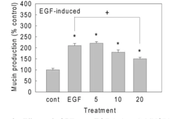
2. EGF로 자극된 mucin생성 증가현상에 미치는 영향 健肺湯은 최종 추출물 20μl/200μl PBS의 투여 농도에서 EGF로 자극된 mucin생성 증가를 유의하 게 감소시켰다(Fig. 2).
Fig. 2. Effect of GPT on EGF-induced MUC5AC mucin production from NCI-H292 cells.
NCI-H292 cells were pretreated with GPT extract 5 - 20μl/200μl media for 30 min and then stimulated with EGF (25ng/ml) for following 24hrs. Cell lysates were collected for quantitation of MUC5AC mucin production by ELISA as described in Materials and Methods. Each bar represents a mean ± S.E.M. of 4 culture wells.
* : significantly different from control (p<0.05).
+ : significantly different from EGF only (p<0.05).
3. TNF-alpha로 자극된 mucin생성 증가현상에 미 치는 영향
健肺湯은 최종 추출물 5 - 20μl/200μl PBS의 투 여 농도범위에서 TNF-alpha로 자극된 mucin생성 증가에 유의한 작용을 나타내지 못하였다(Fig. 3).
Fig. 3. Effect of GPT on TNF-alpha-induced MUC5AC mucin production from NCI-H292 cells.
NCI-H292 cells were pretreated with GPT extract 5 - 20μl/200μl media for 30 min and then stimulated with TNF-alpha (0.2nM) for following 24hrs.
Cell lysates were collected for quantitation of MUC5AC mucin production by ELISA as described in Materials and Methods. Each bar represents a mean ± S.E.M. of 4 culture wells.
* : significantly different from control (p<0.05).
4. PMA로 자극된 mucin 유전자 발현 증가현상에 미치는 영향
健肺湯은 최종 추출물 20μl/200μl PBS의 투여 농도에서 PMA로 자극된 mucin유전자 발현을 감 소시키는 경향을 보여주었다(Fig. 4).
Fig. 4. Effect of GPT on PMA-induced MUC5AC gene expression in NCI-H292 cells.
NCI-H292 cells were pretreated with GPT extract 20μl/200μl media for 30 min and then stimulated with PMA (10ng/ml) for following 24hrs. Total RNA was isolated and MUC5AC mRNA levels were analyzed by RT-PCR. The PCR products were separated on 1.0% agarose gel and stained with ethidium bromide as described in Materials and Methods.
5. EGF로 자극된 mucin 유전자 발현 증가현상에 미치는 영향
健肺湯은 최종 추출물 20μl/200μl PBS의 투여 농도에서 EGF로 자극된 mucin유전자 발현을 감소 시키는 경향을 보여주었다(Fig. 5).
Fig. 5. Effect of GPT on EGF-induced MUC5AC gene expression in NCI-H292 cells.
NCI-H292 cells were pretreated with GPT extract 20μl/200μl media for 30 min and then stimulated with EGF (25ng/ml) for following 24hrs. Total RNA was isolated and MUC5AC mRNA levels were analyzed by RT-PCR. The PCR products were separated on 1.0% agarose gel and stained with ethidium bromide as described in Materials and Methods.
6. TNF-alpha로 자극된 mucin 유전자 발현 증가현 상에 미치는 영향
健肺湯은 최종 추출물 20μl/200μl PBS의 투여 농도에서 TNF-alpha로 자극된 mucin 유전자 발현 을 감소시키는 경향을 보여주었다(Fig. 6).
Fig. 6. Effect of GPT on TNF-alpha-induced MUC5AC gene expression in NCI-H292 cells.
NCI-H292 cells were pretreated with GPT extract 20μl/200μl media for 30 min and then stimulated with TNF-alpha (0.2nM) for following 24hrs.
Total RNA was isolated and MUC5AC mRNA levels were analyzed by RT-PCR. The PCR products were separated on 1.0% agarose gel and stained with ethidium bromide as described in Materials and Methods.
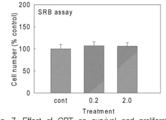
7. NCI-H292 세포의 생존 및 증식에 미치는 영향 健肺湯을 NCI-H292 세포에 0.2 - 2μl/200μl (배 양액)의 투여 농도로 72시간 동안 투여했을 때, 세 포의 생존 및 증식에 유의성 있는 영향을 나타내 지 않았다(Fig. 7).
Fig. 7. Effect of GPT on survival and proliferation of NCI-H292 cells.
NCI-H292 cells were treated with 0.2μl GPT extract/200μl media for 72 hrs. The number of cells survived was counted as described in Materials and Methods.
Ⅳ. 考 察
본 연구의 최종 목표는 임상 한의학에서 호흡기 질환 치료에 사용되고 있는 健肺湯이 호흡기 mucin 의 생성 및 mucin 유전자의 발현에 어떠한 영향을 미치는 지를 검증하는 것이었다.
健肺湯은, 辛溫解表시키고 理氣化痰하는 효능이 있어 신체가 虛弱한 사람이 風寒을 外感하고 內에 痰飮이 있어 惡寒發熱하고 頭痛鼻塞, 咳嗽痰多, 胸 膈滿悶한 증상을 치료하는 蔘蘇飮9과 理氣健脾, 燥 濕化痰의 효능을 강화할 목적으로 貯痰之器인 脾 臟에 대하여 燥濕健脾하고, 이외에 行氣和胃의 효 능으로 脾胃의 濕滯로 인한 脘腹脹滿, 不思飮食, 口淡無味, 惡心嘔吐, 噯氣呑酸, 肢體沈重, 倦怠嗜臥 등의 증상을 치료하는 平胃散9의 合方으로 氣虛한 사람의 外感風寒, 內有痰飮으로 인한 咳嗽多痰, 胸 膈痞滿, 食無味 등의 증상을 치료하는 효과가 있다.
특히 처방 중의 半夏의 燥濕化痰, 降逆止嘔, 消痞 散結하는 효능, 白茯苓의 利水滲濕, 健脾寧心하는 효능, 白朮의 補裨, 益胃, 燥濕, 和中하는 효능, 山 藥의 健脾, 補肺, 固腎, 益精하는 효능, 桑白皮;의 瀉肺平喘, 利水消腫하는 효능, 前胡의 降氣祛痰, 宣 散風熱하는 효능, 陳皮의 理氣, 調中, 燥濕, 化痰하 는 효능, 蒼朮의 燥濕健脾, 祛風散寒하는 효능, 厚 朴의 行氣燥濕, 降逆平喘하는 효능으로31, 호흡기 점액분비에 일정한 효과가 기대된다.
蔘蘇飮에 관한 기존의 연구를 살펴보면, 이 등의 蔘蘇飮의 항알레르기 효과에 대한 연구10, 류 등의 蔘蘇飮 알코올 추출물이 생쥐 면역세포의 시토킨 조절에 미치는 효과에 대한 연구11, 남 등의 蔘蘇飮 과 蔘蘇飮加味方의 抗알레르기효과에 관한 실험적 연구12 및 최 등의 蔘蘇飮과 蔘蘇飮加味方의 항알 레르기효과에 관한 실험적 연구13 등이 있다. 또한 호흡기 점액의 mucin 분비에 대한 연구로는 류 등 의 加味治哮散 및 加味理中湯이 기도 객담 분비에 미치는 영향에 대한 연구14, 박 등의 加味六君子湯 이 호흡기 mucin 분비 및 mucin 유전자 발현에 미
치는 영향에 대한 연구15, 김 등의 加味腎氣湯 및 加味淸肺湯이 기도점액 분비 및 기관평활근 긴장 도에 미치는 영향에 대한 연구16 및 박 등의 紫莞 治哮散과 瓜蔞枳實湯이 일차배양된 설치류 기도 배상세포에서의 mucin 분비에 미치는 영향에 대한 연구17, 김 등의 加味鎭咳湯이 호홉기 점액의 mucin 분비에 미치는 영향에 대한 연구18, 강 등의 杏蘇湯 및 取淵湯이 호홉기 뮤신 유전자의 발현과 점액분 비에 미치는 영향에 대한 연구19, 박 등의 Mucin 분비에 미치는 大靑龍湯 및 <石室秘籙> 急治法方 에 대한 연구20, 심 등의 射干麻黃湯 및 <石室秘籙>
逆醫法方이 호홉기 점액의 분비에 미치는 영향에 대한 연구21, 박 등의 淸燥救肺湯과 理陰煎이 호홉 기 점액분비에 미치는 영향에 대한 연구22, 김 등의 小靑龍湯加石膏 및 <石室秘籙> 小治法方이 호홉기 점액의 분비에 미치는 영향에 대한 연구23 등이 보 고되어 있다. 그러나 현재까지 健肺湯이 호흡기 점 액의 주요 구성요소인 mucin의 생성 및 그 유전자 발현에 미치는 영향에 대한 보고는 찾아볼 수 없 었다. 이에 健肺湯이 호흡기 mucin의 생성 및 유전 자 발현에 어떠한 영향을 미치는 지를 검증하기 위해, 인간의 호흡기 상피세포인 NCI-H292 세포 주를 이용하여 연구를 진행하게 되었다.
PMA는 칼슘의존성 단백질 인산화효소 (Calcium- dependent protein kinase (PKC))를 활성화시키는 물질로 생쥐 피부에서의 강력한 발암 촉진작용, 강 력한 산화질소 생성 촉진작용, 유도형 산화질소 생 성효소의 발현 촉진작용32,33 등을 나타내는 것으로 알려져 있는 물질로, 인간 호흡기 상피세포에서 mucin의 생성을 증가26시키는 것으로도 알려져 있는 물질이다. 또한, EGF는 EGF-R (epidermal growth factor receptor) - MEK-ERK pathway를 경유하 고, TNF-alpha는 TNF-R (tumor necrosis factor receptor)를 매개로 전사인자 결합부위 NF-κB (nuclear factor-kappa B)로 이어지는 신호전달 경 로를 경유하여, MUC5AC의 생성 증가를 유도할 수 있는 것으로 알려져 있다34.
이러한 타 연구자들의 보고에 근거하여 본 연구 에서는, 인간의 호흡기 상피세포인 NCI-H292 세 포에 健肺湯을 먼저 처리하고 30분이 지난 후 각각 PMA, EGF, TNF-alpha를 처리하고 연속되는 24 시간 동안 세포로부터 생성되는 mucin의 양을 측정 한 결과, 健肺湯은 PMA 또는 EGF로 자극된 호흡기 mucin의 생성을 감소(Fig. 1, 2)시켰으며, TNF-alpha 로 자극된 호흡기 mucin의 생성에 대해서는 유의 한 영향을 나타내지 못함을 알게 되었다(Fig. 3).
그렇다면, 같은 조건에서 健肺湯이 PMA, EGF, TNF-alpha에 의해 자극된 MUC5AC mucin 유전 자 발현에는 어떠한 영향을 미치는 지 탐색한 결 과, 健肺湯은 PMA 등 자극 물질(inducer)에 의한 MUC5AC 유전자의 발현을 감소시키는 경향을 보 여주었다(Fig. 4, 5, 6). 이러한 제반 실험결과는, 健肺湯이 호흡기 mucin의 생성 혹은 유전자 발현을 자극(유도)시킬 수 있는 병리적 조건에서 mucin의 생성과 그 유전자의 발현에 유의한 억제효과를 발 휘함으로써, 인간의 호흡기 점액 과다분비 질환의 치료과정에서 확인할 수 있는 健肺湯의 임상적 효 과에 대한 기초과학적 기전을 시사하고 있다 하겠 다. 다만, TNF-alpha 처리 후의 mucin 생성에 대 한 健肺湯의 작용은 비록 MUC5AC 유전자의 발현 수준에서는 감소시키는 경향을 보여주었으나, 호흡 기 mucin의 생성에 대해서는 유의한 영향을 나타 내지 못함으로써 이에 대해서는 향후 추가적인 기 전연구를 통하여 더 자세히 규명되어야 할 것으로 판단된다. 또한, 健肺湯이 약리작용을 발현하는 동 시에 NCI-H292 세포에 대해 독성을 발현하는 지 여부를 검증하기 위해, 세포의 증식과 생존에 미치 는 특정물질의 작용을 평가하는 독성시험 방법인 SRB assay를 시행한 결과, 현저한 세포독성을 발 현하지 않을 가능성을 보여 주었다(Fig. 7).
결론적으로, 이상의 연구결과들은 호흡기 객담 이 과다 분비되는 병리적 상황에서 健肺湯이 객담 (점액)의 주요 구성성분인 mucin의 생성 및 유전 자 발현을 감소시킴으로써 질환의 치유를 촉진할
가능성을 제시하고 있으며, 향후 추가적인 기전연 구를 통하여 개별 호흡기 질환의 다양한 병태생리 학적 특성을 적절히 반영할 수 있는 실험모델을 사용하여 健肺湯의 작용 및 기전에 대한 다각적이 고도 심층적인 후속연구가 요망된다.
Ⅴ. 結 論
健肺湯의 인간 호흡기 상피세포에서의 mucin 생 성 및 유전자 발현에 미치는 영향을 관찰한 결과 다 음과 같은 결론을 얻었다. 健肺湯은 호흡기 mucin의 생성 혹은 유전자 발현을 자극(유도)시킬 수 있는 병리적 조건에서 mucin의 생성과 그 유전자의 발 현에 유의한 억제효과를 발휘함으로써, 호흡기 객 담이 과다분비되는 병리적 상황에서 질환의 치유 를 촉진할 가능성을 제시하고 있으며, 향후 개별 호흡기 질환의 병태생리학적 특성을 적절히 반영 할 수 있는 실험모델을 사용하여 健肺湯의 작용 및 기전에 대한 다각적이고도 심층적인 후속연구 가 요망된다.
參考文獻
1. Newhouse, M.T. and Biennenstock, J. Respiratory tract defense mechanism, In, textbook of pulmonary disease (Baum, G.L. and Wolinsky, E.(eds)), 3rd ed.: Little Brown and Company; 1983.
2. Frigas, E., Loegering, D.A., Solley, G.O., Farrow, G.M. and Gleich, G.J. Elevated levels of the eosinophil granule major basic protein in the sputum of patients with bronchial asthma.
Mayo Clin. Proc. 1981;56:345-53.
3. Culpitt SV, Rogers DF, Traves SL, Barnes PJ, Donnelly LE. Sputum matrix metalloproteases:
comparison between chronic obstructive pulmonary disease and asthma. Respir Med. 2005;99(6)
:703-10.
4. Gleich, G.J. The eosinophil and bronchial asthma:
Current understanding. J. Allergy Clin. Immunol.
1990;85:422-36.
5. Rogers DF. Airway mucus hypersecretion in asthma:
an undervalued pathology? Curr Opin Pharmacol.
2004 Jun;4(3):241-50.
6. Mutschler, E. and Derendorf, H.. Drug actions.
Florida: CRC press, Inc., Boca Raton; 1995, p.
410-1.
7. Rogers DF, Barnes PJ. Treatment of airway mucus hypersecretion. Ann Med. 2006;38(2) :116-25.
8. Rogers DF. Mucociliary dysfunction in COPD:
effect of current pharmacotherapeutic options.
Pulm Pharmacol Ther. 2005;18(1):1-8.
9. 한의과대학 방제학교수 공편저. 방제학. 영림사;
2003, p. 98, 486
10. Lee, SE, Shin, JY, Lee, SH, Anti-allergic effects of Shensuyin. Korean J. Orient. Int. Med.
2005;26(1):119-28.
11. Ryu NH, Park SM, Kang H, Shim BS, Kim SH, Choi SH etc. Effect of Samsoeum on cytokine regulation of mouse T cell. Korean J.
Oriental Physiology&Patholojy. 2008;22(4):856-62.
12. Nam BS, Kim YB. The experimental study on the anti-allergic effects of Samsoeum and Samsoeumgamibang. The Journal of Oriental Medical Surgery, Ophthalmology & Otolaryngology.
2002;15(1):343-55.
13. Choi SB, Rhee HK. A study of effects of Smasoeumon the analgesia, Antipyresis and O3 posisoned lung in rats and mice. K. H.
Univ. O. Med. J. 1985;8:139-48.
14. Ryu IS, Kim YS, Seol IC. Effects ofGamichihyo-san andGamiijung-tangon airway mucus secretion.
Korean J. Oriental Physiology&Pathology. 2004
;18(6):1746-51.
15. Park YC. Effect of Gamiyukgunja-tang on secretion and gene expression of airway mucin.
Korean J. Oriental Physiology&Pathology.
2007;21(1):98-103.
16. Kim YH, Kang TL, Han DS. Effects ofGamisingi -tangandGamichengpye-tangon airway mucus secretion. Korean J. Oriental Physiology&
Pathology. 2006;20(1):156-62.
17. Park JJ, Kim YS, Seol IC. Effect of Jawan -chihyosanandGwaru-jisiltangon secretion of mucin by the first cultivated goblet cells of Rodent's airway. Korean J. Oriental Physiology
&Pathology. 2006;20(1):69-75.
18. Kim YH, Joo YJ, Jung HM, Seo UK. Effects of Gamijinhae-tang(Jiaweizhenke-tang)on Tracheal Smooth Muscle Contraction and Mucin Secretion from Airway Epithemal Cells. J Korean Oriental Med 2008;29(3);63-75.
19. Kang WJ, Seo UK. Effects ofHaengso-tangand Chwiyeon-tang on Expression of Respiratory Mucin Gene and Secretion of Airway Mucus. J Korean Oriental Med 2008;29(3);76-87.
20. Park WY, Seo UK. The Effect ofDaecheongryong -tang and prescription A on airway mucin secretion. Korean J. Orient. Int. Med. 2006;27(1)
;92-101.
21. Sim SH, Lee JI, Jung YJ, Seo WK. The Effect of Saganmahwang-tang and prescription C on airway mucin secretion. Korean J. Orient. Int.
Med. 2006;27(2);295-304.
22. Park WY, Seo WK. Effects of Cheongjogupye -tang and Yieum-jeon on Secretion of Mucin from Respiratory Epithelial Cells. Korean J.
Orient. Int. Med. 2008;29(2);318-33.
23. Kim H, Seo WK. The Effect ofSocheongryongtang -ga-seoggo and prescription D on airway
mucin secretion. Korean J. Orient. Int. Med.
2006;27(4);895-904.
24. Lee, C.J. Specificity in the inhibition of mucin release from airway goblet cells by polycationic peptides. J. Appl. Pharmacol. 2001;9(3):218-23.
25. Ko, K.H., Lee, C.J., Shin, C.Y., Jo, M.-J. and Kim, K.C. Inhibition of mucin release from airway goblet cells by polycationic peptides.
Am. J. Physiol. 1999;277(21):L811-5.
30. Shao MXG, Ueki IF, Nadel JA. Tumor necrosis factor alpha-converting enzyme mediates MUC5AC mucin expression in cultured human airway epithelial cells. Proc Natl Acad Sci USA.2003
;100:11618-23.
31. Song KS, Lee WJ, Chung KC, Koo JS, E. Yang EJ, Choi JY, Yoon JH. Interleukin-1β and Tumor Necrosis factor-alpha Induce MUC5AC overexpression through a mechanism involving ERK/p38 mitogen-activated protein kinases- MSK1-CREB activation in human airway epithelial cells.J Biol Chem.2003;278:23243-50.
32. Karlinsey, J., Stamatoyannopoulos, G., Enver, T. Simultaneous purification of DNA and RNA from small numbers of eukaryotic cells. Anal. Biochem. 1989;180(2):303-6.
33. Kim YD, Kwon EJ, Park DW, Song SY, Yoon SK, Baek SH. Interleukin-1beta induces MUC2 and MUC5AC synthesis through cyclooxygenase-2 in NCI-H292 cells. Mol. Pharmacol. 2002;62(5) :1112-8.
34. Skehan P, Storeng R, Scudiero D, Monks A, McMahon J et. al.. New Colorimetric Cytotoxicity Assay for Anticancer Drug Screening. J. Natl.
Cancer Inst. 1990;82:1107-12.
35. 전국한의과대학 공동교수편집위원회. 本草學. 서 울: 永林社; 2000, p. 289-90, 291-2, 302-3, 347-8, 448, 458-9, 484, 536-8.
36. Schmidt, R. and Hecker, E. Autoxidation of phorbol esters under normal storage conditions.
Cancer Res. 1975;35:1375-7.
37. Saitoh, T. and Dobkins, K.R. Protein kinase C in human brain and its inhibition by calmodulin.
Brain Res. 1986;379:196-9.
38. Li D, Gallup M, Fan N, Szymkowski DE, Basbaum CB. Cloning of the amino-terminal and 5'-flanking region of the human MUC5AC mucin gene and transcriptional up-regulation by bacterial exoproducts. J Biol Chem 1998;
273:6812-20.