287
Immune gene expression following LPS exposure in the gill of rainbow trout, Oncorhynchus mykiss
Suhee Hong†
Faculty of Marine Bioscience and Technology, Kangnung National University, Kangnung 210-702, KOREA
In the present study, immune gene expression of rainbow trout, Oncorhynchus mykiss, against bacterial endotoxin (LPS) was studied in vivo. The expression of proinflammatory cytokine genes (IL-1βand TNF-α) and IFN-related genes (IRF-1, Mx-3) at gill was assessed by RT-PCR at different time point of day1 and day 3 post-injection. It was shown that the proinflammatory cytokine gene expression at gill was induced 1 day after LPS injection but the expression was not sustained until day 3. Meanwhile upregulated expression of IFN-related genes was found to be only at day 3 post injection, indicating indirect effect of LPS on these genes.
Key words: Immune, Oncorhynchus mykiss, Lipopolysaccharide, Gill, Interleukin-1β, TNF-a, Mx-3, IRF-1
Introduction
There is a plenty of information on mammal immune system including the response against bac- terial invasion. However, less information exists on fish immune system, especially at molecular level.
Thus in this study an attempt was tried to elucidate the immune response of fish against LPS (Lipopolysaccharide) by assessing the immune gene expression at gill. LPS is an endotoxic compo- nent of the outer membrane in gram-negative bacte- ria, consisting of three covalently linked regions:
lipid-A-coreoligosaccharide-O-specific chain and known to induce strong inflammatory and immuno stimulatory responses. According to Sergey et al.
(1998), LPS is a potent inducer of endogenous IL-1, a proinflammatory cytokine, and the effects induced by LPS are similar to those observed following IL-1 βadministration. Indeed, LPS-induces effects can be blocked or ameliorated by the IL-1 receptor
antagonist and IL-1β-deficient mice exhibit lower fever following LPS administration.
A model of cytokine release during infection with a pathogen has been proposed (Campos et al., 1993) to explain the sequence of events that result in pathogenesis and immunity to that pathogen. The initial response to pathogens is generally character- ized by early inflammatory indications, followed by infiltration and aggregation of inflammatory cells and then the initiation of specific immune responses mediated by T and B lymphocytes (Campos et al., 1993). These responses appear to be orchestrated by the production of cytokines in a specific manner in response to invading pathogen.
According to their release following infection, the cytokines can be classified into two categories:
early and late cytokines (Hughes and Babiuk, 1998). Generally, early cytokines are produced at the site of infection and are responsible for the early inflammatory response, regulation of adhesion mol-
†Corresponding Author : Suhee Hong, Tel : 033-640-2852, Fax : 033-640-2340, E-mail : [email protected]
287
ecules, and infiltration of lymphoid cells (Hughes and Babiuk, 1998). These cytokines include the proinflammatory cytokines such as IL-1, TNF-α, and IL-6, as well as the chemotactic cytokines such as IL-8, the gro family of cytokines, and other chemoattractants (Hughes and Babiuk, 1998).
Meanwhile, late cytokines, such as IL-2, IL-4, IL-5, and IFN-γare produced by T lymphocytes follow- ing recognition of antigen in association with MHC molecules present on the surface of antigen-present- ing cells (Hughes and Babiuk, 1998). These late cytokines are responsible for the differentiation and clonal expansion of reactive cells, e.g. phagocytes and lymphocytes, as well as the regulation of the immune response (Hughes and Babiuk, 1998).
In this study it was examined the effect of LPS on proinflammatory cytokines (IL-1β, TNF-α) and IFN-related genes (IRF-1, Mx-3) using a semi- quantitative RT-PCR method.
Materials and Methods
Tissue sampling and total RNA isolation
To investigate the effect of LPS on immune gene expression, two groups of 6 fish were anaesthetized in 25 ㎍ ethyl-4-aminobenzoate (benzocaine, BDH, Poole, UK)/ml water, and injected intraperitoneally with 300 ㎕ of PBS or LPS (100 ㎍). Fish were allowed to recover from anaesthesia in an aerated recovery tank, and then moved back to the original tank. At 1 and 3 days post-injection, the gill tissue samples were aseptically taken from killed fish and wrapped individually in aluminium foil, immediate- ly frozen in liquid nitrogen, and stored at -70℃.
The gill tissue was chosen because it is a major immune organ and represents a mucosal site. Tissue samples were homogenized in RNAzol (Biogene- sis, CS-104) using a glass homogeniser on ice, and total RNA was extracted and reverse transcribed
using previously documented methodology (Hong et al., 2001), with the resultant cDNA dissolved in DEPC-treated water and stored at -20 ℃.
RT-PCR analysis
To detect expression (mRNA) of immune genes following injection, PCR was carried out using dif- ferent primer sets (Table 1) and different conditions (Table 2) for β-actin, IL-1β, TNF-α, Mx-3 and IRF- 1 genes. PCR reactions were performed in 25 ㎕ reactions containing 5 ㎕ of cDNA (diluted in water), 1.25 ㎕ (25 pmol) of each primer, 2.5 ㎕ of 10X reaction buffer (160 mM (NH4)2SO4, 670 mM Tris-HCl, 0.1% Tween-20, pH8.8, Bioline), 0.5 ㎕ dNTP mixture (2.5 mM for each base, Bioline), 1.25 ㎕ of MgCl2(50 mM, Bioline) and 0.125 ㎕ (0.625 U) of Taq polymerase (Bioline), using a Techne thermocycler (Genius).
The first PCR in each case was for β-actin, and the amount of cDNA used in each sample was titrated (between 1 and 3 ㎕) to give constant prod- uct yield. The same amount of cDNA was then used for all subsequent PCR’s as a way of normalis- ing the data in order to give a more quantitative result. PCR conditions were preliminary optimised for each gene and the optimum number of cycles was chosen to detect gene expression level in the log phase of amplification.
PCR products were visualised on a 2% agarose gel containing 0.1 ㎍/ml ethidium bromide in TBE buffer. The relative levels of RNA were quantified for each gene by densitometric scanning of agarose gel images using a UVP gel imaging system and UVP Gel-works ID advanced software. The relative ratios of gene product to β-actin product were cal- culated for each of the immune genes and used to quantify inter-group differences in expression lev- els.
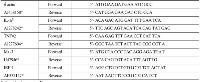
Table 1. Primers for specific PCR
β-actin Forward 5’- ATG GAA GAT GAA ATC GCC
AJ438158* Reverse 5’- CAT GGA GAA GAT CTG GCA
IL-1β Forward 5’- ACA GAC ATG GAT TTT GAA TCA
AJ278242* Reverse 5’- TTC AGC AGT ACA TCA CAG TAT GAG
TNFα2 Forward 5’- CAA GAG TTT GAA CCT CAT TCA
AJ277604* Reverse 5’- GGG TAA TCT ACT TAG CGG GGT A
Mx-3 Forward 5’- ATG CCA CCC TAC AGG AGA TGA T
U47946* Reverse 5’- CCA CAG TGT ACA TTT AGT TG
IRF-1 Forward 5’- AGG CTG TCT GTG CTG TCT ACT AT
AF332147* Reverse 5’- AAT AAC TTC CCG CTC CAT CT
Table 2. The cycling protocol for PCR
Gene Temp. (℃) Time No. of cycles
Denaturising 94 5 min 1
94 45 sec
β-actin Amplification 58 45 sec 24
72 30 sec
Extension 72 10 min 1
Denaturising 94 5 min 1
94 45 sec
IL-1β Amplification 58 45 sec 35
72 30 sec
Extension 72 10 min 1
Denaturising 94 5 min 1
94 30 sec
TNFα2 Amplification 62 20 sec 40
72 20 sec
Extension 72 10 min 1
Denaturising 94 5 min 1
94 45 sec
Mx-3 Amplification 52 45 sec 35
72 45 sec
Extension 72 10 min 1
Denaturising 94 2 min 1
94 30 sec
IRF-1 Amplification 60 30 sec 30
72 20 sec
Extension 72 10 min 1
* : Gene bank accession Nos.
Statistical analysis
Gene expression data were analysed using Kruskal-Wallis ANOVA and Mann-Whitney U-test since the data were not normally distributed.
Results and Discussion
A semi-quantitative RT-PCR method was used to analyse rainbow trout proinflammatory cytokines (IL-1β, TNF-α2) and IFN-related genes (IRF-1, Mx-3) playing an important role in immune regula- tion. Minor differences in input RNA quantity and efficiency of cDNA synthesis were corrected by normalization of the β-actin products and subse- quent expression of the immune gene products as a ratio relative to β-actin.
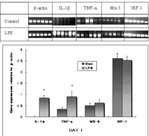
In this study, LPS significantly increased TNFα and IL-1βmRNA level at the doses of 100 ㎍ (Fig.
1). It is known that stimuli that induce NF-κβ translocation to the nucleus and binding to the pro- moter region of TNFαand IL-1βare potent induc- ers of these genes. LPS is a strong inducer of NF-κβ in macrophages, and phorbol myristate acetate (PMA), the classic inducer of proteinase C activity, and TNF itself, also recruit NF-κβin the nucleus (Myers et al., 1994).
Even though, LPS significantly increased TNFα and IL-1βmRNA level at day 1 but the effect was not sustained to day 3 (Fig. 2). This result is consis- tent with other studies. Adarns et al. (1990) have reported that intravenous administration of LPS into both cattle and pigs induces a rapid and transient
Fig. 1. Effect of LPS on immune gene expression in gill at day 1 post-injection. (a) Fish, 6 Rainbow trout for each group, were injected with PBS buffer as a negative control, or LPS (100 ㎍) 24 h before RNA extraction from the gill.
cDNA from 5 ㎍ RNA was titrated to give a constant β- actin product and then used for PCR using specific primers for IL-1β, TNF-α, Mx-3 and IRF-1. (b) Levels of immune gene expression were described as a ratio to that of β-actin gene expression from densitometric-scanned values. The plot represents the mean and S.E for each group. * : signifi- cant difference from the negative control group in the Mann-Whitney test.
Fig. 2. Effect of LPS on immune gene expression in gill at day 3 post-injection. (a) Fish, 6 Rainbow trout for each group, were injected with PBS buffer as a negative control, or LPS 100 ㎍) 72 h before RNA extraction from the gill.
cDNA from 5 ㎍ RNA was titrated to give a constant β- actin product and then used for PCR using specific primers for IL-1β, TNF-α, Mx-3 and IRF-1. (b) Levels of immune gene expression were described as a ratio to that of β-actin gene expression from densitometric-scanned values. The plot represents the mean and S.E for each group. * : signifi- cant difference from the negative control group in the Mann-Whitney test.
elevation of TNFαin the blood as detected in bioas- say. TNFαlevels were proportional to the amount of LPS administered, peaked at about 1-2 h, and were no longer detectable at approximately 4 h. In fish, the TNFα2 gene is also likely to be an early response gene. Zou et al. (2002) demonstrated that TNFα2 expression was increased significantly by LPS from 2 h post-stimulation, reached the highest values at 4 h, decreased from 8 h, and returned to the level in unstimulated cells within 36 h, in head kidney cells.
Both Mx-3 and IRF-1 genes showed a similar pattern of expression when stimulated by LPS in this study (Fig. 1 & Fig. 2). Effects on IFN-related gene (Mx-3 and IRF-1) expression were delayed until day 3, implying an indirect effect of LPS may be occurring. It was reported that the peak expres- sion of the Mx protein in salmon macrophages induced by poly I:C occurred after 48 h whereas peak IFN production was observed by 24 h after addition of poly I:C, suggesting that poly I:C induces the Mx protein indirectly through the activ- ity of type I IFN (Nygaard et al., 2000). Apparently, the increase of those gene expression in LPS inject- ed group at day 3 seem to be due to the lower expression of control group compared to day 1.
However, it is not difficult to compare day 1 and day 3 data since they were detected by semi-quanti- tative method.
The delayed increase of IRF-1 and Mx-3 gene expression is postulated to be due to an indirect effect of LPS, since they are genes known to be induced by other cytokines like IFN. Davidson et al. (1999) also reported the delayed upregulation of another IFN-inducible gene, trout low molecular weight polypeptide (LMP) 2, where there was no difference after stimulation with PHA for 4 h in vitro but upregulation after 24 h, compared to con- trols.
In conclusion, LPS administered intrapenitoneal- ly showed significant stimulatory effects on inflam- matory gene expression i.e. IL-1β, TNF-αand showed a possible indirect stimulatory effect on IFN-related (IRF-1 and Mx-3) gene expression in the gill.
Acknowledgement
This work was supported by Korea Research Foundation Grant (KRF-2004-015-C00613).
References
Adarns, J. L., Semrod, S. D. and Czuprynski, C. J.:
Administration of bacterial lipopolysaccha- ride elicits circulating TNF-a in neonatal calves. J. Clin. Microbiol., 28: 998-1001, 1990.
Campos, M., Godson, D., Hughes, H. P. A. and Babiuk, L. A.: Cytokine applications in infectious disease, In: Ruminant Immunolo- gy (B. M. Goddeeris, ed.), CRC Press, Boca Raton, FL, 229-235, 1993.
Davidson, J., Smith, T. and Martin, S. A. M.:
Cloning and sequence analysis of rainbow trout LMP2 cDNA and differential expres- sion of the mRNA. Fish & Shellfish Immunol., 9: 621-632. 1999.
Hong, S., Zou, J., Crampe, M., Peddie, S., Scapigliati, G., Bols, N., Cunningham, C.
and Secombes, C. J.: The production and bioactivity of rainbow trout (Oncorhynchus mykiss) recombinant IL-1β. Vet. Immunol.
Immunopathol., 81: 1-14, 2001.
Hughes, H. P. A. and Babiuk, L. A.: Potentiation of the immune response by cytokines. In Hand- book of Vertebrate Immunology (P.-P. Pas- toret, P. Griebel, H. Bazin & A. Goaverts,
eds). London: Academic Press. pp. 183-202, 1998.
Myers, M. J. and Murtaugh, M. P.: Biology of Tumour Necrosis Factor. In the Cytokines Handbook, 2nd ed. (ed. A. W. Thomson).
Academic Press, London, 121-151, 1994 Nygaard, R., Husgard, S., Sommer, A. I., Leong, J.
A. C. and Robertsen, B.: Induction of Mx protein by interferon and double-stranded RNA in salmonid cells. Fish & Shellfish Immunol., 10: 435-450, 2000.
Sergey, E. L., Gayle, D., Flynn, M. C. and Plata- Salaman, C. R.: IL-1βsystem (ligand, recep- tor type I, receptor accessory protein and receptor antagonist), TNFα, TGF-β1 and
neuropeptide Y mRNAs in specific brain regions during bacterial LPS-induced anorexia. Brain research Bulletin., 45: 507- 515, 1998.
Zou, J., Wang, T., Hirono, I., Aoki, T., Inagawa, H., Honda, T., Soma, G. I., Ototake, M., Nakan- ishi, T., Ellis, A. E. and Secombes, C. J.: Dif- ferential expression of two tumor necrosis factor genes in rainbow trout, Oncorhynchus mykiss. Dev. Comp. Immunol., 26: 161-172, 2002.
Manuscript Received : November 02, 2005 Revision Accepted : December 10, 2005 Responsible Editorial Member : Jung-Woo Park
(Ulsan Univ.)