Effect of Matrix Metalloproteinases-2 and -9 during IVC-2 on the Development Competence and Gene Expression Profile of Bovine In Vitro-Produced Embryos
Kyeong-Lim Lee1, Jae-Il Bang1, A-Na Ha1, Md. Fakruzzaman1, Chan-Sik Min3 and Il-Keun Kong1,2,†
1Department of Animal Science, Division of Applied Life Science (BK21Plus), Gyeongsang National University, Jinju 660-701, Republic of Korea
2Institute of Agriculture and Life Science, Gyeongsang National University, Jinju 660-701, Republic of Korea
3Gyeongsangnamdo Agriculture Research & Extension Services, Jinju 660-985, Republic of Korea
ABSTRACT
Matrix Metalloproteinases (MMP)-2 and -9 are participated in embryo development, implantation, remodeling of epithelial cell and ovulation. The objective of this study is to evaluate an impact of MMP2 and MMP9 on embryonic developmental competence as well as gene expression profiles of in vitro-produced bovine embryos. After in vitro fertilization, embryos of all groups were transferred into IVC-2 medium treated with MMP2 and MMP9 to check the optimum concentration on the basis of embryo development competence and cell numbers. The optimum concentrations for MMP2 and 9 were 1,200 ng/ml and 300 ng/ml. The blastocyst development competence was not different among 1,200 ng/ml of MMP2 vs. 300 ng/ml of MMP9 vs. combined MMP2 + 9 vs. control groups (41.46 ± 10.66 vs. 37.73
± 8.92 vs. 45.11 ± 11.41% vs. 41.59 ± 11.88, respectively). Furthermore, the developmental competences to hatching and hatched blastocysts were not also different among the same groups (79.84 ± 12.63 vs. 83.3 ± 17.46 vs. 78.55
± 14.48% vs. 72.02 ± 14.09). In addition, total cell number was significantly (p<0.05) greater in blastocyst treated with MMP9 300 ng/ml among all treatment groups. On the other hand, there was no significant difference of ICM vs. TE ratio in all groups. The expression of five out of six genes (i.e., MMP2, MMP9, IFNt, SSLP1 and HNRNPA2B1) was different among the groups. The expression of IFNt and HNRNPA2B1 genes was significantly greater in MMP9 (p<0.05), but there was no difference of MMP9 expression between MMP2 and MMP9 group (p>0.05). The normalized expression of MMP2 and SSLP1 was greater in MMP2 than other groups (p<0.05). In conclusion, MMPs treatment during IVC-2 medium was remarkably effected on blastocyst developmental competence and gene expression profiles that are related to embryo quality and implantation.
(Key words : matrix metalloproteinase, bovine, embryo developmental competence, gene expression)
* This work was partly supported by grant from the Rural Development Administration (Grant No. PJ009321012014) and a scholarship from the BK21 plus program. Kyeong-Lim Lee, A-Na Ha and Md. Fakruzzaman were supported by BK21 plus fellowship in Gyeongsang National University, Republic of Korea.
† Correspondence : E-mail : [email protected]
INTRODUCTION
Matrix Metalloproteinases (MMPs) are able to be controlled exactly its activation by combined with tissue inhibitors of metalloproteinases. MMPs play very pivotal roles for extra- cellular matrix (ECM) remodeling during ovarian follicular development, ovulation and atresia (Imai et al., 2003). Moreover, MMPs is participated in embryo development, implantation, remodeling of epithelial cell and formation of bone, etc. MMP9 expressed most strongly in trophoblast cells of embryo being implanted that had been reported in mouse. A well-timed break- down of ECM composed of structural protein such as collagens, proteoglycans and glucoprotein is essential phenomenon at em-
bryogenesis, morphogenesis, reproduction and absorption and reformation of tissue. Especially, MMPs were denominated matrixins of which perform an important function in this pro- cess of remodeling of ECM, implantation and ovulation (Nagase and Woessner, 1999, McCawley and Matrisian, 2000). Most of MMPs expression is become transcriptional control through cell growth factors, hormones, cytokines, transformation of cell and etc. In goats, MMP2 activity is regulated by co-localized mem- brane-type 1 MMP (MT1-MMP) and tissue inhibitor of metallo- proteinase-2 (TIMP-2), and they control endometrium remodeling during gestation (Uekita et al., 2004). In bovine, MMPs has been played a crucial role during peri-partum, termination of gestation, and post-partum (Walter and Boos, 2001, Takagi et
al., 2007). The detailed expression profiles of gelatinases have not been clarified during implantation; namely, the proteolysis mechanisms of the endometrial ECM are still obscure during implantation in cows (Kizaki et al., 2008). During the implan- tation process, trophoblast cells eliminate epithelial cells, and the epithelium is reorganized (Yamada et al., 2002a, Yamada et al., 2002b).
Several difficulties are arising during implantation process of in vitro-produced embryos. Gelatinases may play a significant role in this process. In situ zymography for gelatinolytic activity established a pattern of activity that corresponded with the localization of MMP-2 and MMP-9 mRNA around developing follicles (Curry et al., 2001). Several researches have been con- ducted about MMP2, 9 regarding their functions related to ovulation, implantation, maturation, and tissue remodeling, etc.
To date, no studies have elucidated the exact role of MMP2 and MMP9 added into medium for development of bovine in vitro-produced embryo. Therefore, this study is the first to find out the influence of MMP2 and MMP9 addition during IVC-2 on embryo developmental competence and further impact on embryo quality as well as gene expression analysis.
MATERIALS AND METHODS
All chemicals and reagents were obtained from Sigma-Aldrich (St. Louis, MO, USA), unless otherwise noted. Experiments were conducted in accordance with Gyeongsang National Uni- versity guidelines for the care and use of laboratory animals (approval no. GAR-110502-X0017).
1. Oocyte Retrieval
Ovaries were obtained from Korean native cows (Hanwoo) at a local abattoir and transported to the laboratory within 2 h in physiological saline (0.9% NaCl) maintained at 35 to 37℃.
Ovaries were washed in fresh Dulbecco’s PBS, and cumulus- oocyte complexes (COCs) were retrieved as described by (Deb et al., 2011). In brief, COCs were recovered from 2 to 8 mm diameter follicles using an 18-G needle attached to a vacuum pump. Only COCs having more than three layers of compact cumulus cells with homogenous cytoplasm were selected in TL- HEPES medium (114 mM sodium chloride, 3.2 mM potassium chloride, 2 mM sodium bicarbonate, 0.34 mM sodium bipho- sphate, 10 mM sodium lactate, 0.5 mM magnesium chloride, 2 mM calcium chloride, 10 mM HEPES, 1 µl/ml phenol red,
100 IU/ml penicillin and 0.1 mg/ml streptomycin) under a ste- reomicroscope.
2. In Vitro Maturation (IVM)
Oocytes were cultured in vitro maturation medium according to (Deb et al., 2011). In brief, collected COCs were washed three times in maturation medium (TCM-199) supplemented with 10% (v/v) fetal bovine serum (FBS), 1 µg/ml estradiol-17 β, 10 µg/ml FSH, 0.6 mM cysteine and 0.2 mM sodium lactate.
The COCs were then incubated in 700 µl IVM medium at 38.5℃ in a humidified atmosphere of 5% CO2 in air for 23 to 24 h.
3. In Vitro Fertilization (IVF) and In Vitro Culture (IVC)
In vitro matured COCs were fertilized with thawed sperm as previously described (Deb et al., 2011). Thawing was per- formed at 36℃ for 1 min, after which sperm were washed and pelleted in Dulbecco’s PBS (D-PBS) by centrifugation at 750
× g for 5 min at room temperature. The pellet was diluted with 500 µl heparin (20 µg/ml) in fertilization (IVF) medium (Tyrode lactate solution supplemented with 6 mg/ml bovine serum albu- min (BSA), 22 µg/ml sodium lactate, 100 IU/ml of penicillin, and 0.1 mg/ml of streptomycin and incubated at 38.5℃ in a humidified atmosphere of 5% CO2 in air for 15 min. Capaci- tated spermatozoa were diluted in IVF medium to 1 to 2 × 10⁶ spermatozoa /ml. Matured oocytes were transfer redin to 700 µl IVF medium containing sperms for 18 to 20 h.
After IVF, cumulus cells of COCs were removed by pipetting and denuded presumed zygotes were placed in 700 µl CR1-aa medium (Hurskainen et al., 1996) supplemented with 44 µg/ml sodium lactate, 14.6 µg/ml glutamine, 10 µl/ml penicillin-strep- tomycin, 3 mg/ml BSA and 310 µg/ml glutathione for 3 days (IVC-I). Cleaved embryos were then cultured until Day 8 of embryonic development (Day 0 = Day of in vitro fertilization) in a medium of the same composition (IVC-I), except that BSA was replaced with 10% (v/v) FBS (IVC-II). Day 8 blastocysts were washed three times in TL-HEPES, transferred into fixative (4% [v/v] paraformaldehyde in 1 M PBS), and stored at 4℃ until cell number determination. For gene expression analysis, Day 8 blastocysts were transferred into a 1.5 ml Eppendorf tube, immediately snap frozen in liquid nitrogen, and stored at —80℃
until use.
4. Differential Staining
Differential staining was performed according to Thouas et
al. (2001) with minor modification. In brief, fixed embryos were washed with 1 mg/ml ployvinylpyrrolidone (PVP) in 0.1 M phosphate buffer saline (PBS) before permeabilization in 0.5%
(v/v) Triton X-100 and 0.1% (w/v) sodium citrate. The stock of propidium iodide (PI) (Sigma, P4170) was prepared by dissol- ving 2.5 mg/ml in PBS and stored at 4℃. For working solution, dilute the stock of PI solution 1:25 in 0.5% Triton X-100 to a concentration of 100 µg/ml. The Hoechst33258 stock was pre- pared by dissolving 25 mg Hoechst 33258 (Sigma, B2883) in 2.5 ml of distilled water (10 mg/ml) and stored at 4℃. On the day of use, the stock solution was diluted of the ratio of 1:1,000 in 4% paraformaldehyde to give a working concentra- tion of 1 µg/ml.
Five hundred µl of PI solution was placed in one well of a SPL (SPL Life Science Co., Cat#30004) 4-well plate and remaining 3 wells were filled with 500 µl of PBS/PVP and placed the four-well plate on a slide warmer at 39℃ for 5∼10 min. After warming, embryos were removed from culture solu- tion as little medium as possible and placed into the PI solution well for 30 sec. After incubation in PI solution, embryos were washed thrice in warm PBS/PVA and moved into a 50 µl droplet of the Hoechst 33258 solution and incubated at room tempera- ture for 15 min. Then the embryos were washed thrice in PBS/
PVA. After washing in PVP-PBS, embryos were mounted onto glass slides and covered with a cover slip and their nuclear configuration was analyzed.
5. RNA Extraction and cDNA Preparation
1) RNA Extraction and Isolation
For RNA extraction, Arcturus picopure RNA isolation kit (ARCTURS; Cat# 12204-01) were used. In brief, 100 µl of extraction buffer was added to sample tube and incubated at 42℃ for 30 min. After incubation, the sample tube was cen- trifuging with 3,000 × g for 2 min, supernatant moved into a new 1.5 ml RNA-free tube. Two hundred µl of conditioning buffer was added into a column tube and incubated at room temperature for 5 min, and then centrifuged at 16,000 × g for 1 min. One hundred µl of 70% ethanol added to extract cells from RNA extraction, and pipetting thoroughly. Then the mix- ture added into a column tube, centrifuged at 100 × g for 2 min, again with 16,000 × g for 30 sec to remove flow through.
Putted 100 µl wash buffer 1 into the column and centrifuged for 1 min at 8,000 × g. DNase I solution (5 µl DNase added
to 35 µl buffer RDD) were prepared and mixed gently. Then 40 µl DNase I mixture were putted directly into the purifica- tion column membrane and incubated at room temperature for 15 min. Again 40 µl wash buffer 1 were placed into the column tube and centrifuged at 8,000 × g for 15 sec. Then 100 µl wash buffer 2 were putted into the column and centrifuged for 2 min at 16,000 × g followed by 1 min centrifuge at 16,000 × g for complete removed of washing buffer. Finally the purification column was transferred into a new 1.5 ml RNasse-free tube and placed 20 µl of elution buffer into center of column and incubated at room temperature for 1 min and centrifuge at 1,000
× g for 1 min followed by centrifuge again at 16,000 × g for 1minute. The RNA samples were used immediately or stored at —80℃ until use.
2) cDNA Synthesis
RNA concentration and purity were checked by NANO drop machine (Thermo Fisher Scientific, NANO DROP 2000c). The mRNA samples were reverse transcribed into first-stand cDNA using Bio-Rad Company. The 15 µl mRNA samples were transferred into a 200-µl eppendorf tube containing 4 µl 5×
iScript Reaction Mixture and 1 µl iScript Reverse Transcrip- tase. The reactions was terminated by heating at 25℃ for 5 min, 42℃ for 30 min, 85℃ for 5 min and finally hold at 4℃.
3) Real Time PCR
The primers were designed using Primer3 software (http://
frodo.wi.mit.edu/) and were presented in Table 1. The qPCR was performed in duplicate in a CFX98 instrument (Bio-Rad Labo- ratories, Hercules, CA) using a 10 µl reaction mixture containing 0.2 mM of each bovine-specific primer, 1× iQ SYBR Green Supermix (iQ SYBR Green Supermix kit, Bio-Rad Labora- tories), and 1.0 µl of cDNA. The cycling conditions were as follows: 95℃ for 3 min followed by 44 cycles of 15 sec at 95℃, 20 sec at 57℃, and 30 sec at 72℃, and a final extension of 5 min at 72℃. Amplification was followed by a melting curve analysis using progressive denaturation, during which the temperature was raised from 65 to 95℃ at a transition rate of 0.2℃ per second. Continuous fluorescence measurements were made during the progressive heating. A negative control was performed for each gene. The PCR products that exhibited only a single fusion temperature, which confirms a unique PCR pro- duct, were retained for further quantitative analysis. The target genes were quantified by the ΔΔ C(t) method using CFX ma-
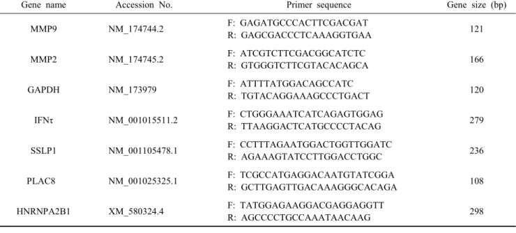
Table 1. Information on primers used for quantitative real-time PCR
Gene name Accession No. Primer sequence Gene size (bp)
MMP9 NM_174744.2 F: GAGATGCCCACTTCGACGAT
R: GAGCGACCCTCAAAGGTGAA 121
MMP2 NM_174745.2 F: ATCGTCTTCGACGGCATCTC
R: GTGGGTCTTCGTACACAGCA 166
GAPDH NM_173979 F: ATTTTATGGACAGCCATC
R: TGTACAGGAAAGCCCTGACT 120
IFNτ NM_001015511.2 F: CTGGGAAATCATCAGAGTGGAG
R: TTAAGGACTCATGCCCCTACAG 279
SSLP1 NM_001105478.1 F: CCTTTAGAATGGACTGGTTGGATC
R: AGAAAGTATCCTTGGACCTGGC 236
PLAC8 NM_001025325.1 F: TCGCCATGAGGACAATGTATCGGA
R: GCTTGAGTTGACAAAGGGCACAGA 108
HNRNPA2B1 XM_580324.4 F: TATGGAGAAGGACGAGGAGGTT
R: AGCCCCTGCCAAATAACAAG 298
nager V1.1 software (Bio-Rad Laboratories, Hercules, CA, USA).
6. Experimental Design
1) Experiment 1 : Find out Optimal Concentration of MMP2, 9 and Embryo Developmental Competence
The first experiment was focused on finding out the optimal concentration of MMP2 and MMP9 during IVC-2 for improve- ment of embryo development competence. To find out the best concentration we added three different concentration of MMP2 (300, 750, 1,200 ng/ml) and MMP9 (100, 300, 600 ng/ml) including control. On the basis of TE cell numbers per blasto- cyst, total cell numbers as well as blastocyst development, we selected optimum concentration for 1,200 ng/ml in MMP2 and 300 ng/ml in MMP9 and used in next step experiment inclu- ding of combined MMP2+MMP9 and control.
2) Experiment 2 : Effect of MMP2 and 9 Additions during IVC-2 on the Gene Expression Profiles of IVP Blastocysts
In this experiment, three treatment groups were used 1,200 ng/ml of MMP2, 300 ng/ml of MMP9, combined MMP2+9 and control groups. The MMP2 and MMP9 were added in IVC2 medium and checked blastocyst developmental competence at D 8. Day 8 blastocysts were washed several times with PBS- PVP and kept in 0.1% pronase for 2∼5 min to dissolve zona pellucida. The blastocysts were putted into a 1.5-ml RNase free
tube with minimum volume of solution, immediately snap frozen in liquid nitrogen, and stored at —80℃ until use.
7. Statistical Analysis
All experiment was performed at least three replications. The embryo development rate (cleavage and blastocyst) and quality (total cell, ICM and TE cell numbers) were expressed as Mean
± SD. Significant differences between groups were detected using Turkey’s and Duncan’s multiple range test (SPSS Inc., Chicago, IL, USA). Differences with p<0.05 were considered significant.
RESULTS
1. Developmental Competence of Blastocysts, Hatching and Cell Number according to Different Concentration of MMP2 and MMP9 during IVC2 Period
The blastocyst development competence of MMP2 and 9 trea- tment during IVC-2 was not different among control vs. 300, 750, 1,200 ng/ml of MMP2 vs. 100, 300, 600 ng/ml of MMP9 groups (43.4 ± 11.9 vs. 51.7 ± 12.1, 44.4 ± 5.9, 47.1 ± 10.2 vs.
33.0 ± 5.5, 36.6 ± 5.4, 40.7 ± 6.1%, p>0.05). Also hatching blastocysts competence was not different among control vs. same MMP2 vs. same MMP9 groups (59.8 ± 19.2 vs. 58.0 ± 15.3, 55.1 ± 17.8, 64.6 ± 12.9 vs. 73.0 ± 24.1, 70.0 ± 12.9, 67.8 ± 17.8%, p>0.05), respectively (Table 2). In addition, total cell
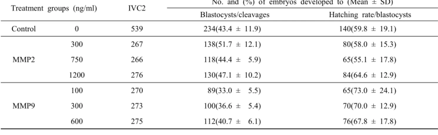
Table 2. Effect of MMP-2 and -9 during IVC-2 on the developmental competence of blastocyst and hatching embryos
Treatment groups (ng/ml) IVC2 No. and (%) of embryos developed to (Mean ± SD) Blastocysts/cleavages Hatching rate/blastocysts
Control 0 539 234(43.4 ± 11.9) 140(59.8 ± 19.1)
MMP2
300 267 138(51.7 ± 12.1) 80(58.0 ± 15.3)
750 266 118(44.4 ± 5.9) 65(55.1 ± 17.8)
1200 276 130(47.1 ± 10.2) 84(64.6 ± 12.9)
MMP9
100 270 89(33.0 ± 5.5) 65(73.0 ± 24.1)
300 273 100(36.6 ± 5.4) 70(70.0 ± 12.9)
600 275 112(40.7 ± 6.1) 76(67.8 ± 17.8)
Table 3. Comparison of cell numbers of Day 8 blastocysts among the groups Treatment groups
(ng/ml)
No. of blastocysts differentially stained
No. of cells counted (Mean ± SD) ICM : TE ratio
Total ICM TE
Control 33 140.6 ± 48.3a,b 31.8 ± 12.9 108.6 ± 47.9 1 : 4.1 ± 2.7
MMP2
300 20 130.5 ± 33.2a,b 40.8 ± 11.9 89.6 ± 24.7 1 : 2.3 ± 0.5
750 14 120.4 ± 26.5a,b 32.3 ± 11.1 88.1 ± 17.7 1 : 3.0 ± 1.0
1,200 12 124.0 ± 34.5a,b 32.2 ± 9.7 91.8 ± 28.5 1 : 3.1 ± 1.3
MMP9
100 13 116.1 ± 24.8a 28.1 ± 10.9 88.0 ± 27.3 1 : 3.9 ± 3
300 11 157.3 ± 39.4b 25.4 ± 8.0 132.4 ± 42.6 1 : 5.9 ± 3.1
600 13 137.6 ± 32.6a,b 27.6 ± 10.7 110.0 ± 33.2 1 : 4.7 ± 2.8
a,b Values with different superscripts in same column denoted were significantly different(p<0.05).
number was significantly (p<0.05) greater in 300 ng/ml of MMP9 treated blastocysts among the all treatment groups, whereas sig- nificantly (p<0.05) lower in 100 ng/ml of MMP9 blastocysts.
On the other hand, there was no significant difference of ICM vs. TE ratio in all groups (Table 3).
We selected optimal concentration of MMP2 and MMP9 from above experiment (Table 1 & 2) and then evaluated blastocyst developmental competence after addition of 1,200 ng/ml of MMP2, 300 ng/ml of MMP9 and 1,200 + 300 of MMP2+9 and control during IVC2. The blastocysts development competence was not significantly different among control vs. 1,200 ng/ml of MMP2 vs. 300 ng/ml of MMP9 vs. MMP2+9 groups (41.59
± 11.88 vs. 41.46 ± 10.66 vs. 37.73 ± 8.92 vs. 45.11 ± 11.41%.
respectively, p>0.05). Furthermore, the development competence of hatching and hatched blastocysts was not also significantly different among control vs. 1,200 ng/ml of MMP2 vs. 300 ng/ml of MMP9 vs. MMP2+9 groups (72.02 ± 14.09 vs. 79.84 ± 12.63
vs. 83.30 ± 17.46 vs. 78.55 ± 14.48%, p>0.05) (Table 4).
2. Gene Expression Profiles of In Vitro-Produced Blastocysts Derived from Different Groups
Table 4. Effect of selected concentration of MMP-2 and -9 on the development of blastocysts and hatching competence
Treatment groups (ng/ml)
IVC2
No. and (%) of embryos developed to (Mean ± SD)
Blastocyst Hatching
rate/blastocysts Control 349 149(41.59 ± 11.88) 104(72.02 ± 14.09) 1,200 MMP2 313 130(41.46 ± 10.66) 103(79.84 ± 12.63) 300 MMP9 349 129(37.73 ± 8.92) 108(83.30 ± 17.46)
1,200+
300 MMP2+9 345 155(45.11 ± 11.41) 118(78.55 ± 14.48)
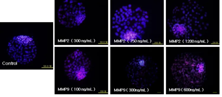
Fig. 1. Representative images of differential stained blastocyst derived from different treatments of MMP2 and MMP9 and control.
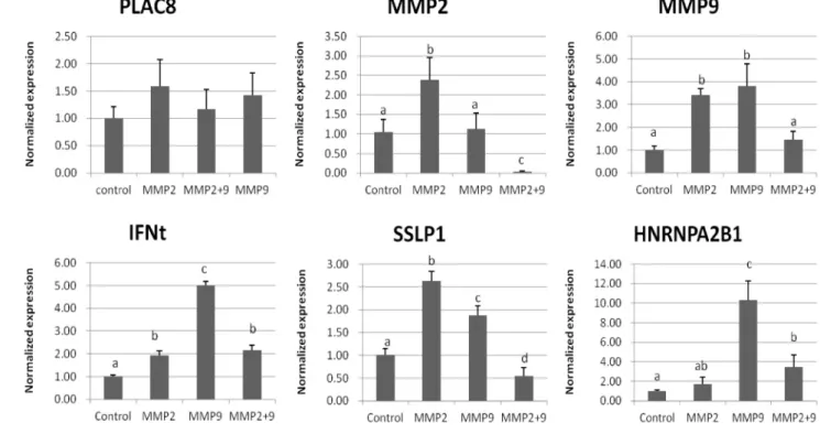
The normalized expression of six embryo biomarker genes namely matrix metalloproteinase 2 (MMP2), matrix metallo- proteinase 9 (MMP9), placenta-specific 8 (PLAC8), interferon-t (IFNt), secreted seminal-vesicle Ly-6 protein 1 (SSLP1) and heterogeneous nuclear ribonucleoprotein A2/B1 (HNRNPA2B1) were investigated. The relative amounts of the studied genes were calculated following ΔΔ C(t) method that normalized against expression of GAPDH reference gene (Fig. 2). The expression of five out of six genes (i.e., MMP2, MMP9, IFNt, SSLP1 and HNRNPA2B1) was different among the groups.
The expression of IFNt and HNRNPA2B1 genes were greater in MMP9 compared to control, MMP2 and MMP9+MMP2 embryos (p<0.05), but there was no significant difference of MMP9 expression between MMP2 and MMP9 embryos (p>
0.05). The normalized expression of MMP2 and SSLP1 were significantly greater in MMP2 than other groups (p<0.05).
DISCUSSION
MMPs play very important role of the embryos hatching and implantation. Process of embryo implantation was invasion and adhesion that takes place between the embryo and the endome- trium (Kim et al., 2002). In human, MMP2 and MMP 9 made in trophoblast and the cultured embryos secreted MMP2 (Puis- tola et al., 1989; Unemori et al., 1991). In the first trimester of human trophoblast was producing MMP2 and MMP9, and also
the cultured embryos secreted MMP2. Fibronectin and laminin were secreted from the embryos that promoted the formation of MMP2 during implantation (Turpeenniemi et al., 1995). MMP9 was highly expressed in mouse blastocysts, and inhibited of extracellular matrix degradation. Extracellular proteases such as serine proteases and MMPs are thought to play pivotal roles for extensive tissue remodeling during both follicular develop- ment and the breakdown of the follicular wall at the time of ovulation (Liu et al., 1998). Another report showed that an ex- tracellular matrix degrading metalloproteinases and their inhibitor are expressed during early mammalian development (Brenner et al., 1989).
MMP2 and MMP9 had a crucial impact on ovulation, im- plantation, remodeling and hatching (Alexander et al., 1996;
Huppertz et al., 1998; Xu et al., 2002; Isaka et al., 2003). In current study, there was no significant difference in blastocyst development and cell numbers among the treatment groups. But several biomarkers gene expression has shown significant diffe- rences among the groups. In our research, IFNτ gene was over- expressed in MMP treatment groups and this was consistent of other researches. Interferon-t originally named trophoblastin or trophoblast protein-1, is the best known specific pregnancy per- ception signal involved in the establishment of early pregnancy in ruminants (Wang et al., 2003). Interferon-t was found to be over-expressed in the hatched blastocyst when compared with the expanded and early stages (Rekik et al., 2011). IFNτ pro-
Fig. 2. Normalized expression levels in Control, MMP2, MMP9 and MMP2+9 embryos by RT-PCR.
duction by bovine embryo begins at the blastocyst stage, and then increases as the conceptus starts to extend (Ealy et al., 2001). HNRNPA2/B1 was over-expressed in MMP treatment groups. The main functions of HNRNPA2B1 included transcrip- tion, alternative pre-mRNA splicing, cytoplasmic trafficking of mRNA and translation (Hutchison et al., 2002; Rekik et al., 2011). HNRNPA2/B1 was confirmed to be down-regulation in the expanded and the hatched blastocysts by 4.8-fold (Rekik et al., 2011). SSLPI gene was up-regulation in MMP treatment groups in this study. SSLP1 was over-expressed in hatched blastocysts (Rekik et al., 2011) and expressed in fetal tissues (Wright et al., 1990), and also be involved in the remodeling of the extracellular matrix and the organization of the mesen- chymal villi of ruminant cotyledons (Ushizawa et al., 2009).
In bovine, PLAC8 gene was reported to be up-regulated in biopsies from blastocysts that led to calf delivery when com- pared with imbibition. PLAC8 is substantially expressed in trophectoderm in preimplantation embryos, and in the tropho- blast giant cells and spongiotrophoblast layer at later stages in development. It was also reported that PLAC8 be up-regulated in the endometrium of pregnant compared to non-pregnant cows (Galaviz-Hernandez et al., 2003). PLAC8, like IFNt, was up- regulated in hatched compared to early blastocysts, which might
confirmed the importance of these two marker genes for embryo apposition and pregnancy induction (Rekik et al., 2011). In our result showed that although there was no significant difference among the groups but the fold changes was higher in MMP2 and MMP9 groups than others.
In conclusion, the present study showed that there was no significant difference of embryo development and cell numbers of embryo, but the gene expression profiles related to pregnancy were up-regulated in addition of MMP2 and 9 during IVC-2.
So, MMPs addition during IVC-2 has positive effect on gene expression profiles.
REFERENCES
Alexander CM, Hansell EJ, Behrendtsen O, Flannery ML, Kish- nani NS, Hawkes SP and Werb Z. 1996. Expression and function of matrix metalloproteinases and their inhibitors at the maternal-embryonic boundary during mouse embryo im- plantation. Development 122: 1723-1736.
Brenner CA, Adler RR, Rappolee DA, Pedersen RA and Werb Z. 1989. Genes for extracellular-matrix-degrading metallo- proteinases and their inhibitor, TIMP, are expressed during early mammalian development. Genes Dev. 3: 848-859.
Curry TE Jr, Song L and Wheeler SE. 2001. Cellular localiza-
tion of gelatinases and tissue inhibitors of metalloproteinases during follicular growth, ovulation, and early luteal formation in the rat. Biol. Reprod. 65: 855-865.
Deb GK, Dey SR, Bang JI, Cho SJ, Park HC, Lee JG and Kong IK. 2011. 9-cis retinoic acid improves developmental competence and embryo quality during in vitro maturation of bovine oocytes through the inhibition of oocyte tumor necrosis factor-alpha gene expression. J. Anim. Sci. 89:
2759-2767.
Ealy AD, Larson SF, Liu L, Alexenko AP, Winkelman GL, Kubisch HM, Bixby JA and Roberts RM. 2001. Polymor- phic forms of expressed bovine interferon-tau genes: Relative transcript abundance during early placental development, promoter sequences of genes and biological activity of pro- tein products. Endocrinology 142: 2906-2915.
Galaviz-Hernandez C, Stagg C, de Ridder G, Tanaka TS, Ko MS, Schlessinger D and Nagaraja R. 2003. Plac8 and Plac9, novel placental-enriched genes identified through microarray analysis. Gene 309: 81-89.
Huppertz B, Kertschanska S, Demir AY, Frank HG and Kauf- mann P. 1998. Immunohistochemistry of matrix metallopro- teinases (MMP), their substrates, and their inhibitors (TIMP) during trophoblast invasion in the human placenta. Cell Tissue Res. 291: 133-148.
Hurskainen T, Hoyhtya M, Tuuttila A, Oikarinen A and Autio- Harmainen H. 1996. mRNA expressions of TIMP-1, -2, and -3 and 92-kD type IV collagenase in early human placenta and decidual membrane as studied by in situ hybridization.
J. Histochem. Cytochem. 44: 1379-1388.
Hutchison S, LeBel C, Blanchette M and Chabot B. 2002. Dis- tinct sets of adjacent heterogeneous nuclear ribonucleoprotein (hnRNP) A1/A2 binding sites control 5' splice site selec- tion in the hnRNP A1 mRNA precursor. J. Biol. Chem.
277: 29745-29752.
Imai K, Khandoker MA, Yonai M, Takahashi T, Sato T, Ito A, Hasegawa Y and Hashizume K. 2003. Matrix metallopro- teinases-2 and -9 activities in bovine follicular fluid of different-sized follicles: Relationship to intra-follicular inhibin and steroid concentrations. Domest. Anim. Endocrinol. 24:
171-183.
Isaka K, Usuda S, Ito H, Saqawa Y, Nakamura H, Nishi H, Suzuki Y, Li YF and Takayama M. 2003. Expression and activity of matrix metalloproteinase 2 and 9 in human tro- phoblasts. Placenta 24: 53-64.
Kim JH, Hong SH, Nah HY, Lee JY, Chae HD, Kim CH, Kang BM and Bae IH. 2002. Influence of transforming growth factor-α on expression of matrix metalloproteinase-2 and matrix metalloproteinase-9 mRNA in mouse embryos. Korean Society of Obstetrics and Gynecology 45: 443-449.
Kizaki K, Ushizawa K, Takahashi T, Yamada O, Todoroki J, Sato T, Ito A and Hashizume K. 2008. Gelatinase (MMP-2 and -9) expression profiles during gestation in the bovine endometrium. Reprod. Biol. Endocrinol. 6: 66.
Liu K, Wahlberg P and Ny T. 1998. Coordinated and cell- specific regulation of membrane type matrix metalloproteinase 1 (MT1-MMP) and its substrate matrix metalloproteinase 2 (MMP-2) by physiological signals during follicular develop- ment and ovulation. Endocrinology 139: 4735-4738.
McCawley LJ and Matrisian LM. 2000. Matrix metallopro- teinases: Multifunctional contributors to tumor progression.
Mol. Med. Today 6: 149-156.
Nagase H and Woessner JF, Jr. 1999. Matrix metalloproteina- ses. J. Biol. Chem. 274: 21491-21494.
Puistola U, Roennberg L, Martikainen H and Turpeenniemi- Hujanen T. 1989. The human embryo produces basement membrane collagen(type IV) degrading protease activity.
Hum. Reprod. 4: 309-311.
Rekik W, Dufort I and Sirard MA. 2011. Analysis of the gene expression pattern of bovine blastocysts at three stages of development. Mol. Reprod. Devel. 78: 226-240.
Takagi M, Yamamoto D, Ohtani M and Miyamoto A. 2007.
Quantitative analysis of messenger RNA expression of matrix metalloproteinases (MMP-2 and MMP-9), tissue inhibitor-2 of matrix metalloproteinases (TIMP-2), and steroidogenic enzymes in bovine placentomes during gestation and post- partum. Mol. Reprod. Dev. 74: 801-807.
Thouas GA, Korfiatis NA, French AJ, Jones GM and Troun- son AO. 2001. Simplified technique for differential staining of inner cell mass and trophectoderm cells of mouse and bovine blastocysts. Reprod. Biomed. Online 3: 25-29.
Turpeenniemi-Hujanen T, Feinberg RF, Kauppila A and Puis- tola U. 1995. Extra-cellualr matrix interactions in early human embryos: Implications for normal implantation events. Fertil Steril 64: 132-138.
Uekita T, Yamanouchi K, Sato H, Tojo H, Seiki M and Tachi C. 2004. Expression and localization of matrix metallopro- teinases (MT1-MMP, MMP-2) and tissue inhibitor of metal- loproteinase-2 (TIMP-2) during synepitheliochorial placen-
tation of goats (Capra hircus). Placenta 25: 810-819.
Unemori EN, Hibbs MS and Amento EP. 1991. Constitutive expression of a 92-kD gelatinase (type V collagenase) by rheumatoid synovial fibroblasts and its induction in normal human fibroblasts by inflammatory cytokines. J. Clin. In- vest. 88: 1656-1662.
Ushizawa K, Takahashi T, Hosoe M, Kizaki K and Hashizume K. 2009. Characterization and expression analysis of SOLD1, a novel member of the retrotransposon-derived ly-6 super- family, in bovine placental villi. PLoS. One. 4: e5814.
Walter I and Boos A. 2001. Matrix metalloproteinases (MMP-2 and MMP-9) and tissue inhibitor-2 of matrix metallopro- teinases (TIMP-2) in the placenta and interplacental uterine wall in normal cows and in cattle with retention of fetal membranes. Placenta 22: 473-483.
Wang B, Xiao C and Goff AK. 2003. Progesterone-modulated induction of apoptosis by interferon-tau in cultured epithelial cells of bovine endometrium. Biol. Reprod. 68: 673-679.
Wright RM, John E, Klotz K, Flickinger CJ and Herr JC.
1990. Cloning and sequencing of cDNAs coding for the human intra-acrosomal antigen SP-10. Biol. Reprod. 42:
693-701.
Xu P, Alfaidy N and Challis JR. 2002. Expression of matrix metalloproteinase (MMP)-2 and MMP-9 in human placenta and fetal membranes in relation to preterm and term labor.
J. Clin. Endocrinol. Metab. 87: 1353-1361.
Yamada O, Todoroki J, Kizaki K, Takahashi T, Imai K, Patel OV, Schuler LA and Hashizume K. 2002a. Expression of prolactin-related protein I at the fetomaternal interface during the implantation period in cows. Reproduction 124: 427- 437.
Yamada O, Todoroki J, Takahashi T and Hashizume K. 2002b.
The dynamic expression of extracellular matrix in the bovine endometrium at implantation. J. Vet. Med. Sci. 64: 207-214.
(Received: 2014. 2. 27/ Reviewed: 2014. 5. 10/ Accepted: 2014. 5. 19)