J Korean Soc Pediatr Nephrol 2013;17:149-153 DOI: http://dx.doi.org/10.3339/jkspn.2013.17.2.149
Copyright © 2013 The Korean Society of Pediatric Nephrology ISSN 1226-5292 (print) ISSN 2234-4209 (online)
비전형적 혈전성 미세병증 1례
연세대학교 의과대학 소아청소년과, 아주대학교 의과대학 소아청소년과*, 연세대학교 의과대학 임상병리학과†
국민건강보험 일산병원 임상병리과‡, 국민건강보험 일산병원 소아청소년과§
오지영ᆞ박세진*ᆞ김기환ᆞ임범진†ᆞ정현주†ᆞ기정혜‡ᆞ김기혁§ᆞ신재일
A Case of Atypical Thrombotic Microangiopathy
We report the case of a 14-year-old girl, diagnosed with atypical thrombotic microangiopathy (TMA). The patient presented with persistent fever, nausea, and newly developed peripheral edema. Her laboratory findings indicated chronic anemia with no evidence of hemolysis, thrombocytopenia, or elevated serum creatinine level. A few days after hospitalization, acute renal failure and fever worsened, and proteinuria developed. On day 40 of hospitalization, she experienced a generalized tonic seizure for 5 min, accompanied by renal hypertension. Brain magnetic resonance imaging revealed posterior reversible leukoencephalopathy syndrome. After steroid pulse therapy, a renal biopsy was performed because of delayed recovery from thrombocytopenia. The biopsy findings showed features of thrombotic microangiopathic hemolysis with fibrinoid change restricted. Current diagnostic criteria for TMA have focused on thrombotic thrombocytopenic purpura and hemolytic uremic syndrome, and diagnosis is based on the clinical presentation and etiology, with the consequence that idiopathic and atypical forms of TMA can be overlooked.
Developing effective tools to diagnose TMA, such as studying levels of ADAMTS13 or testing for abnormalities in the complement system, will be the first step to improving patient outcomes.
Key words: Fever, Anemia without hemolysis, Thrombocytopenia, Atypical throm- botic microangiopathy (TMA), Renal biopsy
Ji Young Oh, M.D., Se Jin Park, M.D.*, Ki Hwan Kim, M.D., Beom Jin Lim, M.D.
†, Hyeon Joo Jeong, M.D.
†, Jung Hye Ki, M.D.
‡, Kee Hyuck Kim, M.D.
§, and Jae Il Shin, M.D.
Department of Pediatrics, Severance Children's Hospital, Yonsei University College of Medicine Department of Pediatrics*, Ajou University Hospital, Ajou University School of Medicine, Department of Pathology†, Yonsei University College of Medicine, Department of Pathology,‡ National Health Insurance Corporation Ilsan Hospital, Department of Pediatrics§, National Health Insurance Corporation Ilsan Hospital, Goyang, Korea
Corresponding Author: Jae Il Shin Department of Pediatrics, Yonsei University College of Medicine, Seoul, Korea
Tel: 02-2228-2050, Fax: 02-393-9118 E-mail: [email protected]
Received: 20 September 2013 Revised: 11 October 2013 Accepted: 11 October 2013
This is an open-access article distributed under the terms of the Creative Commons Attribu- tion Non-Commercial License (http:// crea- tivecom mons.org/licenses/bync/3.0/) which permits unrestricted non-commercial use, distribution, and reproduction in any medium, provided the original work is properly cited.
Introduction
Traditionally, the classical form of thrombotic microangiopathy (TMA) has been diagnosed on clinical criteria which include microangiopathic hemolytic anemia with schistocytes, generalized microvascular occlusion induced by circulated thrombus, and thrombocytopenia caused by platelet consumption [1]. Thrombotic thrombocytopenic purpura (TTP) and hae
molytic uraemic syndrome (HUS) are the most classical forms in the TMA categories; these show different the manifestations but have the same pathological findings [2]. There has been a tendency to consider that only TTP and HUS can be identified as TMA.
Rrecently, however a few cases reported identifying atypical form of TMAs in nonhemolytic anemia or non
thrombocytopenic patients with renal failure [35].
Here, we describe a 14yearold girl who presented as the first case of TMA diagnosed without hemolytic anemia in Korea.
Case report
A 14yearold girl was transferred to Yonsei Medical Center with fever and nausea lasting for 3 weeks. Despite treatment with multiple antibiotics for 2 weeks at her prior hospital, she did not improve, and her symptoms were worsened. In addition, peripheral edema had de
veloped 3 days before admission to our hospital. On physical examination, she had a fever of 38.5℃, blood pres
sure of 118/80 mmHg, heart rate of 90 beats/minute, a respiratory rate of 20 breaths/minute. She was acutely illlooking, but was not pale. Liver and spleen were not palpable, and no definite tenderness was noticed on her abdomen. She had no lymphadenopathy. Her lung sound was decreased at both lung field and heart beat was regular without murmur. No skin rash was found on her body, and peripheral pitting edema was shown. Her neurologic examination presented normal findings.
The laboratory findings on her first day in our hospital were hemoglobin 12.4 g/dL, hematocrit 35.9%, white blood cell count 9,420/mm3, and platelet count 93,000/
mm3. No erythrocyte fragments or schistocytes were seen on a peripheral blood smear, and reticulocyte count was 1.24%. Direct and indirect coombs were ne
gative. Albumin level was 2.1 g/dL, and the other liver function tests were normal. Blood urea nitrogen (BUN) was 12.8 mg/dL and creatinine was 1.16 mg/dL. Lactate dehydrogenase level was 222 IU/L and total bilirubin was 0.4 mg/dL. Disseminated intravascular coagulation profile were fibrinogen 461 mg/dL; ddimer 4,206 ng/
mL; antithrombin III 66%; F.D.P. 32.9 µg/mL. Erythrocyte sedimentation rate was 70 mm/hr and Creactive protein was 189 mg/L. Plasma C3 and C4 level was normal, and antinuclear antibody serologic test was negative.
On urinalysis, hematuria and pyuria were present.
A protein of 24hour urine showed 1,622 mg (43 mg/
m2/hr). Urine β2microglobulin was 0.45 mg/L (normal range: 00.25), and cystatin C was 2.2 mg/L. Glomerular filtration rate based on the cystatin C was 30 ml/min/
1.73m2.
Chest Xray showed bilateral pleural effusions without focal infiltrations. The abdominopelvic ultrasound showed splenomegaly (13.1 cm) and diffusely increased cortical echogenicity of both kidneys without corticomedullary junction obliteration.
Due to persistent fever and thrombocytopenia from initial lab findings, her first diagnosis was considered idiopathic thrombocytopenic purpura due to severe infection of unknown organism. Under the first impres
sion, intra venous immunoglobulin (IVIG, dose; 1 g/
kg) was administered for 3 days; however, fever and thrombo cytopenia persisted. Proteinuria was started at 5 days after admission, and the patient’s urination was gradually decreased despite administration of diuretics.
Anemia and thrombocytopenia proceeded over time;
thus, on the 20th hospital day, her hemoglobin level was 7.7 g/dL and platelet count was 23,000/mm3. At that time, reti culocyte was 2.56% and no erythrocyte fragments or schistocytes were seen on a peripheral blood smear.
Lactate dehydrogenase level was 292 IU/L and total bilirubin was 0.4mg/dL. Renal biopsy was shown to be helpful in the diagnosis but was delayed because of thrombocytopenia. On 17th19th hospital days, steroid pulse therapy (dose; 20mg/kg/day) was done; a week later, fever started to subside and urination was increased. A protein of 24hour urine also decreased from 1,622 mg to 557 mg (14 mg/m2/hr) but still noted. However, in the week following the steroid therapy, thrombocytopenia had developed again, and we administered steroid pulse therapy again. After the second steroid therapy, thrombocytopenia improved; however, on the 37th hospital day, 7 days after the last steroid pulse therapy, there was sudden elevation of systolic blood pressure,
115/80 mmHg to 180/100 mmHg despite administration of hypertensive medication. On the 40th hospital day, she had generalized tonic seizure for 5 minutes, and a brain MRI was performed. The MRI findings suggested posterior reversible leukoencephalopathy syndrome, and an antiepileptic drug was started (Fig. 1).
On the 41st hospital day, her platelet count rose to 213,000/mm3 and hemoglobin rose to 9.8 g/dL. At the 42nd hospital day, renal biopsy was performed. Renal biopsy findings presented features of thrombotic micro
angiopathic hemolysis with fibrinoid change (Fig. 24).
On the 50st hospital day, her platelet count and hemog
lobin level were stable, and BUN and creatinine fell to 17.2 mg/dL and 0.57 mg/dL, respectively. Steroid were tapered and she was discharged. There was no more proteinuria on urinalysis 3 months later at the outpatient clinic. Also she tapered out hypertensive drugs after 6 months later and her blood pressure was 110/65 mmHg without drugs.
Discussion
We report a 14yearold patient with non hemolytic anemia, thrombocytopenia, and acute renal failure who only on the renal biopsy showed an evidence of hemolysis, one of the findings of atypical microangiopathy.
In 1998, Fogo et al. reported an atypical form of TMA at American journal of kidney disease that was very similar to our case [7]. A 50yearold woman was admitted to hospital with a threeweek history of oliguria, and her laboratory findings indicated thrombocytopenia and anemia without evidence of hemolysis on peripheral blood. Only renal biopsy findings showed diffuse loss
Figure 1.
Fig. 1. Multiple cortical or subcortical areas of hyperintense signal in bilateral parietal and occipital lobes.
Figure 2.
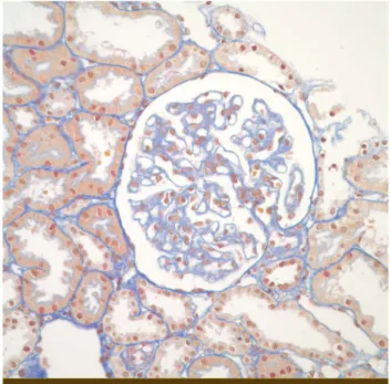
Fig. 2. Glomeruli compacted with impacted red cells (periodic acid-Schiff stain, ×400).
Figure 3.
Fig. 3. Light microscopic features of TMA; The glomerular basement membrane is diffusely thickened with a double contour feature.
Segmented red cells is within glomerulus (Mallory's trichrome stain, ×400).
of endothelial cells and erythrocyte fragmentation, which strongly suggested a microangiopathic hemolytic process.
These two cases showed that, despite microangiopathic hemolytic anemia being one of the typical finding in TMA, hemolysis could occur only in the kidney, without any evidence of peripheral smear. Some other cases also emphasized that lack of the evidence of hemolysis on per
ipheral blood does not exclude the possibility of TMA [6].
Because mortality of TMA is still high, early diagnosis of this disease leads to optimal management and improved outcomes [1]. Current diagnostic criteria of TMAs are fo cused on TTPHUS, based on the clinical presentation and etiology, with the consequence that idiopathic and atypical form of TMAs are overlooked and may not be considered for timely therapeutic interventions [8]. There fore, atypical form of TMAs are started to draw attention that pathologic findings were consistent with TMA but complete hematologic criteria of TMA are not satisfied [8].
Recently, diagnostic markers such as ADAMTS13 or abnormal mechanism of the complement system have been studied to improve the accuracy of diagnosis of TMA [9].
To improve clinical outcomes, developing more effective diagnostic tools for TMA should be a first step.
한글요약
응고성 미세혈관병증은 빠른 진단이 예후에 중요한 인자 이나, 현재의 진단 기준에 따라서는 thrombotic thrombo
cytopenic purpura, haemolytic uremic syndrome 외의 비전형적인 응고성 미세혈관병증의 진단이 늦어짐에 따라 나쁜 예후를 초래하게 되는 경우가 많다고 보고되어 있다.
본 저자들은 시행한 혈액 검사상 용혈의 증거가 없는 빈혈, 혈소판 감소증 그리고 급성 신부전을 보인 소아 환아에서 신조직 검사를 통해 비특이적 응고성 미세혈관병증을 진단 받은 1증례를 보고하고자 한다.
14세 여자 환아는 3주간 지속된 발열, 구역과 전신 부종 을 주소로 본원으로 전원되었다. 내원하여 시행한 혈액 검 사상 빈혈과 혈소판 감소증을 보였으나, 용혈의 증거는 없 었으며, 혈정 크레아티닌이 증가되어 있었다. 내원 이후 급 성 신부전과 발열은 지속적으로 진행되었으며, 소변 검사 상 단백뇨가 발생하였다. 환아는 내원 40일경 신고혈압과 동반된 전신 경련이 5분간 있어 뇌 자기 공명 영상을 촬영 하였으며, 가역성 후백질 뇌병증 증후군의 양상을 보여 항 경련제 투여를 시작하였다. 이후 지속되는 혈소판 감소증 및 발열은 고용량 스테로이드 치료를 진행한 후 호전되었 으나, 급성 신부전 및 단백뇨가 지속되어 신장 조직 검사를 진행하였으며, 검사 결과상 혈전성 미세혈관병증의 소견을 보였다. 이와 같이 조직검사상에서는 응고성 미세혈관병증 을 보이나 전형적인 응고성 미세혈관병증의 혈액학적인 진 단 기준이 충족되지 않는 비특이적 형태의 응고성 혈관병 증의 효과적인 진단을 위하여 보체 기전이나 ADAMTS 13 와 같은 유전자 범위의 보다 활발한 연구를 통한 효과적인 진단 기준의 마련이 되어야 할 것으로 보인다.
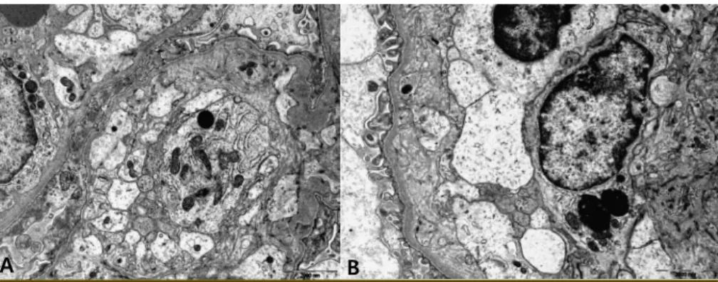
Figure 4.
A B
Fig. 4. Electron microscopic features of TMA ; The glomerular basement membrane presents diffuse expansion of endothelial zone with deposition of electron lucent material. Capillary loops are narrowing compacted with fibrinogen thrombus (×400).
References
1) Bahloul M, Dammak H, Kallel H, Khlaf-Bouaziz N, Ben Hamida C, Chaari A, et al. [Thrombotic microangiopathies. Incidence, pathogenesis, diagnosis, treatment and prognosis]. J Mal Vasc 2007;32:75-82.
2) Coppo P, Veyradier A, Durey MA, Fremeaux-Bacchi V, Scrobohaci ML, Amesland F, et al. [Pathophysiology of thrombotic micro- angiopathies: current understanding]. Ann Med Interne (Paris) 2002;153:153-66.
3) Morel-Maroger L, Kanfer A, Solez K, Sraer JD, Richet G. Prog- nostic importance of vascular lesions in acute renal failure with microangiopathic hemolytic anemia (hemolytic-uremic syndrome): clinicopathologic study in 20 adults. Kidney Int 1979;15:548-58.
4) Akashi Y, Yoshizawa N, Oshima S, Takeuchi A, Kubota T, Kondo S, et al. Hemolytic uremic syndrome without hemolytic anemia: a case report. Clin Nephrol 1994;42:90-4.
5) Brilliant SE, Lester PA, Ohno AK, Carlon MJ, Davis BJ, Cushner HM. Hemolytic-uremic syndrome without evidence of micro- angiopathic hemolytic anemia on peripheral blood smear.
South Med J 1996;89:342-5.
6) De Serres SA, Isenring P. Athrombocytopenic thrombotic microangiopathy, a condition that could be overlooked based on current diagnostic criteria. Nephrol Dial Transplant 2009;
24:1048-50.
7) Goral S, Horn R, Brouillette J, Fogo A. Fever, thrombocytopenia, anasarca, and acute renal failure in a 50-year-old woman. Am J Kidney Dis 1998;31:890-5.
8) De Serres SA, Isenring P. Renal thrombotic microangiopathy revisited: when a lesion is not a clinical finding. Saudi J Kidney Dis Transpl 2010;21:411-6.
9) Chapman K, Seldon M, Richards R. Thrombotic microangio- pathies, thrombotic thrombocytopenic purpura, and ADAMTS- 13. Semin Thromb Hemost 2012;38:47-54.