298
Prolonged Extreme Thrombocytosis in a Postsplenectomy Patient with Hereditary Spherocytosis
Yae Min Park, M.D., Jinny Park, M.D., Ph.D., Yunjeong Jo, M.D., Sei-Hyun Kim, M.D., Kwen-Chul Shin, M.D., In Sik Won, M.D., Sun Jin Sym, M.D., Eun Kyung Cho, M.D.,
Ph.D., Dong Bok Shin, M.D., Ph.D. and Jae Hoon Lee, M.D., Ph.D.
Department of Internal Medicine, Gachon University of Medicine and Science, Gil Hospital, Incheon, Korea
We report a case of prolonged extreme reactive thrombocytosis in a post-splenectomy patient with heredi- tary spherocytosis. A 29-year-old female patient presented with gall stones detected incidentally by ab- dominal ultrasonography. Her laboratory findings showed hemolytic anemia with spherocytosis on the peripheral blood smear and increased osmotic fragility. She was diagnosed with hereditary spherocytosis and underwent a laparoscopic cholecystectomy and splenectomy. After undergoing surgery, the hemolytic anemia was resolved but thrombocytosis was newly detected. Nineteen months after the splenectomy, the thrombocytosis was still persistent and extremely high. To our knowledge, this is the first report of a prolonged extreme reactive thrombocytosis after a splenectomy in Korea. (Korean J Hematol 2009;44:298-303.)
Key Words: Reactive thrombocytosis, Postsplenectomy, Hereditary spherocytosis
접수:2009년 8월 29일, 수정:2009년 12월 7일 승인:2009년 12월 10일
교신저자:박진희, 인천시 남동구 구월동 1198
405-780, 가천의과학대학교 길병원 내과 Tel: 032-460-8209, Fax: 032-432-4355 E-mail: [email protected]
Correspondence to:Jinny Park, M.D., Ph.D.
Department of Internal Medicine, Gachon University of Medicine and Science, Gil Hospital
1198, Guwol-dong, Namdong-gu, Incheon 405-780, Korea Tel: +82-32-460-8209, Fax: +82-32-432-4355
E-mail: [email protected] INTRODUCTION
Thrombocytosis is frequently encountered as an incidental laboratory finding.1) The common etiologies of reactive thrombocytosis are infec- tion, trauma, surgery, and occult malignancy.
Splenectomy was found to be one of the main causes of thrombocytosis. The probability of thrombocytosis in patients who have had splenec- tomy is about 75∼82% and about 9% of all re- active thrombocytosis occurrences are caused by this procedure. The platelet count in reactive thrombocytosis is expected to normalize after the resolution of the underlying condition.2)
We experienced a patient of prolonged extreme
thrombocytosis for more than 19 months after splenectomy with hereditary spherocytosis with- out any complications such as bleeding or throm- bosis.
CASE REPORT
In June 2007, a 29-year-old female patient pre- sented with incidentally detected gall stones by an ultrasound of the abdomen. She had no sig- nificant past medical or surgical history and de- nied any medications. Her family history was unremarkable. Physical examination showed no tenderness or rebound tenderness in the pal- pation on right upper quadrant but her spleen was palpable. Laboratory values were significant
Fig. 1. Peripheral blood smear shows normocytic normo- chromic anemia with spherocytes which are small hyper- chromatic cells without the usual clear area in the center (Wright stain, ×400).
Fig. 2. Abdominopelvic computed tomography shows sub- tle high density in gall bladder suggesting GB stone and splenomegaley of 155 mm.
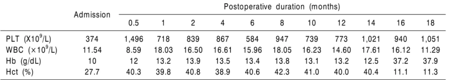
Table 1. Characteristics of blood count in relation to hospital course
Admission Postoperative duration (months)
0.5 1 2 4 6 8 10 12 14 16 18
PLT (X109/L) 374 1,496 718 839 867 584 947 739 773 1,021 940 1,051
WBC (×109/L) 11.54 8.59 18.03 16.50 16.61 15.96 18.05 16.23 14.60 17.61 16.12 11.29
Hb (g/dL) 10 12 13.2 13.9 13.5 13.4 13.8 13.1 13.2 12.5 37.2 37.9
Hct (%) 27.7 40.3 39.8 40.8 38.9 40.6 42.3 41.0 40.0 40.4 11.1 11.3
Abbreviations: PLT, platelet; WBC, white blood cell; Hb, hemoglobine; Hct, hematocrete.
for a hemoglobin (Hb) level of 10.0 g/dL, a hema- tocrit (Hct) of 27.7%, white blood cell count (WBC) of 11.54×10⁹/L (neutrophil 69.5%, lym- phocyte 26%) and a platelet count of 374×109/L.
The reticulocyte count was 14.9% and corrected reticulocyte count was 9.2%. The peripheral blood smear (PBS) showed spherocytosis (Fig. 1). The total bilirubin was 4.7 mg/dL with 3.6 mg/dL of indirect bilirubin and AST, ALT were 17 U/L and 22 U/L. The LDH was 366 IU/L, the alka- line phosphatase 39 IU/L, and the gamma-GT 23 IU/L. And direct and indirect Coombs’ tests and anti-nuclear antibody test were negative but the patient’s osmotic fragility was increased. Com- puted tomography (CT) of the abdomen showed multiple gall stones and splenomegaly of largest diameter 155 mm (Fig. 2). She was diagnosed as
hereditary spherocytosis and underwent laparo- scopic cholecystectomy and splenectomy.
Her platelet count was 266×109/L immediately after the splenectomy but abnormally elevated to 1,496×109/L two weeks after the surgery. Other laboratory findings showed Hb level of 12.8 g/dL, a Hct of 40.3%, WBC of 8.59×10⁹/L and we did not detect the spherocytes on the PBS any more.
Hemolytic anemia is improving but thrombocy- tosis, which was not present prior to splenectomy, was noted and persistent. There is no evidence of infection or trauma by the medical history and physical examination.
Four months after the splenectomy, her platelet count was still elevated at 867×109/L with Hb level of 13.5 g/dL. She hadn’t taken any medi- cation or show any symptoms, signs of trauma, infection or inflammation (Table 1). We per-
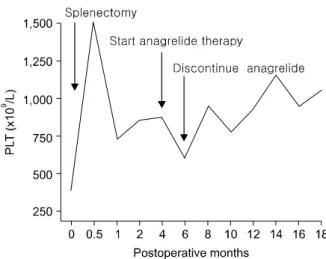
Fig. 3. The platelet count was increased after splenectomy.
After the anagrelide therapy, the count was decreased, but after the discontinuation of the anagrelide therapy, it began to increase again. 19 months after having underwent sple- nectomy, extreme thrombocytosis is persistent.
formed chest X-ray, chest CT and abdomi- nopelvic CT to rule out any other causes of sec- ondary thrombocytosis such as acute or chronic inflammatory conditions including post operative inflammatory lesions, infectious focuses, or occult malignant lesions. But the test results did not show any other specific abnormal findings.
Therefore, we performed bone marrow aspiration and biopsy to rule out myeloproliferative dis- orders. However, variable cellularity for her age as 20∼50% (overall 30%), with normal mega- karyocytic count and morphology was revealed.
Also, the erythroid and granulocytic elements were normal in proportion and maturation. It re- vealed adequate storage of iron without any pathologic ringed sideroblasts.
At the first period of thrombocytosis, she com- plained of headaches, dizziness, and a digital tin- gling sensation, and consequently, she received anagrelide 0.5 mg qid per day for cytoreduction with an aspirin 100 mg per day for antiplatelet agent. After the anagrelide therapy, platelet count was decreased and it showed 584×109/L after 2 months receiving anagrelide (Table 1). The sym- ptoms were resolved and anagrelide treatment was discontinued but the antiplatelet agent was continued. After the discontinuation of the ana-
grelide therapy, the platelet count began to in- crease again (Table 1, Fig. 3). At 8 months after the splenectomy, we performed second bone mar- row aspiration and biopsy because thrombocy- tosis was persistent but there were no abnormal findings.
Now it is 19 months after undergoing splenec- tomy, and extreme thrombocytosis is persistent.
But now, with only the help of the antiplatelet agent, she has been asymptomatic and working full-time with no complications, despite her pe- ripheral blood platelet count being over 1,000×
109/L for more than 12 months.
DISCUSSION
The definition of thrombocytosis varies among authors but is most commonly defined as a plate- let count >500×109/L.2) and extreme thrombocy- tosis defined as platelet counts >1,000×109/ L.3) Thrombocytosis generally either is a reactive process (secondary thrombocytosis) or is caused by a clonal bone marrow (myeloproliferative) dis- order; the latter category includes essential throm- bocythemia.1)
Reactive thrombocytosis is a common cause of thrombocytosis, the response to infection, trauma, or surgery.4) In one study of patients with throm- bocytosis, reactive thrombocytosis was diagnosed in 70% and primary thrombocytosis in only 22%.5) Similarly, in patients with extreme throm- bocytosis, reactive thrombocytosis is a more com- mon cause of thrombocytosis than primary or es- sential thrombocytosis. In another study of 280 patients with a platelet count >1,000×109/L, re- active thrombocytosis was the cause in more than 80% and myeloproliferative disorder in only 14%.3) In that study the etiologies of extreme re- active thrombocytosis are infection (31%), post- splenectomy or hyposplenism (19%), malignancy (14%), trauma (14%), non infectious inflamma- tion (9%), blood loss (6%).3) Therefore, splenec- tomy was found to be one of the main causes of extreme reactive thrombocytosis. Another study
shows 75% of individuals without myeloprolifer- ative disorders developed thrombocytosis after splenectomy.6)
The spleen plays a major role in platelet regu- lation, as it is the primary site of destruction of platelets, which is why thrombocytosis is seen with hyposplenism.7,8) Reactive thrombocytosis is a predictable finding after splenectomy, with the platelet count peaking at 1 to 3 weeks and return- ing to normal levels in weeks, months, and rarely, years.2)
Regardless of cause, a high platelet count has the potential to be associated with vasomotor (headache, visual symptoms, lightheadedness, atypical chest pain, acral dysesthesia, erythrome- lalgia), thrombotic, or bleeding complication.9) The association of thrombocytosis with thrombo- sis and hemorrhage appear to be related to qual- itative rather than quantitative platelet abnor- malities.10) Clonal involvement of megakaryocyto- poiesis is regarded as the main origin of throm- boembolism in myeloproliferative (MPD) dis- order and results in abnormal platelet production.
These platelets show increased size heterogeneity and ultrastructural abnormalities, and their func- tion in vitro is in many ways impaired with a high degree of individual variability. Elevated levels of platelet-specific proteins, increased thromboxane generation, and expression of acti- vation-dependent epitopes on the platelet surface are common on chronic MPD, and may reflect an inappropriate state of platelet activation. Al- though a variety of platelet receptor deficiencies and some defects of intracellular signaling path- ways have been identified, the different platelet defects in MPD could not be traced back to an underlying general pathogenetic mechanism. On progression of chronic MPD to more advanced stages of the disease, the number of platelet ab- normalities tend to increase.11)
Postsplenectomy venous thrombosis is usually associated with platelet counts >600×109/L to > 800×109/L,12) and occurs in approximately 5% of patients.13) Less commonly, postsplenectomy
thrombocytosis results in arterial thrombosis that leads to stroke or myocardial infarction.10) But these hemostatic events infrequently occur in pa- tients with reactive thrombocytosis.3,14) This is presumably due to the fact that the interaction of platelets with the vessel wall remains qual- itatively normal in secondary thrombocytosis.1) Neurologic complications including chronic head- ache or dizziness, or focal neurologic sign occur in about 25 percent of patients with essential thrombocythemia, and may be manifested as non- specific symptoms.15) Neurologic complications are presumably caused by platelet-mediated cere- brovascular ischemia.1)
The first step in managing a patient who pres- ents with elevated platelet count is to determine if the etiology is a primary process or a reactive response,1) because the platelet count in reactive thrombocytosis is expected to normalize after res- olution of the underlying condition.7) Furthermore because their abnormal platelet count itself does not place them at risk for hemostatic or vascular events, patients with reactive thrombocytosis gen- erally do not require platelet-lowering or anti- platelet treatment.1) If it is a primary process in- cluding essential thrombosis, the immediate risk to the patient from the increased platelet count and additional risk factors for thrombotic compli- cations include advanced age, a history of throm- bosis, hypercholesterolemia and cigarette smok- ing should be assessed.1) Thereafter, management of the thrombocytosis and prevention of compli- cations should be initiated. Some pharmacologic agents used for this purpose are acetyl salicylic acid, ticlopidine, enoxaparin, hydroxyurea, ana- grelide, interferon alpha along with associated adverse effects such as hemorrhage, myelofib- rosis, leukemic transformation.7) Anagrelide is a newer platelet-lowering agent, an orally ad- ministered quinazoline derivative that inhibits megakaryocyte proliferation and differentiation.
It has been approved in patients with essential thrombocytosis, and now has been established as alternative first-line therapy to reduce the plate-
let count.1,7) Anagrelide is nonleukemogenic and is therefore a particularly reasonable initial op- tion in young patients who require long term pla- telet count control.1) Common side effects include fluid retention, palpitations, and arrhythmias, heart failure, headaches, and anemia.1) Long-term data on the side effects and complications of ana- grelide are lacking, however, preliminary data suggest it is well tolerated, with mild to moderate anemia as a frequent side effect.
So after weighing the benefits versus the risks of various treatment plans, determine whether re- duction of platelet numbers or simple observation is indicated. Although the degree of elevation in the platelet count does not correlate with the risk of thrombosis, control of the platelet count by cy- toreduction does reduce the frequency of throm- bosis in some patients.1) Our patient showed ex- treme thrombocytosis after the splenectomy and the first period of postsplenectomy thrombocy- tosis, she complained of headaches, dizziness and a digital tingling sensation, and consequently she received cytoreductive therapy and antiplatelet therapy.
But 2 months after cytoreductive therapy, it was discontinued because the symptoms were resolved. Now it is 19 months after the splenec- tomy, and she has been asymptomatic and work- ing full-time without any complications, despite her peripheral blood platelet count being over 1,000×109/L for a long duration. To our knowl- edge, this is the first report of a prolonged ex- treme reactive thrombocytosis in postsplenectomy patient in Korea.
요 약
혈소판증가증은 흔히 관찰되는 검사소견이다.
특히 극도의 혈소판증가증의 경우 이차성 혈소판 증가증이 대부분을 차지하며 이의 흔한 원인으로 는 감염, 외상, 수술, 종양 등이 있다. 이차성 혈소 판증가증은 원인 질환이 해결되면 대부분 호전되 어 치료를 요하는 경우는 흔하지 않다. 비장절제 술 또한 이차성 혈소판증가증의 주된 원인이며
비적출술을 받은 환자들 중 약 75∼82%에서 나 타날 수 있지만 수년간 지속되는 경우는 매우 드 물다. 저자는 우연히 발견된 담석과 용혈성빈혈 을 주소로 내원한 환자에서 유전구형적혈구증을 진단하고 이에 대한 치료로 비장절제술을 시행한 후 용혈빈혈은 호전되었으나 19개월 이상의 장기 간 극도의 혈소판증가증이 지속되는 환자를 경험 하였기에 문헌고찰과 함께 보고한다.
REFERENCES
1) Schafer AI. Thrombocytosis. N Engl J Med 2004;
350:1211-9.
2) Greer JP, Foerster J, Rodgers GM, et al. Wintrobe's Clinical Hematology: Philadelphia: Lippincott Willi- ams & Wilkins, 2008:1128-34.
3) Buss DH, Cashell AW, O'Connor ML, Richards F 2nd, Case LD. Occurrence, etiology, and clinical sig- nificance of extreme thrombocytosis: a study of 280 cases. Am J Med 1994;96:247-53.
4) Santhosh-Kumar CR, Yohannan MD, Higgy KE, al-Mashhadani SA. Thrombocytosis in adults: analy- sis of 777 patients. J Intern Med 1991;229:493-5.
5) Tefferi A, Ho TC, Ahmann GJ, Katzmann JA, Greipp PR. Plasma interleukin-6 and C-reactive pro- tein levels in reactive versus clonal thrombocytosis.
Am J Med 1994;97:374-8.
6) Boxer MA, Braun J, Ellman L. Thromboembolic risk of postsplenectomy thrombocytosis. Arch Surg 1978;
113:808-9.
7) Khan PN, Nair RJ, Olivares J, Tingle LE, Li Z.
Postsplenectomy reactive thrombocytosis. Proc (Bayl Univ Med Cent) 2009;22:9-12.
8) Bullen AW, Losowsky MS. Consequences of im- paired splenic function. Clin Sci (Lond) 1979;57:
129-37.
9) Johnson M, Gernsheimer T, Johansen K. Essential thrombocytosis: underemphasized cause of large-ves- sel thrombosis. J Vasc Surg 1995;22:443-7.
10) Daya SK, Gowda RM, Landis WA, Khan IA.
Essential thrombocythemia-related acute ST-segment elevation myocardial infarction. A case report and literature review. Angiology 2004;55:319-23.
11) Wehmeier A, Südhoff T, Meierkord F. Relation of platelet abnormalities to thrombosis and hemor- rhage in chronic myeloproliferative disorders. Semin Thromb Hemost 1997;23:391-402.
12) Hayes DM, Spurr CL, Hutaff LW, Sheets JA.
Post-splenectomy thrombocytosis. Ann Intern Med
1963;58:259-67.
13) Stamou KM, Toutouzas KG, Kekis PB, et al.
Prospective study of the incidence and risk factors of postsplenectomy thrombosis of the portal, mesen- teric, and splenic veins. Arch Surg 2006;141:663-9.
14) Buss DH, Stuart JJ, Lipscomb GE. The incidence of
thrombotic and hemorrhagic disorders in association with extreme thrombocytosis: an analysis of 129 cases. Am J Hematol 1985;20:365-72.
15) Kesler A, Ellis MH, Manor Y, Gadoth N, Lishner M.
Neurological complications of essential thrombocy- tosis (ET). Acta Neurol Scand 2000;102:299-302.