J o u r n a l o f R h e u m a t i c D i s e a s e s V o l . 1 9 , N o . 3 , J u n e , 2 0 1 2
http://dx.doi.org/10.4078/jrd.2012.19.3.160 □ Case Report □
160
<Received:July 29, 2011, Revised:(1st: September 28, 2011, 2nd: September 29, 2011), Accepted:September 30, 2011>
Corresponding to:Jinseok Kim, Division of Rheumatology, Department of Internal Medicine, Jeju National University Hospital, 1753-3, Ara 1-dong, Jeju 690-121, Korea, E-mail: [email protected]
pISSN: 2093-940X, eISSN: 2233-4718
Copyright ⓒ 2012 by The Korean College of Rheumatology
This is a Free Access article, which permits unrestricted non-commerical use, distribution, and reproduction in any medium, provided the original work is properly cited.
An Atypical Case of Plasmodium vivax Malaria after Initiating Adalimumab Therapy
Sang Yop Shin1, Gil Myeong Seong1, Young Ree Kim2, Jin Woo Kang3, Jinseok Kim1
Departments of Internal Medicine1 and Laboratory Medicine2, Jeju National University Hospital, Department of Medicine, Jeju National University School of Medicine3, Jeju, Korea
We report an unusual case of Plasmodium vivax malaria that occurred in a 22-year-old ankylosing spondylitis pa- tient after initiating adalimumab therapy. P. falciparum malaria was initially included as a possible differential di- agnosis due to hyperparasitemia and similar features in
the peripheral blood smear. The patient was successfully treated with conventional therapy for P. vivax malaria.
Key Words. Adalimumab, Malaria vivax, Spondylitis anky- losing
Introduction
Indigenous malaria was thought to have been eliminated from South Korea until a case reemerged in 1993. Most of the cases have been due to Plasmodium vivax; P. falciparum malaria has not been reported as an indigenous case. Known endemic areas for P. vivax malaria in Korea include some northern regions of Kyunggi province and a sector of the de- militarized zone (DMZ) (1). The diagnosis of malaria is estab- lished using a peripheral blood smear and morphological char- acteristics of individual parasites are often helpful in confirm- ing the diagnosis. We report a case of P. vivax malaria in a patient receiving adalimumab that had similar blood smear findings as P. falciparum malaria.
Case Report
A 22-year-old male was admitted because of fever and chills.
The patient had been previously diagnosed with ankylosing spondylitis (AS) 1-year previously and adalimumab had been initiated 1-month prior to the current admission. The patient had completed military service 6 months previously in Yeunchun, Kyunggi province, which is an endemic area for P. vivax malaria. After military service, the patient moved
back to his hometown, Jeju Island, and did not travel outside the island for the following 6 months. The patient had been in typical health until 7 days prior to the current admission, when subjective fever, chills, fatigue, and malaise developed.
Seven days after symptom development, the patient came to the outpatient clinic. He denied cough, sputum, gastro- intestinal symptoms, shortness of breath, or night sweats. The patient reported a fluctuating fever over the previous 7 days.
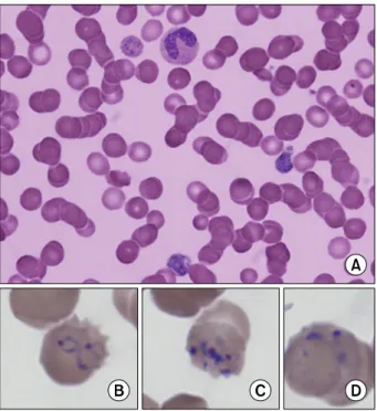
On examination, vital signs showed body temperature of 40.0°C, blood pressure 130/80 mmHg, pulse rate 100 beats per minute, and respiratory rate 18 breaths per minute. Lung and heart sounds were clear. Examination of the abdomen re- vealed no palpable mass or hepatosplenomegaly. Initial labo- ratory findings were white blood cell count 3,900/mm3 (neutrophils 48.1%, eosinophils 11.7%), hemoglobin 12.3 g/dL, platelet count 45,000/mm3, AST/ALT 30/19 IU/L, crea- tinine 1.1 mg/dL, erythrocyte sedimentation rate 39 mm/hour, and C-reactive protein 16.27 mg/dL. A peripheral blood smear revealed numerous trophozoites, schizonts, and ring forms in red blood cells (RBC). Hyperparasitemia was also observed (parasites per white blood cell: 180 per 100). Of note, there were multiple ring forms in one RBC and some were located
Atypical Plasmodium vivax Infection with Adalimumab 161
Figure 1. Peripheral blood smear findings show many trophozoites, schizonts and ring forms (×1,000) (A). Multiple ring forms are present in a red blood cell (B, C), located peripherally (D).
peripherally (Figure 1). On the basis of these findings, adali- mumab therapy was stopped and chloroquine was begun under the suspicion of malaria. By the second day after treatment the patient became afebrile and parasitemia was improved (parasites per white blood cell: 5 per 100). By the fifth day after treatment, parasitemia disappeared in the peripheral blood smear. Polymerase chain reaction-based molecular anal- ysis showed a positive result for P. vivax. Primaquine was ad- ministered for 14 days. After being discharged, the patient had no relapse or adverse event during a 2-year follow-up.
Discussion
We report a case of P. vivax malaria that presented with atypical features in a peripheral blood smear after beginning adalimumab therapy. The blood smear showed marked para- sitemia and multiple ring forms located peripherally in the RBCs. These features are commonly seen in the blood smear of P. falciparum malaria, whereas they are rarely evident in P. vivax malaria (2). However, several clues pointed the diag- nosis towards P. vivax malaria. First, the patient was stationed in a P. vivax-endemic area during military service and had not traveled abroad before service. Second, after military service the patient returned to Jeju Island, a non-endemic area for malaria. Thirdly, the patient had never received a blood transfusion. Furthermore, P. falciparum malaria has never been reported in South Korea. Therefore, it was reasonable
to believe that the P. vivax parasites had infected the subject during the military service, remained dormant as hypnozoites in the liver, and reactivated after a latent period of more than 6 months. Interestingly, it was only a month after the subject commenced adalimumab therapy that the symptoms manifested. These two events may have occurred by chance, or the adalimumab treatment may have triggered reactivation of P. vivax malaria.
Adalimumab is a tumor necrosis factor-alpha (TNF-α) in- hibitor that is now broadly used in patients with autoimmune diseases like rheumatoid arthritis or ankylosing spondylitis.
Systematic reviews have reported that TNF-α inhibitors in- crease the risk of serious infection by 2-4-fold (3).
Furthermore, the risk is particularly relevant to intracellular organisms like tuberculosis, histoplasmosis, and listeriosis be- cause of its immunosuppressive effect on cell-mediated im- munity (3,4). No clear association has been reported between TNF-α inhibitors and malaria infection, although an anti- malarial effect of TNF-α has been suggested (5). According to an ex vivo study on P. vivax malaria, TNF-α plays a vital part in aggregating monocytes/macrophages, activating mono- cytes to secrete reactive nitrogen intermediates, and para- site-killing materials (6). Therefore, TNF-α inhibitors may at- tenuate the antimalarial effect and increase the risk of malaria infection.
Our case presents a circumstance where a latent infection may have reemerged a month after commencing adalimumab therapy. The agent may have influenced the host immune sys- tem to be more susceptible for the multiplication of parasites, resulting in hyperparasitemia and atypical features in the pe- ripheral blood smear.
In contrast, it has been reported that TNF-α inhibitor may help treating cerebral malaria caused by P. falciparum. The mechanistic explanation is that TNF-α inhibitor can suppress overwhelming production of TNF-α seen in the infection (7).
Excessive production of TNF-α induced by P. falciparum in- fection plays an important role in pathogenesis of severe malaria. The local production of cytokines by activated mono- cytes and macrophages leads to the activation of endothelial cells, followed by up-regulation of endothelial cell adhesion molecules. This process finally results in sequestration of par- asite erythrocytes within small vessels of major organs, one of the major complications of P. falciparum malaria (8).
Geraghty et al. reported a case of cerebral malaria in a 45-year-old woman undergoing treatment with infliximab for rheumatoid arthritis (9). Overwhelming parasitemia (estimated to be > 50%) was present in the case even though there ap- peared to be a lack of severe manifestations, and the patient
162 Sang Yop Shin et al.
made a salutary recovery. This seems to be a result of block- ing two conflicting actions of TNF-α simultaneously. In other words, although blocking antiplasmodial effects of TNF-α re- sulted in enhanced multiplication of P. falciparum malaria, it also may have restricted excessive TNF-α production, con- tributing to a comparatively quick recovery without any complication.
In our case, the final diagnosis was P. vivax malaria. To our knowledge there has been no report regarding atypical features shown in P. vivax infected patients using a TNF-α inhibitor.
Within a day of chloroquine treatment, clinical symptoms and signs dramatically improved, as did parasitemia. This suggests that blood features similar to P. falciparum in this setting do not always require early antifalciparum treatment, and that conventional treatment is sufficient in treating P. vivax malaria. This may be because P. vivax malaria is relatively benign and chloroquine-sensitive (10). Ahn et al. reported a case of P. vivax malaria in a renal transplant patient after the commencement of immunosuppressant therapy, which showed atypical findings in the peripheral blood smear similar to our case; the patient was treated successfully with conventional antimalarial therapy (11).
Conclusion
TNF-α inhibitor therapy could result in atypical features of peripheral blood smears in P. vivax infection. Therefore, ob- taining relevant clinical information is important to make the proper diagnosis. Further research would be needed to better understand the atypical features of malarial infection under an- ti-TNF-α treatment.
References
1. Ahn MH. Traveling and imported parasitic diseases. J Korean Med Assoc 2007;51:993-1004.
2. Wassmer SC, Cianciolo GJ, Combes V, Grau GE.
Inhibition of endothelial activation: a new way to treat cerebral malaria? PLoS Med 2005;2:e245.
3. Bongartz T, Sutton AJ, Sweeting MJ, Buchan I, Matteson EL, Montori V. Anti-TNF antibody therapy in rheumatoid arthritis and the risk of serious infections and malig- nancies: systematic review and meta-analysis of rare harmful effects in randomized controlled trials. JAMA 2006;295:2275-85.
4. Curtis JR, Patkar N, Xie A, Martin C, Allison JJ, Saag M, et al. Risk of serious bacterial infections among rheu- matoid arthritis patients exposed to tumor necrosis factor alpha antagonists. Arthritis Rheum 2007;56:1125-33.
5. Cush JJ. Safety overview of new disease-modifying anti- rheumatic drugs. Rheum Dis Clin North Am 2004;
30:237-55.
6. Karunaweera ND, Carter R, Grau GE, Kwiatkowski D, Del Giudice G, Mendis KN. Tumour necrosis factor-de- pendent parasite-killing effects during paroxysms in non-immune Plasmodium vivax malaria patients. Clin Exp Immunol 1992;88:499-505.
7. Richards AL. Tumour necrosis factor and associated cyto- kines in the host's response to malaria. Int J Parasitol 1997;27:1251-63.
8. Muniz-Junqueira MI. Immunomodulatory therapy asso- ciated to anti-parasite drugs as a way to prevent severe forms of malaria. Curr Clin Pharmacol 2007;2:59-73.
9. Geraghty EM, Ristow B, Gordon SM, Aronowitz P.
Overwhelming parasitemia with Plasmodium falciparum infection in a patient receiving infliximab therapy for rheumatoid arthritis. Clin Infect Dis 2007;44:e82-4.
10. Kager PA, Wetsteyn JC. Malaria and drug resistance. Ned Tijdschr Geneeskd 1996;140:151-5.
11. Ahn JH, Heo YS, Kang ET, Yu SH. A case of malaria in the kidney transplanted patients with maintenance immunosuppression. Korean J Med 2000;59:540-3.