Needling Procedures for Calcific Tendinitis Performed by Orthopedic Surgeons
Chae Hyun Pang, Dong Ho Kum, Jeung Yeol Jeong, Seung Min Park, Jae Chul Yoo
Department of Orthopedic Surgery, Samsung Medical Center, Sungkyunkwan University School of Medicine, Seoul, Korea
Background: Common and effective treatments for calcific tendinitis involve needling procedures. However, it has been widespread practice to refer patients with calcific tendinitis, which is a predominantly orthopedic condition, to radiology department. The purpose of this study was to compare clinical and radiological outcomes after ultrasound-guided needling for calcific tendinitis between the ortho- pedics and radiology department.
Methods: Seventy-seven shoulders (Group 1) and 38 shoulders (Group 2) treated in the radiology and orthopedic department, respec- tively. A fellowship-trained orthopedic surgeon and a musculoskeletal radiologist each performed the procedure of ultrasound-guided needle decompression with subacromial steroid injection. Clinical outcomes was evaluated using the visual analogue scale for pain (pVAS) and the American Shoulder and Elbow Surgeons (ASES) shoulder score before treatment and at each follow-up. The pre- and post- needling size and shape of the calcific deposits were compared between the two groups.
Results: We analyzed a total of 56 shoulders for Group 1 and 32 shoulders for Group 2. The mean age and sex ratio of the patients no significantly different. We found that the mean decrease in the diameter of calcification between pre- and post-needling was 9.0 mm for Group 1 and 13.1 mm for Group 2; the difference was significantly larger in Group 2 than in Group 1. Both groups showed improved pVAS and ASES scores after needling but the extent of these improvements did not differ with the type of operator.
Conclusions: Needling decompression performed by orthopedic surgeons could a viable option for the treatment of calcific tendinitis.
(Clin Shoulder Elbow 2017;20(2):84-89)
Key Words: Shoulder joint; Rotator cuff; Tendinopathy; Ultrasonography; Interventional; Calcific tendinitis Clinics in Shoulder and Elbow Vol. 20, No. 2, June, 2017
https://doi.org/10.5397/cise.2017.20.2.84
Received January 15, 2017. Revised April 2, 2017. Accepted April 16, 2017.
Correspondence to: Jae Chul Yoo
Department of Orthopedic Surgery, Samsung Medical Center, Sungkyunkwan University School of Medicine, 81 Irwon-ro, Gangnam-gu, Seoul 06351, Korea
Tel: +82-2-3410-3501, Fax: +82-2-3410-0061, E-mail: [email protected] IRB approval (No. 2016-12-069).
Financial support: None. Conflict of interests: None.
Introduction
Calcific tendinitis of the shoulder is one of the most common diseases among the middle-aged population. The incidence of calcific tendinitis has been reported to range between 2.7%
and 54%.1-5) Recently, the prevalence of calcific tendinitis in the rotator cuffs of patients with subacomial pain has been shown to reach 42.5%, compared to just 7.8% in individuals without sub- acromial pain.6) The treatment of symptomatic calcific tendinitis with functional restriction can be broadly divided into conserva- tive treatment and surgical treatment. Primary treatment usu- ally includes injection of non-steroidal anti-inflammatory drugs
(NSAIDs) or other drugs, such as Cimetidine.7) If symptoms do not resolve with primary treatment, secondary treatment, such as extracorporeal shock wave therapy and ultrasound (US)-guid- ed needling are performed. If symptoms still remain recalcitrant, then surgical treatment, such as arthroscopic surgery may be performed whereby calcific deposits are surgically excised.8)
Several papers concerning the methods and outcomes of US- guided needling have been published.9-15) But despite the fact that calcific tendinitis is principally an orthopedic disease, to the best of our knowledge, no study has investigated the outcomes of calcific tendinitis after US-guided needling performed at an orthopedic department. Rather most studies have used findings
that are derived from treatments performed by skilled radiolo- gists.9-13,15-17) Even though the results of US examinations have been shown to be highly operator-dependent, these examina- tions when conducted by a skilled orthopedic have been report- ed to show similar outcomes to when they are conducted by a radiologist.18,19)
We hypothesized that the outcomes of US-guided needling performed at the orthopedic department would be similar to the outcomes of the same procedure performed at the radiology department. To this end, we comparatively analyzed the clinical and the radiologic outcomes of US-guided needle decompres- sion with subacromial steroid injection in patients with calcific tendinitis performed at the two departments (radiography vs.
orthopedic) and determined the relative effectiveness and supe- riority of US-guided needling performed at each department.
Methods
Patient Selection
We retrospectively analyzed the outcomes of patients with calcific tendinitis who had undergone US-guided needle de- compression with subacromial steroid injection between June 2011 and December 2013. Whether treated at Samsung Medi- cal Center or in a different hospital, patients who had not shown clinical or radiological improvement despite administration of more than three months of drug therapy, for which they had received US-guided needle decompression with subacromial steroid injection, were recruited in our study. The following inclusion criteria were applied: 1) involvement of the calcific tendinitis in either the supraspinatus muscle or the infraspinatus muscle; 2) calcific tendinitis that could be classified as either Type A (dense homogenous calcification with clear contours) or Type B (dense fragmented calcification with clear contours) based on the French Arthroscopic Society classification (FAS) system20); and 3) deposits with diameters larger than 5 mm (measured on radiographs; the threshold value is based on find- ings of a previous study21)). We excluded patients who had a full thickness rotator cuff tear, diagnosed using US; a preexisting history of extracorporeal shock wave therapy or needling on the affected side; calcific tendinitis caused by surgery or fracture; in- fection; concomitant inflammatory arthropathy or arthritis of the humeral joint; and diffused or fragmented calcification without clear contours. A total of 73 patients (77 shoulders) were recruit- ed between June 2011 and June 2012 and treated by a muscu- loskeletal radiologist with over 10 years of experience (Group 1), and a total of 38 patients (38 shoulders) were recruited between July 2012 and December 2013 and treated by an orthopedic surgeon trained as fellowship for one year (Group 2). Amongst the recruited patients, only the patients who participated in at least a six-month follow-up were enrolled in the study, leading to a final study population of 56 shoulders in Group 1 and 32
shoulders in Group 2.
Measurement of Clinical and Radiologic Parameters With the patients’ shoulders in neural rotation, plain radio- graphs were taken in the anteroposterior view, the true antero- posterior view, axial and lateral views, and the arch view. We measured the size of calcific deposits at the point of the largest diameter on radiographs and classified them according the FAS classification system.20) Clinical outcome was measured using two parameters—the visual analogue scale for pain (pVAS) and the American Shoulder and Elbow Surgeons (ASES) shoulder score. Both radiologic and clinical scores were measured before needling and at every post-needling follow-up. The first follow- up was performed after a month of the treatment, and the number of subsequent follow-ups until the final follow-up varied depending on the severity of each patient’s symptoms. The final follow-up of the patient was conducted using a telephone sur- vey, through which we obtained patients’ final pVAS and ASES scores.
Ultrasound Therapy
The same course of US therapy was administered by both departments. The head of the bed was raised, the patient was inclined in Fowler’s position, and the affected shoulder was exposed for diagnostic US scan. The area of calcification was depicted by the US scan. First, the affected area was sterilized using betadine and alcohol. Then, over the affected area, an US probe covered with a sterilized film and a sterilized US gel were used to generate the scans. We injected local anesthesia (10 ml of 1% lidocaine, a 23-gauge needle) into the skin, subcutaneous tissues, and the subacromial bursa. Guided by continuous US imaging, we next performed aspiration of the calcific deposits by using an 18-gauge needle (10 ml syringe). If the calcific de- posits could not be fully aspired at the first attempt, two or three more attempts of aspirations were made. After decompression, we performed multiple punctures until the needle reached the whole range of the calcification. Once the needling was per- formed, we injected a mixture of 40 ml of 1% lidocaine and 1 ml of steroid (triamcinolone acetonide, 40 mg/ml) at the sub- acromion, under direct US guidance. Post-needling, the patients were prescribed 3 days of NSAIDs.
Statistical Analysis
We performed all statistical analyses using the SAS ver. 9.4 (SAS Institute, Cary, NC, USA). Continuous demographic vari- ables (age, follow-up period, pre- post-needling pVAS, ASES, and deposit size) were analyzed using the t-test and the Mann- Whitney U-test. Categorical variables (gender, involved side, dominant arm, and FAS classification) were analyzed using the chi-square test, the Fisher’s exact test, and univariate analysis.
Those found to show a meaningful association through the uni-
variate analysis were further analyzed using multivariate analysis and multiple linear regression analysis. Statistical level was set to p<0.05.
Results
The final study population comprised of 56 patients in Group 1 and 32 patients in Group 2. The average age of the patients at the time of treatment was 57.1 years in Group 1 and 54.0 years in Group 2. We found that the average follow-up period was significantly longer in Group 1 than in Group 2 (46.7 months vs. 39.8 months). The male-to-female gender ratios were 13:43 and 11:21, respectively. Generally, calcific tendinitis involved the right shoulder, and the dominant arm was more often affected than the non-dominant arm. The calcific deposits were classified according to the FAS classification, as either Type A or Type B.
We observed that the ratios of FAS classification (ratio of Type A to Type B) significantly differed between the groups: Group 1 showed a significantly higher proportion of patients with Type A calcific tendinitis than Group 2 (55:1 vs. 27:5) (Table 1).
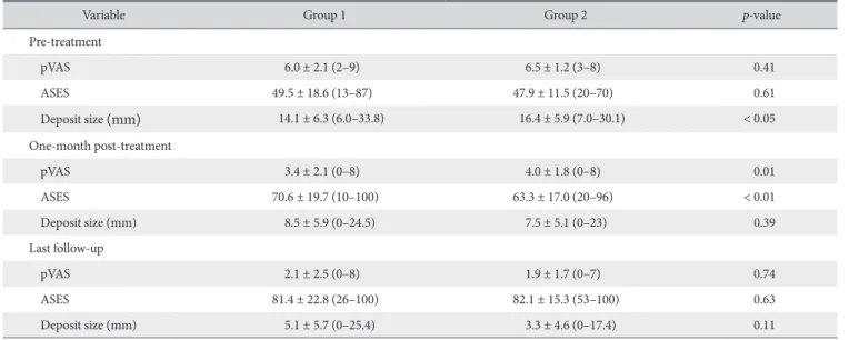
We found the pre-needling pVAS and ASES did not signifi- cantly differ between the two groups, but the size of calcific deposits was significantly larger in the latter group (14.1 mm vs.
16.4 mm). At one-month follow-up, we found that the pVAS was significantly lower and the ASES, significantly higher in Group 1 than in Group 2. However, the size of calcific deposits did not differ between the groups. At the final follow-up, no significant between-group differences were observed (Table 2).
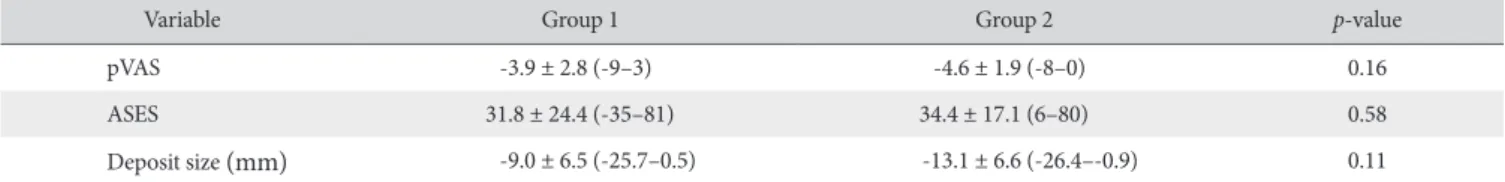
Compared to Group 1, Group 2 was associated with a signifi- cantly larger change in deposit size between the pre-needling and the final follow-up time points. Significant differences were not observed in other variables across these time points (Table 3).
Table 1. Demographic Data of Patients
Vaariable Group 1 Group 2 p-value
Age (yr) 57.1 ± 8.2 (40–79) 54.0 ± 9.6 (34–81) 0.12
Follow-up duration (mo) 46.7 ± 19.0 (6–64) 39.8 ± 8.6 (9–49) <0.0001
Ratio of sex (male:female) 13:43 11:21 0.26
Ratio of involved side (right:left) 38:18 22:10 0.93
Ratio of dominant arm (right:left) 54:2 32:0 0.53
Dominant side affected 40 33 0.79
Ratio of FAS classification (Type A: Type B)* 55:1 27:5 0.02
Values are presented as mean ± standard deviation (range) or number only.
Group 1: radiologist performed, Group 2: fellowship-trained orthopedic surgeon performed, FAS: French Arthroscopic Society.
*Type A: dense homogenous calcification with clear contours, Type B: dense fragmented calcification with clear contours.
Table 2. Univariate Analysis of Results between Groups
Variable Group 1 Group 2 p-value
Pre-treatment
pVAS 6.0 ± 2.1 (2–9) 6.5 ± 1.2 (3–8) 0.41
ASES 49.5 ± 18.6 (13–87) 47.9 ± 11.5 (20–70) 0.61
Deposit size (mm) 14.1 ± 6.3 (6.0–33.8) 16.4 ± 5.9 (7.0–30.1) < 0.05
One-month post-treatment
pVAS 3.4 ± 2.1 (0–8) 4.0 ± 1.8 (0–8) 0.01
ASES 70.6 ± 19.7 (10–100) 63.3 ± 17.0 (20–96) < 0.01
Deposit size (mm) 8.5 ± 5.9 (0–24.5) 7.5 ± 5.1 (0–23) 0.39
Last follow-up
pVAS 2.1 ± 2.5 (0–8) 1.9 ± 1.7 (0–7) 0.74
ASES 81.4 ± 22.8 (26–100) 82.1 ± 15.3 (53–100) 0.63
Deposit size (mm) 5.1 ± 5.7 (0–25.4) 3.3 ± 4.6 (0–17.4) 0.11
Values are presented as mean ± standard deviation (range).
Group 1: radiologist performed, Group 2: fellowship-trained orthopedic surgeon performed, pVAS: visual analogue scale for pain, ASES: American Shoulder and Elbow Surgeons shoulder score.
Multivariate analysis revealed a statistically significant be- tween-department difference in pVAS measured at one-month follow-up but not in ASES. Comparing the change in calcific de- posits between the pre-needling and final follow-up time points, we observed there was a between-department difference that was statistically significant (Table 4–6).
Discussion
Until now, studies on US-guided needling for calcific ten- dinitis have generally used outcomes of treatments performed by radiologists.9-13,15-17) To the best of our knowledge, there are no studies that have used the outcomes of US-guided needling
performed by orthopedic surgeons. Yet US and US-guided treatment have been reported to be effective for other diseases whether they are performed by a radiologist or by an orthopedic surgeon. For instance, an US study on cuff tears reported that the results between a radiologist and an orthopedic surgeon with one year’s experience did not significantly differ.18,19) Even more, for the US diagnosis of foot and ankle diseases, another study showed that US and US-guided treatments performed by a skilled orthopedic surgeon yielded better results in terms of patient satisfaction, the promptness of treatment, and the medi- cal expense incurred by the patient.22) Thus, we investigated whether this treatment even when performed by a fellowship- trained orthopedic surgeon with relatively less experience than a skilled radiologist can give comparable clinical and radiological results. We compared the between-department differences in pain, functional scores, and size of calcific deposits after US- guided needling and performed univariate and multivariate analyses to determine whether the differences were associated with the type of operator or whether there were any confound- ing variables.
We found that the average change in size of the calcification was significantly larger in Group 2 than in Group 1 for which association the multivariate analysis revealed there were no confounding factors. But given that the calcific deposits were significantly larger in Group 2 than in Group 1 before needling and the same at the final follow-up, it implies that the reduction in calcification is independent of the operator. At one-month Table 3. Univariate Analysis of the Change in Variables between Pre-treatment and Final Follow-up between Group
Variable Group 1 Group 2 p-value
pVAS -3.9 ± 2.8 (-9–3) -4.6 ± 1.9 (-8–0) 0.16
ASES 31.8 ± 24.4 (-35–81) 34.4 ± 17.1 (6–80) 0.58
Deposit size (mm) -9.0 ± 6.5 (-25.7–0.5) -13.1 ± 6.6 (-26.4–-0.9) 0.11
Values are presented as mean ± standard deviation (range).
Group 1: radiologist performed, Group 2: fellowship-trained orthopedic surgeon performed, pVAS: visual analogue scale for pain, ASES: American Shoulder and Elbow Surgeons shoulder score.
Table 4. Multivariate Analysis Using Logistic Regression of Factors Associated with US-guided Needle Decompression with Subacromial Steroid Injection and the pVAS Measured at 1-month Post-treatment
Variable Exp(B) 95% CI p-value
Age -0.003 -0.056–0.05 0.91
Follow-up duration -0.001 -0.03–0.028 0.94
Involved side -0.009 -1.034–1.016 0.99
Dominant arm -2.06 -5.109–0.989 0.18
FAS classification -0.548 -2.352–1.256 0.55
US: ultrasound, pVAS: visual analogue scale for pain, Exp(B): exponentiation of the B coefficient,CI: confidence interval, FAS: French Arthroscopic Soci- ety.
Table 5. Multivariate Analysis Using Logistic Regression of Factors Associated with US-guided Needle Decompression with Subacromial Steroid Injection and the ASES Score Measured at 1-month Post-treatment
Variable Exp(B) 95% CI p-value
Age 0.099 -0.389–0.586 0.69
Follow-up duration 0.113 -0.153–0.379 0.40
Involved side 4.198 -5.274–13.669 0.38
Dominant arm 19.943 -8.221–48.107 0.16
FAS classification 1.139 -15.528–17.806 0.89 US: ultrasound, ASES: American Shoulder and Elbow Surgeons, Exp(B):
exponentiation of the B coefficient,CI: confidence interval, FAS: French Ar- throscopic Society.
Table 6. Multivariate Analysis Using Logistic Regression of Factors Associated with US-guided Needle Decompression with Subacromial Steroid Injection and the Calcific Deposit Change between the Pre-treatment and Final Follow-up
Variable Exp(B) 95% CI p-value
Age -0.111 -0.282–0.061 0.20
Follow-up duration 0.023 -0.07–0.117 0.62
Involved side -1.527 -4.859–1.804 0.36
Dominant arm 1.153 -8.752–11.059 0.82
FAS classification -2.65 -8.512–3.212 0.37
US: ultrasound, Exp(B): exponentiation of the B coefficient,CI: confidence interval, FAS: French Arthroscopic Society.
follow-up, the measured pVAS and ASES scores showed that US-guided needling performed at the radiography department was associated with better relief of acute pain. Further, given our observation of the change in size of calcific deposits, we do not think that the extent of calcific excision correlates with pain or function, as described in previous studies.23) We found that with the US-guided needling procedures calcification was completely excised in around only 36% of our patient sample, which is low in comparison to the rates observed in other studies (56%–
78%).13,24,25) This is probably because Type A calcific tendinitis, which is the type of calcific tendinitis most difficult to remove through needling, was found in the vast majority of patients.13,17)
Since the initial report by Farin et al.17) on US-guided needling and irrigation for calcific tendinitis, several studies that investigate ways to enhance the clinical outcomes of US-guided needling have been published. For instance, in 431 patients with calcific tendinitis, Oudelaar et al.9) found that simple aspiration leads to the complete resolution of symptoms in 74% of patients by the six-month follow-up. However, despite an average 4.4-point im- provement in the numeric rating scale for pain at the 2nd week of treatment, because they assessed general outcome (rather than pain) on a dichotomous symptom scale for all evaluations thereafter, their results and ours are not comparable. In another study, Bazzocchi et al.16) reported that US-guided percutane- ous fragmentation and irrigation with a double needle leads to symptom alleviation in 70% of the patients. Also, we previously reported that within six months of US-guided needling with sub- acromial steroid injection 71.4% of patients showed a positive outcome in response to the treatment.26) One study showed that US-guided needling with subacromial steroid injection was asso- ciated with better outcomes than US-guided needling alone.13)
Serafini et al.15) performed a 10-year follow-up study that examined the outcomes of US-guided needling and irrigation in 219 patients against 68 non-treated controls. They found that the treatment was associated with a significant reduction in pain and restored shoulder function within one year of treatment.
On the basis of their findings from patients with calcific tendinitis who had complained of at least six months of pain, Ogon et al.27) recommended that needling, extracorporeal shock wave therapy, or arthroscopic surgery should be used in patients with bilateral involvement of the shoulders; with calcific deposition found at the anterior acromion or encroaching the subacro- mion; or with a greater than 1,550 mm3 of deposition volume, because these risk factors suggest a poor prognosis after conser- vative treatment. Therefore, in this study we evaluated the clini- cal outcomes of US-guided needling in patients with calcific de- posits of diameters 5 mm or greater and with distinct contours.
Our findings show that clinical improvements were observed in patients treated by either department.
In this study, we comparatively analyzed the clinical and functional outcomes of US-guided needling for calcific tendinitis
of the rotator cuff muscles, performed at two different depart- ments (either the orthopedic department or the radiology de- partment). As well being the first study to compare outcomes for US-guided needling by department, our study is novel in that its reveals that the clinical outcomes of US-guided needling performed by an orthopedic surgeon are comparable to those performed by a skilled musculoskeletal radiologist with more than 10 years of experience. However, limitations of this study include a retrospective study design; a relatively small sample size, which means that statistically meaningful differences may not have been detected28); the different pre-needling size of calcific deposits between groups; and uncalculated volumes of calcific deposits. Thus, prospective studies should involve, for instance, investigating the volumetric changes in calcific deposits over a follow-up period of at least one year so that a more accu- rate comparative analysis can be performed using our data.
Conclusion
We conclude that US-guided needle decompression with subacromial steroid injection, as an ideal treatment method for calcific tendinitis of the rotator cuff, can be performed with good clinical outcomes at not only the radiology department but also the orthopedic department.
References
1. Diehl P, Gerdesmeyer L, Gollwitzer H, Sauer W, Tischer T. Cal- cific tendinitis of the shoulder. Orthopade. 2011;40(8):733- 46.
2. Rupp S, Seil R, Kohn D. Tendinosis calcarea of the rotator cuff.
Orthopade. 2000;29(10):852-67.
3. Uhthoff HK, Sarkar K. Calcifying tendinitis. Baillieres Clin Rheumatol. 1989;3(3):567-81.
4. Bosworth BM. Calcium deposits in the shoulder and sub- acromial bursitis: a survey of 12,122 shoulders. JAMA. 1941;
116(22):2477-82.
5. Friedman MS. Calcified tendinitis of the shoulder. Am J Surg.
1957;94(1):56-61.
6. Louwerens JK, Sierevelt IN, van Hove RP, van den Bekerom MP, van Noort A. Prevalence of calcific deposits within the ro- tator cuff tendons in adults with and without subacromial pain syndrome: clinical and radiologic analysis of 1219 patients. J Shoulder Elbow Surg. 2015;24(10):1588-93.
7. Yokoyama M, Aono H, Takeda A, Morita K. Cimetidine for chronic calcifying tendinitis of the shoulder. Reg Anesth Pain Med. 2003;28(3):248-52.
8. Rubenthaler F, Ludwig J, Wiese M, Wittenberg RH. Prospective randomized surgical treatments for calcifying tendinopathy.
Clin Orthop Relat Res. 2003;(410):278-84.
9. Oudelaar BW, Schepers-Bok R, Ooms EM, Huis In ‘t Veld R,
Vochteloo AJ. Needle aspiration of calcific deposits (NACD) for calcific tendinitis is safe and effective: six months follow-up of clinical results and complications in a series of 431 patients.
Eur J Radiol. 2016;85(4):689-94.
10. Vignesh KN, McDowall A, Simunovic N, Bhandari M, Choudur HN. Efficacy of ultrasound-guided percutaneous needle treatment of calcific tendinitis. AJR Am J Roentgenol.
2015;204(1):148-52.
11. Lanza E, Banfi G, Serafini G, et al. Ultrasound-guided percu- taneous irrigation in rotator cuff calcific tendinopathy: what is the evidence? A systematic review with proposals for future reporting. Eur Radiol. 2015;25(7):2176-83.
12. Sconfienza LM, Viganò S, Martini C, et al. Double-needle ultrasound-guided percutaneous treatment of rotator cuff cal- cific tendinitis: tips & tricks. Skeletal Radiol. 2013;42(1):19-24.
13. de Witte PB, Selten JW, Navas A, et al. Calcific tendinitis of the rotator cuff: a randomized controlled trial of ultrasound- guided needling and lavage versus subacromial corticosteroids.
Am J Sports Med. 2013;41(7):1665-73.
14. Sconfienza LM, Bandirali M, Serafini G, et al. Rotator cuff cal- cific tendinitis: does warm saline solution improve the short- term outcome of double-needle US-guided treatment? Radiol- ogy. 2012;262(2):560-6.
15. Serafini G, Sconfienza LM, Lacelli F, Silvestri E, Aliprandi A, Sardanelli F. Rotator cuff calcific tendonitis: short-term and 10-year outcomes after two-needle us-guided percutaneous treatment–nonrandomized controlled trial. Radiology. 2009;
252(1):157-64.
16. Bazzocchi A, Pelotti P, Serraino S, et al. Ultrasound imaging- guided percutaneous treatment of rotator cuff calcific ten- dinitis: success in short-term outcome. Br J Radiol. 2016;
89(1057):20150407.
17. Farin PU, Jaroma H, Soimakallio S. Rotator cuff calcifications:
treatment with US-guided technique. Radiology. 1995;195(3):
841-3.
18. Jeyam M, Funk L, Harris J. Are shoulder surgeons any good at diagnosing rotator cuff tears using ultrasound?: a comparative analysis of surgeon vs radiologist. Int J Shoulder Surg. 2008;
2(1):4-6.
19. Bowen CJ, Dewbury K, Sampson M, et al. Musculoskeletal ul- trasound imaging of the plantar forefoot in patients with rheu- matoid arthritis: inter-observer agreement between a podiatrist and a radiologist. J Foot Ankle Res. 2008;1(1):5.
20. Molé D, Kempf JF, Gleyze P, Rio B, Bonnomet F, Walch G. Re- sults of endoscopic treatment of non-broken tendinopathies of the rotator cuff. 2. Calcifications of the rotator cuff. Rev Chir Orthop Reparatrice Appar Mot. 1993;79(7):532-41.
21. Yoo JC, Park WH, Koh KH, Kim SM. Arthroscopic treatment of chronic calcific tendinitis with complete removal and rota- tor cuff tendon repair. Knee Surg Sports Traumatol Arthrosc.
2010;18(12):1694-9.
22. Thomason K, Cooke PH. The use of surgeon-performed ultra- sound assessment in a foot and ankle clinic. Foot Ankle Surg.
2012;18(3):213-5.
23. Seil R, Litzenburger H, Kohn D, Rupp S. Arthroscopic treat- ment of chronically painful calcifying tendinitis of the supraspi- natus tendon. Arthroscopy. 2006;22(5):521-7.
24. del Cura JL, Torre I, Zabala R, Legórburu A. Sonographically guided percutaneous needle lavage in calcific tendinitis of the shoulder: short- and long-term results. AJR Am J Roentgenol.
2007;189(3):W128-34.
25. Pfister J, Gerber H. Chronic calcifying tendinitis of the shoul- der-therapy by percutaneous needle aspiration and lavage:
a prospective open study of 62 shoulders. Clin Rheumatol.
1997;16(3):269-74.
26. Yoo JC, Koh KH, Park WH, Park JC, Kim SM, Yoon YC. The outcome of ultrasound-guided needle decompression and steroid injection in calcific tendinitis. J Shoulder Elbow Surg.
2010;19(4):596-600.
27. Ogon P, Suedkamp NP, Jaeger M, Izadpanah K, Koestler W, Maier D. Prognostic factors in nonoperative therapy for chronic symptomatic calcific tendinitis of the shoulder. Arthritis Rheum. 2009;60(10):2978-84.
28. Kim J, Bang H. Three common misuses of P values. Dent Hy- potheses. 2016;7(3):73-80.