218
A 47-year-old man with squeezing nature chest pain visited the emergency department. His heart sounds were normal without murmurs, and no history of cardiovascular risk factors or previous heart disease. Initial electrocardiogram showed si- nus tachycardia with elevation of ST segment in pre-cordial leads, and he complained of continuous chest pain. Emergent coronary angiography was performed but showed no signifi- cant coronary artery stenosis. Transthoracic echocardiography (TTE) showed normal left ventricular dimension and systolic function with relaxation abnormality in mitral inflow Dop- pler. TTE also showed a round shaped echolucent heteroge- nous mass lesion, approximately 4.2 × 3.6 cm in size, com- pressing the left atrium (Fig. 1A). The color Doppler image focusing on the mass lesion revealed an absence of flow. Be- cause of the anatomic location and appearance of the mass le- sion, a dilated and extended esophagus and other external car- diac tumor were included in differential diagnosis. TTE was performed after drinking of a liquid containing carbon diox- ide to differentiate esophagus from tumor or vascular struc- ture. As result, this mass was identified as the esophagus (Fig.
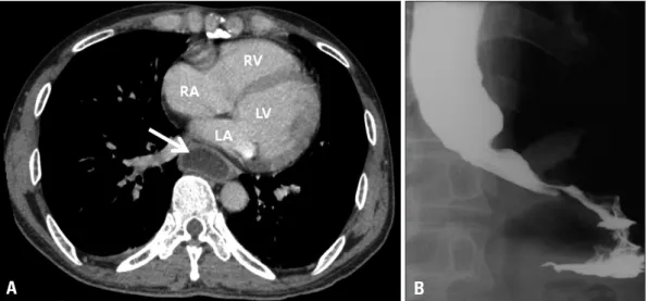
1B). Chest computed tomography scan revealed diffuse esoph- ageal dilatation compressing the left atrium (Fig. 2A). Gastro- grafin esophagography showed esophageal dilatation with narrowing of lower esophagus, so called ‘bird’s beak appear- ance’ (Fig. 2B). High-resolution esophageal manometry dem- onstrated lack of normotensive lower esophageal sphincter
with incomplete relaxation after swallowing consistent with achalasia. Balloon dilatation was performed successfully and patient’s symptom was improved. During admission, serial electrocardiograms showed persistent ST elevations with sad- dle back pattern and Brugada syndrome was suspicious.
Achalasia is a motility disorder characterized by dilatation of distal part of the esophagus with impaired relaxation of the lower esophageal sphincter. Compression of the left atrium
pISSN 1975-4612/ eISSN 2005-9655 Copyright © 2012 Korean Society of Echocardiography www.kse-jcu.org http://dx.doi.org/10.4250/jcu.2012.20.4.218
IMAGES IN CARDIOVASCULAR ULTRASOUND J Cardiovasc Ultrasound 2012;20(4):218-219
A Case of Esophageal Achalasia
Compressing Left Atrium Diagnosed by Echocardiography in Patient
with Acute Chest Pain
Hancheol Lee, MD, Seung-Hyun Lee, MD, Jin Ho Kim, MD, Dong-Jun Lee, MD, Jae-Sun Uhm, MD, PhD, Chi Young Shim, MD, PhD, Hyuck-Jae Chang, MD, PhD, Gue-Ru Hong, MD, PhD, Jong-Won Ha, MD, PhD and Namsik Chung, MD, PhD
Division of Cardiology, Severance Cardiovascular Hospital, Yonsei University College of Medicine, Seoul, Korea
KEY WORDS: Achalasia · Left atrium · Echocardiography.
• Received: August 2, 2012 • Revised: September 7, 2012 • Accepted: November 21, 2012
• Address for Correspondence: Gue-Ru Hong, Division of Cardiology, Severance Cardiovascular Hospital, Yonsei University College of Medicine, 50 Yonsei-ro, Seodaemun-gu, Seoul 120-752, Korea Tel: +82-2-2228-8460, Fax: +82-2-2227-7732, E-mail: [email protected]
• This is an Open Access article distributed under the terms of the Creative Commons Attribution Non-Commercial License (http://creativecommons.org/licenses/by-nc/3.0) which permits unrestricted non-commercial use, distribution, and reproduction in any medium, provided the original work is properly cited.
Fig. 1. A: Parasternal long axis view of transthoracic echocardiography showed round shape mass lesion compressing the left atrium (white arrow). B: After drinking a liquid containing carbon dioxide, the esophagus was filled with air contrast (white arrow). LA: left atrium, LV:
left ventricle, RV: right ventricle.
A B
Achalasia Mimicking Acute Coronary Syndrome | Hancheol Lee, et al.
219 leads to various symptoms and signs, which include from mild
chest discomfort to hemodynamic instability. There are no previous reports about achalasia compressing left atrium and causes acute chest pain mimicking acute coronary syndrome.
TTE is the test of choice for diagnosing the left atrial com- pression by various etiology.1-3) Furthermore, the esophagus can be identified by the appearance of air contrast during the ingestion of liquid containing carbon dioxide.3) Echocardiog- raphy enables clinicians to successfully make the differential
diagnosis in patients with chest pain.
References
1. D’Cruz IA, Feghali N, Gross CM. Echocardiographic manifestations of mediastinal masses compressing or encroaching on the heart. Echocardiogra- phy 1994;11:523-33.
2. van Rooijen JM, van den Merkhof LF. Left atrial impression: a sign of extra-cardiac pathology. Eur J Echocardiogr 2008;9:661-4.
3. Goel K, Rishikant N, Chadha DS. Left atrial compression caused by achalasia. J Am Coll Cardiol 2009;54:955.
Fig. 2. A: Dilatation of the esophagus compressing the left atrium was revealed in chest computed tomography (white arrow). B: Esophagography with gastrografin identified achalasia. LA: left atrium, LV: left ventricle, RV: right ventricle, RA:
right atrium.
A B