427
Copyrights © 2015 The Korean Society of Radiology
INTRODUCTION
In embryology, two renal veins (ventral and dorsal) are formed by the fusion of the supra- and sub-cardinal veins. Over time, the dorsal vein degenerates and the ventral vein usually remains as the single renal vein (1). Therefore, the most common varia- tion of the renal vein is additional renal veins, which are usually clinically silent and detected incidentally during an operation or autopsy (2). However, the absence of a renal vein is a very rare condition, and only two cases in the English literature have been reported (3, 4). We report a case of an absence of the renal vein without any damage to the renal parenchyma. This study was approved by the Institutional Review Board.
CASE REPORT
A 49-year-old female presented with a complained symptom
of upper respiratory infection, and a contrast-enhanced chest CT scan was done. The CT scan incidentally revealed a tortuous vas- cular structure in the right suprarenal space. For further evalua- tion, a contrast-enhanced abdominal CT scan was done (64-sec- tion scanner, Lightspeed VCT; GE Medical Systems, Milwaukee, WI, USA) with two phases (corticomedullary and nephrogenic phases, intravenous injection of Iopamidol; Pamiray, Dongkuk Pharm., Seoul, Korea). The tortuous vessels were intertwined with each other around the right adrenal gland (Fig. 1A). The de- gree of contrast enhancement was similar to that of the abdomi- nal aorta, and we thought it was an arterio-venous malforma- tion. The enhancement pattern of both renal parenchyma was normal, and no hydronephrosis was observed in either kidneys.
The anterior volume-rendered image from CT data (Fig. 1B) showed the tortuous vascular structure at the right renal hilum and the right suprarenal space. Bilateral normal renal arteries and the left renal vein were seen, but the right renal orthopedic
Case Report
pISSN 1738-2637 / eISSN 2288-2928 J Korean Soc Radiol 2015;72(6):427-430 http://dx.doi.org/10.3348/jksr.2015.72.6.427
Received October 25, 2014 Accepted December 13, 2014
Corresponding author: Yong Sun Jeon, MD Department of Radiology, Inha University Hospital, 27 Inhang-ro, Jung-gu, Incheon 400-711, Korea.
Tel. 82-32-890-2773 Fax. 82-32-890-2743 E-mail: [email protected]
This is an Open Access article distributed under the terms of the Creative Commons Attribution Non-Commercial License (http://creativecommons.org/licenses/by-nc/3.0) which permits unrestricted non-commercial use, distri- bution, and reproduction in any medium, provided the original work is properly cited.
A CT scan of a 49-year-old female incidentally revealed a tortuous vascular struc- ture in the right suprarenal space. According to angiographic evaluation of the right renal vessels, the right renal artery was single with normal diameter, and there was no venous drainage through the main right renal vein (orthotopic renal vein).
The venous drainage of the right kidney flowed through the tortuous suprarenal vascular structure into the inferior vena cava. The color Doppler ultrasound revealed the monophasic waveform in that vascular structure without flow disturbance. The renal function and the result of urinalysis of the patient were normal, and any other congenital malformation was not found. Absence of the orthotopic renal vein and aberrant suprarenal venous drainage is a very rare congenital anomaly, and it should be discriminated from the other pathologic conditions.
Index terms
Orthotopic Renal Vein Congenital Anomaly Renal Vein Varices
Absence of Orthotopic Renal Vein with Aberrant Suprarenal Venous Drainage: A Case Report
1정상위치의 신장정맥 부재와 동반된 비정상적 콩팥 위 정맥 환류: 증례 보고1
Eugene Kim, MD
1, Yong Sun Jeon, MD
1, Soon Gu Cho, MD
1, Kee Chun Hong, MD
2, Keun-Myung Park, MD
2, Tack Lee, MD
3Departments of 1Radiology, 2General Surgery, 3Urology, Inha University Hospital, Incheon, Korea
Absence of Orthotopic Renal Vein with Aberrant Suprarenal Venous Drainage
428
J Korean Soc Radiol 2015;72(6):427-430 jksronline.orgparenchyma had been stained (Fig. 2A). Then, the suprarenal tortuous vascular structure with large diameter appeared in the delayed phase (Fig. 2B). This tortuous vascular structure contin- ued inferiorly to the level of right renal hilum and ultimately drained into the inferior vena cava (IVC). In the right renal hi- lum, there was no staining of the normal right renal vein (ortho- topic vein) at its expected location. The angiogram finding showed an absence of the orthotropic renal vein and an aberrant formation of suprarenal collateral venous flow.
vein was not seen in the reconstructed image.
To confirm the arterio-venous malformation and to ensure the proper treatment, we decided to perform an angiography.
We punctured the right common femoral artery and selected the right renal artery via a cobra catheter (Cook, Bloomington, MN, USA). The right renal angiogram showed normal contour of a single renal artery and normal renal parenchymal perfu- sion. After staining the right renal parenchyma, the tortuous vascular structure with small diameter appeared after the renal
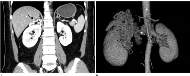
Fig. 1. Coronal reformatted CT image (A) and volume rendered image (B) of the aberrent right renal venous drainage.
A. Contrast-enhanced coronal reformatted abdominal CT showed tortuous dilated vascular structure (black arrow) in the right suprarenal space and a normal parenchymal enhancement of both kidneys.
B. Anterior volume-rendered image from CT data showed tortuous dilated vascular structure (open arrowheads) in the right renal hilum and su- prarenal space. Whereas normal left renal vein (black arrow) is seen at hilar portion, the orthotopic right renal vein is not seen. The aorta (*) is seen and the right renal artery (white arrow) is normally seen.
Fig. 2. Digital subtraction angiography (DSA) image showing the aberrent right renal venous drainage.
A. In DSA image of the right renal arteriogram, the tortuous dilated vascular structure (black arrow) is visualized at hilar portion after the renal parenchyma was stained. The orthopedic renal vein is not observed at the expected location.
B. In DSA image of the right renal arteriogram, the tortuous dilated vascular structure (black arrow) is visualized at the right suprarenal space just after the staining of hilar tortuous vessel. Still the orthopedic renal vein is not observed at the expected location.
A
A
B
B
Eugene Kim, et al
429
jksronline.org J Korean Soc Radiol 2015;72(6):427-430
could also be applied to our case. Another case of the absence of the right renal vein had a venous varicose complex, which drained into the right gonadal (ovarian) complex (4). We sug- gest that our case may be similar to venous collateral complex, which developed during embryogenesis.
Unlike the left renal vein, the right renal vein has less promi- nent collateral flow. In the left renal vein, several venous collat- eral complexes such as the lumbar veins and the retroaortic and prevertebral venous plexuses are commonly found (6). In an an- atomical variation of the right renal vein, the additional renal vein is more commonly found than in the left renal vein (3). Our hypothesis is that congenital or prenatal insult of the right main renal vein would have created the pre-existing additional right renal vein development, which finally became the only aberrant right renal vein. Also, we hypothesize that if a prenatal insult to the left renal vein occurred, the venous collateral complexes of the left renal vein would develop; but, those collateral flows may not have been sufficient for the normal development of the left kidney, in contrast to the additional vein of the right kidney.
Other than the absence of the renal venous drainage, as in our case, there are many acquired causes of renal vein occlusion, such as IVC thrombosis with secondary involvement of the re- nal vein, hypovolemia, primary renal disease, thrombosis, and trauma (7). Renal vein thrombosis is one of the common causes of renal vein occlusion, which presents with insidious symptoms or frequently without any symptoms (7). The well-established renal venous collateral pathways, other than the main renal vein, For the confirmation of the venous flow, a Doppler ultra-
sound (5–8 MHz Curved array probe of iU22 xMATRIX Ultra- sound System, Philips, Bothell, WA, USA) was performed. The ultrasound showed an intertwined anechoic tubular structure in the right suprarenal space below the liver capsule. The color Doppler ultrasound revealed vascularity, and the monophasic waveform with a peak velocity of 23.4 cm/s was shown by the spectral Doppler ultrasound (Fig. 3). The suprarenal anechoic tubular structure continued to the renal hilum and showed a unilateral biphasic waveform, which was efferent from the right kidney with a very low resistance (data not shown). These find- ings confirmed that the vascular structure was composed of di- lated veins which drained into the IVC. The peak velocities and resistive index of the right renal artery were normal.
From the CT, angiographic, and ultrasonographic findings, we concluded that there was a developed venous drainage from the right kidney due to the absence of the right orthotopic renal vein. The serum creatinine and BUN levels of the patient were normal. A regular follow-up for the patient was planned.
DISCUSSION
During the embryonic period, both right and left renal veins are formed by the same pattern–anastomosis of the supracardi- nal and subcardinal veins (1). Anomalies of renal veins have been investigated by many studies, and the most frequent con- genital anomaly is an additional renal vein, which is more fre- quently found on the right side (2). The additional renal vein re- sults in a separate venous drainage of the kidney from the main renal vein, and the flow goes into the IVC independently (5).
Compared to the additional renal vein, the absence of a renal vein is a very rare finding, and only two cases have been report- ed in the English literature (3, 4).
Bozlar et al. (3) reported the aplasia of a right renal vein with a diverted venous drainage, which was a very similar finding to our case. As in our case, any fibrous bands or remnant stalks were not observed at the expected renal vein level of the IVC, and there was no evidence of the parenchymal damage in the right kidney (3). The researchers suggested that a prenatal insult of the renal vein might be the cause of the development of the aberrant venous drainage, prior to the damage of renal paren- chyma, which formed the venous varicosity. This explanation
Fig. 3. Color and spectral Doppler ultrasound of vascular structure lo- cated at the right suprarenal space showed in the monophasic wave- form, suggesting continuous venous flow.
Absence of Orthotopic Renal Vein with Aberrant Suprarenal Venous Drainage
430
J Korean Soc Radiol 2015;72(6):427-430 jksronline.org43-52
3. Bozlar U, Ugurel MS, Bedir S, Ors F, Coskun U, Aydur E.
Right renal vein aplasia associated with diverted renal ve- nous drainage through lower pole. Cardiovasc Intervent Radiol 2008;31 Suppl 2:S140-S143
4. Pinggera GM, Spranger R, Frauscher F, Eder R, Smekal A, Bartsch G. Congenital absence of the right renal vein. J Urol 2003;170:914-915
5. Satyapal KS. Classification of the drainage patterns of the renal veins. J Anat 1995;186(Pt 2):329-333
6. James EC, Fedde CW, Khuri NT, Gillespie JT. Division of the left renal vein: a safe surgical adjunct. Surgery 1978;83:
151-154
7. Witz M, Korzets Z. Renal vein occlusion: diagnosis and treatment. Isr Med Assoc J 2007;9:402-405
8. Diniz GV, Pereira WJ, Moreira AC, Santos BM, Drumond DA, Petroianu A. Kidney function after left renal vein liga- tion in the dog. Rev Hosp Clin Fac Med Sao Paulo 2001;56:
1-4 take some part of the venous drainage. But, the occluded main
renal venous drainage gradually causes renal parenchymal fi- brosis histologically, and the other kidney with preserved ve- nous drainage makes up for the abnormal function of the kid- ney (8). However, our patient had no clinical symptoms or any abnormal lab findings of renal dysfunction, including blood urea nitrogen and serum creatinine. The Doppler ultrasound of the aberrant renal vein showed normal venous drainage into the IVC. Thus, we can assume that the developed aberrant venous drainage could compensate for the absence of the main venous drainage without causing kidney dysfunction. In conclusion, we report an absence of the orthotopic right renal vein with an ab- errant renal venous drainage system.
REFERENCES
1. Mathews R, Smith PA, Fishman EK, Marshall FF. Anomalies of the inferior vena cava and renal veins: embryologic and surgical considerations. Urology 1999;53:873-880
2. Satyapal KS. The renal veins: a review. Eur J Anat 2003;7:
정상위치의 신장정맥 부재와 동반된 비정상적 콩팥 위 정맥 환류:
증례 보고1
김유진
1· 전용선
1· 조순구
1· 홍기천
2· 박근명
2· 이 택
349세 여자 환자의 전산화단층촬영 스캔에서 우측 콩팥 위 공간에서 구불구불한 혈관 구조물이 우연히 발견되었다. 혈관 조영술로 평가하였을 때, 우측 신장 동맥은 한 개였으며 정상 지름을 보였지만, 정상 위치의 콩팥문에 보이는 우측 신장 정맥이 없었다. 대신, 구불구불한 우측 콩팥 위 혈관 구조물을 통해 우측 신장 정맥혈이 대정맥으로 환류되었다. 색도플러 초음파에서 이 혈관 구조물의 내부에 저항없는 단상성의 파형이 관찰되었다. 환자의 요검사 결과는 정상이었고 다른 선천 기형은 발견되지 않았다. 정상위치 신장정맥의 부재 및 동반된 콩팥 위 정맥 환류는 매우 드문 선천기형으로 병적인 상태 와 구분되어 다른 치료를 요하지 않는 정상 변이이다.
인하대학교병원 1영상의학과, 2외과, 3비뇨기과