955 https://e-kcj.org
A previously healthy 34-year-old man presented with progressive dyspnea and chest pain after viral infection. Initial transthoracic echocardiography (TTE) depicted massive pericardial effusion and he underwent pericardiocentesis of 900 mL serosanguinous fluid without identifiable malignant cells. Pericarditis-associated pericardial effusion was initially Korean Circ J. 2020 Oct;50(10):955-956
https://doi.org/10.4070/kcj.2020.0109 pISSN 1738-5520·eISSN 1738-5555
Images in
Cardiovascular Medicine
Received: Mar 16, 2020 Revised: Apr 25, 2020 Accepted: May 13, 2020
Guan-Yi Li , MD
1, and Fa-Po Chung , MD, PhD
2,31
Department of Medicine, Taipei Veterans General Hospital, Taipei, Taiwan
2
Heart Rhythm Center and Division of Cardiology, Department of Medicine, Taipei Veterans General Hospital, Taipei, Taiwan
3
Department of Medicine, National Yang-Ming University School of Medicine, Taipei, Taiwan
Primary Pericardial Angiosarcoma:
an Insidious and Aggressive Malignant Tumor
A B
D E
C TM
TM
RA RA SVC
IVC
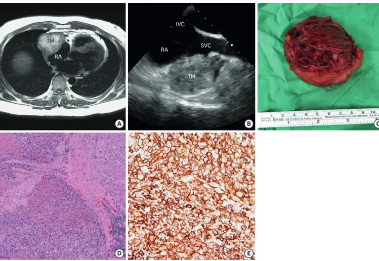
Figure 1. Primary pericardial angiosarcoma. (A) CMR image revealed a huge hyperintense T1-weighted mass at pericardial space with right atrial compression.
(B) TEE demonstrated the tumor, located adjacent to RA and SVC. (C) The excised tumor, in size of 5.5×4.5 cm, showed prominent necrosis and hemorrhage. (D) Pericardial soft tissue with infiltration of malignant endothelial cells (H&E, ×40). (E) The tumor cells were positive for CD34 immunohistochemical staining (×200).
CMR = cardiac magnetic resonance; H&E = hematoxylin and eosin stain; IVC = inferior vena cava; RA = right atrium; SVC = superior vena cava; TEE = transoesophageal
echocardiography; TM = tumor mass.
Correspondence to Fa-Po Chung, MD, PhD
Heart Rhythm Center and Division of Cardiology, Department of Medicine, Taipei Veterans General Hospital, No. 201, Sec. 2, Shih-Pai Road, Taipei 112, Taiwan.
E-mail: [email protected]
Copyright © 2020. The Korean Society of Cardiology
This is an Open Access article distributed under the terms of the Creative Commons Attribution Non-Commercial License (https://
creativecommons.org/licenses/by-nc/4.0) which permits unrestricted noncommercial use, distribution, and reproduction in any medium, provided the original work is properly cited.
ORCID iDs Guan-Yi Li
https://orcid.org/0000-0002-0550-8837 Fa-Po Chung
https://orcid.org/0000-0001-8960-1301 Conflict of Interest
The authors have no financial conflicts of interest.
Author Contributions
Conceptualization: Chung FP; Data curation:
Li GY; Investigation: Li GY; Methodology:
Chung FP; Project administration: Chung FP;
Resources: Li GY; Software: Li GY; Supervision:
Chung FP; Visualization: Chung FP; Writing - original draft: Li GY; Writing - review & editing:
Chung FP.
impressed. Nevertheless, follow-up TTE 3 months later demonstrated recurrent pericardial effusion without identifiable pericardial mass. Cardiac magnetic resonance (CMR) imaging revealed a huge mass, located at pericardial space with right atrial compression (Figure 1A), which was also revealed by transoesophageal echocardiography (Figure 1B). A firm tumor in size of 5.5×4.5 cm, which severely adhered to the pericardium and extended to right atrial wall, superior vena cava and aorta, was excised (Figure 1C). The pathologic examination confirmed the diagnosis of primary pericardial angiosarcoma, given the presence of malignant endothelial cells mainly in pericardial soft tissue (Figure 1D and E). Therapy with pembrolizumab and pazopanib was administered. Unfortunately, CMR revealed the recurrence of tumor 4 months after the surgery, and salvage radiotherapy was performed.
Primary pericardial angiosarcoma is extremely rare, and clinical prognosis of this disease is unfavorable.
1)It is particularly difficult to diagnose pericardial malignancy at the early stage by TTE alone.
2)Sequential imaging studies, such as computed tomography or CMR, are warranted to prevent erroneous diagnosis, particularly for those with undetermined and recurrent pericardial effusion.
3)Furthermore, a positron emission tomography scan, which was not performed in our case, plays a role on evaluating the extent of primary tumor, as well as the possibility of distant metastasis.
4)ACKNOWLEDGMENTS
This work was supported by the Center for Dynamical Biomarkers and Translational Medicine, Ministry of Science and Technology (grant nos. 107-2314-B-010-061-MY2, MOST 106-2314-B-075-006-MY3, MOST 106-2314-B-010-046-MY3, and MOST 106-2314-B-075-073- MY3), Research Foundation of Cardiovascular Medicine, Szu-Yuan Research Foundation of Internal Medicine, and Taipei Veterans General Hospital (grant nos. V106C-158, V106C-104, V107B-014, V107C-060, V107C-054, V108C-107, and V109C-113).
REFERENCES
1. Timóteo AT, Branco LM, Bravio I, et al. Primary angiosarcoma of the pericardium: case report and review of the literature. Kardiol Pol 2010;68:802-5.
PUBMED
2. Meng Q, Lai H, Lima J, Tong W, Qian Y, Lai S. Echocardiographic and pathologic characteristics of primary cardiac tumors: a study of 149 cases. Int J Cardiol 2002;84:69-75.
PUBMED | CROSSREF
3. Holtan SG, Allen RD, Henkel DM, et al. Angiosarcoma of the pericardium presenting as hemorrhagic pleuropericarditis, cardiac tamponade, and thromboembolic phenomena. Int J Cardiol 2007;115:e8-9.
PUBMED | CROSSREF
4. Shi X, Li F. Primary pericardial angiosarcoma shown on FDG PET/CT. Clin Nucl Med 2017;42:973-5.
PUBMED | CROSSREF