• 검색 결과가 없습니다.

Subarachnoid Hemorrhage Misdiagnosed as an Acute ST Elevation Myocardial Infarction

N/A
N/A
Protected

Academic year: 2022

Share "Subarachnoid Hemorrhage Misdiagnosed as an Acute ST Elevation Myocardial Infarction"

Copied!
4
0
0

로드 중.... (전체 텍스트 보기)

전체 글

(1)

216 Copyright © 2012 The Korean Society of Cardiology Korean Circulation Journal

Introduction

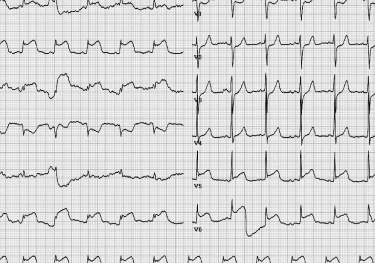
Although, electrocardiographic change is not uncommon in pa- tients with cerebrovascular disease including subarachnoid hemor- rhage, subdural hematoma, and ischemic stroke,

1-3)

typical ST-sge- ment elevation is rare. The precise mechanism is not clear, but it is widely accepted that excess releasing of cathecholamine may in- fluence myocardial contractility and necrosis.

2)4)5)

Here, we intend to emphasize that careful attention of neurologic abnormality can en- able better prognosis.

Case

A 55-year-old female patient presented with severe headache and loss of consciousness. She was previously healthy and had no symp-

Case Report

http://dx.doi.org/10.4070/kcj.2012.42.3.216 Print ISSN 1738-5520 • On-line ISSN 1738-5555

Subarachnoid Hemorrhage Misdiagnosed

as an Acute ST Elevation Myocardial Infarction

Woon Je Heo, MD 1 , Jin Ho Kang, MD 1 , Woo Shin Jeong, MD 1 , Mi Yeon Jeong, MD 1 , Sang Hyuk Lee, MD 1 , Jeong Yeun Seo, MD 1 , and Sang Won Jo, MD 2

1

Departments of Internal Medicine and

2

Radilology, Kangbuk Samsung Medical Center, Seoul, Korea

Without significant coronary artery stenosis, ischemic electrocardiographic change including ST segment elevation, segmental wall motion abnormality and elevated serum cardiac-specific markers (creatine kinase-MB, Troponin-T) may develop after central nervous system injuries such as subarachnoid, intracranial or subdural hemorrhage. Misdiagnosing these patients as acute myocardial infarction may result in catastrophic outcomes. By reporting a case of a 55-year old female with subarachnoid hemorrhage mimicking acute ST elevation myocardial infarction, we hope to underline that careful attention of neurologic abnormality is critical in making better prognosis. (Korean Circ J 2012;42:216-219) KEY WORDS: Myocardial infarction; Subarachnoid hemorrhage.

Received: July 30, 2011

Revision Received: August 31, 2011 Accepted: September 5, 2011

Correspondence: Jin Ho Kang, MD, Department of Internal Medicine, Kangbuk Samsung Medical Center, 29 Saemunan-ro, Jongno-gu, Seoul 110-746, Korea

Tel: 82-2-2001-2050, Fax: 82-2-2001-2049 E-mail: [email protected]

• The authors have no financial conflicts of interest.

This is an Open Access article distributed under the terms of the Creative Commons Attribution Non-Commercial License (http://creativecommons.

org/licenses/by-nc/3.0) which permits unrestricted non-commercial use, distribution, and reproduction in any medium, provided the original work is properly cited.

toms. On arrival, she was drowsy and her blood pressure, pulse rate, respiratory rate and body temperature were 73/50 mm Hg, 80 beats per minute, 20 per minute and 36.0°C, respectively. There was no head trauma, cardiac murmur or abnormal respiration sound. Elec- trocardiogram on arrival presented ST segment elevation in lead II, III, aVF, V 5, 6 and depression in V 1, 2 (Fig. 1). Laboratory examination showed her creatine kinase-MB was 1.21 ng/mL and troponin-T

<0.010 ng/mL. Assuming acute myocardial infarction, we conduct- ed emergency coronary angiography but there was no significant stenosis (Fig. 2). Echocardiogram exhibited hypokinesia on septum and apex and left ventricular ejection fraction was 43%. During coronary angiography, the patient vomited once and became stu- porous. We then checked brain computed tomography immediate- ly, and found a large amount of subarachnoid hemorrhage and small intracranial hemorrhage in the medial temporal lobe base (Fig. 3). Upon cerebral angiographic computed tomography, a small saccular aneurysm in the right posterior communicating artery area was detected (Fig. 3). After taking right pterional craniotomy and aneurysmal neck clipping, the patient improved and was dis- charged in 30 days without any neurological sequela.

Discussion

It has been reported that abnormalities of electrocardiogrphy, echo-

cardiography and serum cardiac specific markers are associated with

cerebrovascular disease.

4-8)

The most common cause is subarachnoid

hemorrhage,

9-11)

but additional causes include head injury, meningi-

(2)

217 Woon Je Heo, et al.

http://dx.doi.org/10.4070/kcj.2012.42.3.216 www.e-kcj.org

Fig. 1. Twelve-lead surface electrocardiogram showing extensive ST elevation (II, III, aVF) and ST depression (V

1,2

).

Fig. 2. There is no significant stenosis in coronary angiography.

(3)

218 Subarachnoid Hemorrhage Misdiagnosed as Acute Myocardial Infarction

http://dx.doi.org/10.4070/kcj.2012.42.3.216 www.e-kcj.org

diagnosed as acute ST elevation myocardial infarction, we should consider a cerebral cause if there is no significant stenosis in coro- nary angiography, and act promptly to save the life of a patient. It is well known that stress induced cardiomyopathy may develop by a cerebrovascular accident, but typical ST elevation is rare. By re- porting this case, we wish to share our uncommon experience and hope that it may be helpful in future cases.

References

1. Oppenheimer S, Norris JW. Cardiac manifestations of acute neuro- logical lesions. In: Aminoff MJ, editor. Neurology and General Medi- cine: the Neurological Aspects of Medical Disorder. 2nd ed. New York, NY: Churchill-Livingstone;1995. p.183-200.

2. IAC Van der Bilt, Visser FC. Neurogenic induced myocardial dysfunc- tion. Heart Metab 2004;24:27-30.

3. Zaroff JG, Rordorf GA, Newell JB, Ogilvy CS, Levinson JR. Cardiac out- come in patients with subarachnoid hemorrhage and electrocardio- graphic abnormalities. Neurosurgery 1999;44:34-9; discussion 39-40.

4. Elrifai AM, Bailes JE, Shih SR, Dianzumba S, Brillman J. Characteriza- tion of the cardiac effects of acute subarachnoid hemorrhage in dogs.

Stroke 1996;27:737-41; discussion 741-2.

5. Yuki K, Kodama Y, Onda J, Emoto K, Morimoto T, Uozumi T. Coronary va- sospasm following subarachnoid hemorrhage as a cause of stunned myocardium: case report. J Neurosurg 1991;75:308-11.

6. Zaroff JG, Rordorf GA, Newell JB, Ogilvy CS, Levinson JR. Cardiac out- come in patients with subarachnoid hemorrhage and electrocardio- graphic abnormalities. Neurosurgery 1999;44:34-9; discussion 39-40.

7. Chen YL, Yu TH, Fu M. Takotsubo cardiomyopathy: transient left ven- tricular apical ballooning mimicking acute myocardial infarction. J For- mos Med Assoc 2006;105:839-43.

tis and brain tumor. Previous investigators has suggested that severe stress such as ischemic stroke and cerebral hemorrhage may pro- voke high catecholamine induced neurogenic-stunned myocardi- um.

2)4-5)

Increased release of catecholamines from local nerve end- ings in the heart may mediate these cardiac abnormalities. Trans- ient severe coronary vasoconstriction leads to ischemia followed by postischemic ventricular failure and subendocardial myocardial da- mage. In addition, a direct cardiotoxic effect of catecholoamine may cause the development of subendocardial damage.

4)5)

Autopsies have revealed areas of characteristics subendocardial myocardial lesions, called contraction band necrosis, in the hearts of patients with sub- arachnoid hemorrhage (SAH).

14-16)

The myocardial damage resem- bles lesions produced in animal experiments by infusion of norepi- nephrine.

4)

The characteristic pattern of myocardial lesions has suggested that damaging catecholamines are released from intra- myocardial nerve endings rather than from the general circula- tion.

17)

Despite all these studies, the precise mechanism of action is still unclear and future studies are required.

When unconscious cerebral hemorrhage patients with electro- cardiograms showing ST segment elevation come to the emergency room, we have a tendency to misdiagnose as acute myocardial in- farction and treat with multiple antiplatelet and anticoagulate agents, which can cause harmful effects. Furthermore, delaying ac- curate diagnosis may result in catastrophic outcome.

12)13)

Therefore, any neurological abnormalities including paralysis or severe head- ache should be dealt with careful attention and the possibility of ce- rebrovascular disease should be considered, prompting the need to be prudent with the use of antiplatelet agents. Even if a patient is mis-

Fig. 3. A large amount of the SAH with a small ICH in the medial temporal lobe base. Small saccular aneurysm in the right posterior communicating artery area (arrow).

SAH: subarachnoid hemorrhage, ICH: intracranial hemorrhage.

(4)

219 Woon Je Heo, et al.

http://dx.doi.org/10.4070/kcj.2012.42.3.216 www.e-kcj.org

8. Tsuchihashi K, Ueshima K, Uchida T, et al. Transient left ventricular api- cal ballooning without coronary artery stenosis: a novel heart syn- drome mimicking acute myocardial infarction. Angina Pectoris-Myo- cardial Infarction Investigations in Japan. J Am Coll Cardiol 2001;38:

11-8.

9. Burch GE, Meyers R, Abildskov JA. A new electrocardiographic pattern observed in cerebrovascular accidents. Circulation 1954;9:719-23.

10. Hersch C. Electrocardiographic changes in subarachnoid haemorrhage, meningitis, and intracranial space-occupying lesions. Br Heart J 1964;

26:785-93.

11. Yamour BJ, Sridharan MR, Rice JF, Flowers NC. Electrocardiographic changes in cerebrovascular hemorrhage. Am Heart J 1980;99:294-300.

12. Hahn TH, Doo YC, Seo YM, Park TR, Choi HY, Rim CY. A case of ST-seg- ment elevation in a patient with subarachnoid hemorrhage. Korean Circ J 1995;25:106-9.

13. Cropp GJ, Manning GW. Electrocardiographic changes simulating myocardial ischemia and infarction associated with spontaneous in- tracranial hemorrhage. Circulation 1960;22:25-38.

14. Brouwers PJ, Wijdicks EF, Hasan D, et al. Serial electrocardiographic recording in aneurysmal subarachnoid hemorrhage. Stroke 1989;20:

1162-7.

15. Doshi R, Neil-Dwyer G. Hypothalamic and myocardial lesions after subarachnoid hemorrhage. J Neurol Neurosurg Psychiatry 1977;40:

821-6.

16. Greenhoot JH, Reichenbach DD. Cardiac injury and subarachnoid hem- orrhage: a clinical, pathological and physiological correlation. J Neu- rosurg 1969;30:521-31.

17. Baroldi G. Pathologic evidence of myocardial damage following acute

brain injuries. In: Di Pasquale G, Pinelli G, editors. Heart-Brain Interac-

tions. New York, NY: Springer-Verlag;1992. p.43-7.

수치

Fig. 1. Twelve-lead surface electrocardiogram showing extensive ST elevation (II, III, aVF) and ST depression (V 1,2 ).
Fig. 3. A large amount of the SAH with a small ICH in the medial temporal lobe base. Small saccular aneurysm in the right posterior communicating artery area (arrow)

참조

관련 문서

Therefore, cTnT is a recommended biomarker for use in the detection of myocardial infarction (MI) and in acute coronary syndromes.[8] Indeed, several authors

The positive change pattern of CK-MB according to time on acute myocardiac infarction patient.. The positive change pattern of troponin-T according to time on

Frequency and clinical significance of the expression of the multidrug resistance proteins MDR1/P-glycoprotein, MRP1, and LRP in acute myeloid leukemia: a

Approved clinical use of bone marrow stem cells for myocardial infarction treatment... Cardiac

Through a review of electronic medical records, we analyzed the applied BCSs, perioperative hematologic changes, and morbidity and mortality in JW patients

(Background) Gallbladder wall thickening(GWT) and gallbladder contraction are often observed in patients with acute hepatitis.. The incidence of acute hepatitis A

The risk of complicated acute appendicitis should be evaluated in terms of medical factors, including psychi- atric disease entity, psychotropic prescription, and

The main findings of this study indicate that early arrival from symptom onset, multiple infarctions reflected in less than six pc-ASPECTS, high initial systolic