ISSN 0378-6471 (Print)⋅ISSN 2092-9374 (Online)
http://dx.doi.org/10.3341/jkos.2016.57.1.134
Case Report
결절성경화증에서 망막 성상세포과오종의 안저자가형광, 형광안저혈관조영 및 스펙트럼영역 빛간섭단층촬영 소견
Fundus Autofluorescence, Fluorescein Angiography and Spectral Domain Optical Coherence Tomography Findings of Retinal Astrocytic Hamartomas in
Tuberous Sclerosis
권영기⋅강동진⋅임종찬⋅김종호⋅박동호⋅신재필
Young Ki Kwon, MD, Dong Jin Kang, MD, Jong Chan Lim, MD, Jong Ho Kim, MD, Dong Ho Park, MD, Jae Pil Shin, MD
경북대학교 의학전문대학원 안과학교실
Department of Ophthalmology, Kyungpook National University School of Medicine, Daegu, Korea
Purpose: To analyze the structural and morphological characteristics of retinal astrocytic hamartomas in tuberous sclerosis pa- tients using fundus autofluorescence, fluorescein angiography and spectral-domain optical coherence tomography.
Case summary: Fundus examination, fundus autofluorescence, fluorescein angiography and spectral-domain optical coherence tomography were performed in three patients with tuberous sclerosis and the morphological and structural characteristics of reti- nal astrocytic hamartomas were analyzed. In the fundus autofluorescence, type 1 retinal astrocytic hamartoma showed hypo- fluorescence and type 3 showed central hyperfluorescence and surrounding hypofluorescence. Spectral domain optical coher- ence tomography showed dome-shaped hyper-reflectivity within the nerve fiber layer and focal adhesion of the vitreous cortex in the type 1 retinal astrocytic hamartoma. No abnormalities were observed in the outer retinal layer and retinal pigment epithelium.
In the type 3 retinal astrocytic hamartoma, optical coherence tomography showed disorganization of retinal tissue and posterior shadowing. Intratumoral cavitation and moth-eaten appearance caused by intratumoral calcification were observed and the vit- reous cortex adhered to the top of the tumor and showed traction. Retinal arterial sheathing was observed in all cases and hy- per-reflectivity of the arterial wall was noted on optical coherence tomography.
Conclusions: Fundus autofluorescence, fluorescein angiography and spectral-domain optical coherence tomography are helpful for the classification and diagnosis of retinal astrocytic hamartomas found in tuberous sclerosis patients as well as for differ- entiation from other lesions.
J Korean Ophthalmol Soc 2016;57(1):134-140
Key Words: Fluorescein angiography, Fundus autofluorescence, Optical coherence tomography, Retinal astrocytic hamartoma, Tuberous sclerosis
■Received: 2015. 6. 26. ■ Revised: 2015. 10. 6.
■Accepted: 2015. 10. 15.
■Address reprint requests to Jae Pil Shin, MD
Department of Ophthalmology, Kyungpook National University Hospital #130 Dongdeok-ro, Jung-gu, Daegu 41944, Korea Tel: 82-53-200-5817, Fax: 82-53-426-6552
E-mail: [email protected]
* This study was presented as an e-poster at the 110th Annual Meeting of the Korean Ophthalmological Society 2013.
ⓒ2016 The Korean Ophthalmological Society
This is an Open Access article distributed under the terms of the Creative Commons Attribution Non-Commercial License (http://creativecommons.org/licenses/by-nc/3.0/) which permits unrestricted non-commercial use, distribution, and reproduction in any medium, provided the original work is properly cited.
결절성경화증은 눈, 중추신경, 피부와 내장을 침범하는 대표적인 눈 신경피부증후군으로, 인구 10,000-25,000명당 한 명꼴로 발생하는 상염색체 우성 유전 질환이며 TSC1 (9q34)과 TSC2 (16p13.3) 유전자의 변이로 인해 발병한 다.1,2 주로 간질발작, 안면의 피지선종, 지능저하의 3대 증 상을 특징으로 하며 대뇌 피질의 과오종(tuber), 뇌실막밑결 절(subependymal nodules), 뇌실막밑 거대세포성상세포종
본 증례에서는 3명의 환자에서 안저검사, 안저자가형광, 형 광안저혈관조영술, 스펙트럼영역 빛간섭단층촬영을 시행하 고 각각의 증례에서 발견된 망막 성상세포과오종의 형태적, 구조적 특징을 분석하였다. 국내에도 결절성경화증 환자에 서 망막 성상세포과오종의 증례가 이미 여러 차례 보고되 었으나 안저자가형광 및 스펙트럼영역 빛간섭단층촬영을 이용하여 분석한 보고는 없어서 이에 보고하고자 한다.
증례보고
증례1
33세 남자로 20세 때 결절성경화증으로 진단 받고 항경 련제를 복용 중인 환자로 양안 나안 시력은 20/20이었고 안 저검사에서 양안의 황반부 주위에 0.5-1.0 유두직경 크기의 편평하고 반투명의 옅은 회색 병변이 보여 1형 망막 성상 세포과오종으로 진단하였다(Fig. 1A). 안저자가형광에서 저형광으로 나타났고(Fig. 1B) 형광안저혈관조영술에서 초 기에는 저형광으로 나타났으나 일부 병변은 초기부터 잘 발달된 모세혈관망으로 인한 과형광을 나타냈고 후기에는 약한 과형광 소견을 보였다. 일부 병변은 검안경으로 발견 되지 않던 병변이 형광안저혈관조영술에서는 쉽게 발견되 었다(Fig. 1C, D). 빛간섭단층촬영에서는 병변이 망막의 신경섬유층 내에 위치하고 있었으며 망막외층 및 망막색 소상피층은 정상 소견을 보였고 후유리체 피질이 병변의 표면에 붙어 있었다. 병변 내 석회화를 의심할 만한 고반 사 음영은 보이지 않았으나 망막동맥의 혈관벽이 두꺼워 져 있고 이로 인한 부분적인 고반사 음영이 보였다(Fig.
1E). 망막동맥의 이상은 안저검사 및 형광안저혈관조영술 에서는 부분적인 동맥의 혈관초(sheathing) 및 협착으로 나 타났다(Fig. 1A, C, D).
증례 2
31세 여자로 경련이나 지능저하는 없었으나 14살 때 결 절성경화증으로 진단 받은 환자로, 2013년 6월 측두부의
및 망막색소상피층은 정상 소견을 보였으나 병변의 중심 부분에서는 부분적인 후방음영(posterior shadowing)이 보였 다. 병변 내 석회화로 인한 고반사 음영은 보이지 않았으나 망막동맥의 벽이 두꺼워져 있고 이로 인한 부분적인 고반사 음영이 보였으나 망막정맥은 정상소견을 보였다(Fig. 2E). 이 러한 망막동맥의 이상은 안저검사 및 형광안저혈관조영술 에서 부분적인 동맥의 혈관초 및 협착으로 나타났다(Fig.
2A, C, D).
증례3
31세 남자로 1살 때부터 경련이 있었고 3살 때 다낭포성 신장질환으로 결절성경화증으로 진단 받은 환자로 안과에 내원하여 시행한 검사에서 양안 최대 교정시력이 20/25였 으며 안저검사에서 융기되고 회백색의 불투명한 병변이 둘 러싸고 있는 뽕나무열매 형태의 병변과 회백색의 반투명한 병변이 여러 개 발견되어 망막 성상세포과오종 1형 및 3형 으로 진단하였다(Fig. 3A). 안저자가형광에서 1형 망막 성 상세포과오종은 저형광으로 나타났고 3형 망막 성상세포과 오종은 중심부분이 과형광으로, 주변 부분은 저형광의 고 리 모양으로 나타났다(Fig. 3B). 형광안저혈관조영에서는 두 가지 형태 모두 초기에는 저형광으로 나타났으나 후기 에는 1형의 경우 약한 과형광으로 나타났고 3형의 경우 과 형광의 점들이 병변의 중심부에서 보였고 주위로 고리 모 양의 과형광이 둘러싼 형태로 보였다(Fig. 3C, D). 빛간섭 단층촬영에서는 3형의 경우 망막전층에 종양으로 인한 고 반사 음영과 망막조직의 해체(disorganization) 및 이로 인 한 후방 음영(posterior shadowing)이 나타났다. 종양 내의 석회화로 인한 고반사음영과 종양 내 공동(cavitation)으로 인한 좀먹은 형태(moth-eaten appearance)를 보였고 이로 인해 망막외층 및 색소상피층은 잘 관찰되지 않았으며 종 양 위에 유리체가 부착되어 견인을 보이고 있었다(Fig. 3E).
하이측에서 약 5 유두직경 크기의 맥락막망막위축 병변도 발견되었는데 빛간섭단층촬영에서는 외과립층을 포함한 외측 망막과 망막색소상피층, 맥락막모세혈관층의 위축 소
A B
C D
E
Figure 1. Case 1. (A) Fundus photography shows a faintly gray-colored lesion below the macular (arrow) and arterial sheathing (as-
terisk). (B) Fundus autofluorescence shows hypofluorescence (arrow). (C) Early phase of fluorescein angiography shows hypofluor- escence (white arrow) and hyperfluorescence (red arrow) with well developed capillary network in the tumor which is not visible in the fundus photography. (D) Late phase of fluorescein angiography shows hyperfluorescence (both white and red arrow) due to leakage. (E) Spectral domain optical coherence tomography shows two hyperreflective dome-shaped retinal tumors localized within the nerve fiber layer with a gradual transition from the surrounding normal retina. One of the tumor is not visible in the fundus pho- tography (red arrow). The vitreous cortex shows focal adhesions to the surface of the tumor (arrowhead). Retinal artery (asterisk) passing through the tumor has wall of high reflectivity and shows back shadowing.C D
E
Figure 2. Case 2. (A) Fundus photography shows a faint semi-translucent lesion close to the optic disc in the right eye and arterial
sheathings (asterisks). (B) Fundus autofluorescence shows hypofluorescence due to tumor (arrow). (C) Early phase of fluorescein angiography shows hypofluorescence (white arrow) and arterial narrowing (asterisks). (D) Late phase of fluorescein angiography shows mild hyperfluorescence due to leakage (arrow). (E) Spectral domain optical coherence tomography shows hyperreflective dome-shaped retinal tumor localized within the nerve fiber layer with a gradual transition from the surrounding normal retina. The vitreous cortex shows focal adhesions to the surface of the tumor (arrowhead). Retinal arteries (white asterisks) have walls of high reflectivity and show back shadowing, while the retinal vein (black asterisk) is indiscernible.A B
C D
E
F
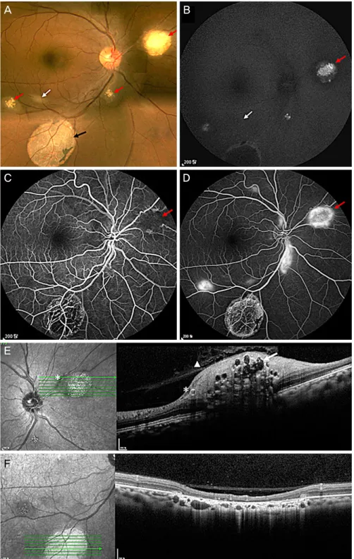
Figure 3. Case 3. (A) Fundus photography shows multiple retinal hamartomas with calcified nodular central core surrounded by a
relatively flat, smooth-surfaced, semitransparent periphery (red arrows) and faint semi-translucent lesion (white arrow). A large area of chorioretinal depigmentation is also seen (black arrow). (B) Fundus autofluorescence shows hypofluorescence (white arrow) and pinpoint hyperfluorescence in the center and surrounding hypofluorescence (red arrow). Chorioretinal depigmented lesion shows central hypofluorescence and surrounding hyperfluorescence. (C) Early phase of fluorescein angiography shows hypofluor- escence (red arrow). (D) Late phase of fluorescein angiography shows dot hyperfluorescence in the center and surrounding hyper- fluorescence (red arrow). (E) Spectral domain optical coherence tomography shows gradual transition from normal retina to the hy- perreflective intraretinal mass with loss of retinal organization and posterior shadowing. Note the moth-eaten optically empty spaces representing intralesional calcification (arrow). The vitreous cortex shows focal adhesions to the surface of the tumor (arrowhead).Retinal arteries (asterisks) have walls of high reflectivity. (F) Spectral domain optical coherence tomography of chorioretinal de- pigmentation shows focal loss of outer retinal layer, retinal pigment epithelial layer and choriocapillary layer.
화된 종괴의 형태로 망막신경섬유층 내에 위치하고 망막 혈관이나 동맥의 표층에 존재하며, 2형은 고전적인 뽕나무 열매(mulberry) 또는 물고기알 형태를 보이며 융기되고 석 회화된 종양이 후극부나 시신경 주위 또는 바로 위에 위치 하며, 3형은 앞의 두 가지 형태가 혼합된 형태로 종양 내 석회화가 있으며 주변부에 반투명의 불규칙한 테두리가 있 는 형태이다.3-5
망막 성상세포과오종은 조직 검사를 하지 않아도 안저검 사, 형광안저혈관조영술, 안초음파 등을 통해 진단하는 것 은 어렵지 않다. 본 연구에서는 3명의 증례를 통해 여러 가 지 안과검사에서 나타나는 망막 성상세포과오종의 특징에 대해서 알아보고자 하였다. 먼저 안저자가형광에서 망막 성상세포과오종 1형은 저형광을, 3형은 중심부의 과형광과 주변부의 고리모양의 저형광을 나타냈다. Mennel et al9은 이러한 안저자가형광 소견이 특히 2형 망막 성상세포과오 종의 진단에 도움이 된다고 하였다.
형광안저혈관조영술에서 초기에는 저형광을 보였고 후 기에는 과형광과 약한 형광누출을 보였다. Mennel et al9은 초기 저형광의 원인을 종양이 망막혈관 위에 위치하기 때 문에 이로 인한 형광 차단 때문이라고 보고하였다. 본 증례 에서는 증례1의 일부 병변에서 초기부터 잘 발달된 모세혈 관망으로 인한 과형광이 발견되었다. 또한 증례1에서 안저 사진이나 검안경 검사에서는 발견되지 않던 병변이 형광안 저혈관조영술에서 발견되었는데 이는 특히 1형의 망막 성 상세포과오종의 경우 안저검사만으로는 전체 병변을 다 발 견하기 어려우며 형광안저혈관 조영술이 보조적인 진단 방 법으로 유용함을 보여주는 것이다.
빛간섭단층촬영에서 나타나는 망막 성상세포과오종의 소견은 이전의 보고와 일치하였다.10-14 본 증례에서도 1형 은 망막신경섬유층에 한정된 돔 형태의 고반사층으로 나타 나며 외망막층과 망막색소상피층은 정상적인 소견을 보였 다. 표면에 유리체 피질이 붙어 있는 형태를 보이며 종양 내의 석회화로 인한 고반사음영이나 공동(cavitation)은 보 이지 않았고 인접한 망막에 부종도 나타나지 않았으며 정
영으로 검사하여 망막동맥의 석회화라고 보고하였다. 본 증례에서도 모든 증례의 환자(1형과 3형)에서 이러한 망막 동맥의 이상 소견이 발견되었으며 형광안저혈관조영에서 는 동맥의 국소적인 협착으로 나타났다. 그러나 이러한 동 맥이상이 석회화로 인한 것인지 종양세포의 침윤으로 인한 것인지에 대해서는 아직 논란의 여지가 있다. 본 증례3에서 3형의 망막 성상세포과오종은 Shields et al10의 2형 망막 성 상세포과오종과 같은 소견을 보였으나 주변에 1형의 망막 성상세포과오종이 둘러싸고 있는 부분이 차이점이었다.
Rowley et al4은 망막 성상세포과오종 환자의 39%에서 망막의 중간주변부에서 맥락망막의 탈색소 병변이 나타나 며 이는 일반인 대조군에 비해 유의하게 높게 나타난다고 하였다. 본 증례에서도 증례1과 2에서 이러한 맥락망막의 탈색소병변이 보였으며 빛간섭단층촬영에서 외망막층, 망 막색소상피층 및 맥락막모세혈관층의 소실을 나타내었다. 이러한 맥락망막의 탈색소병변이 망막 성상세포과오종과 의 직접적인 연관은 알려져 있지 않으나 Shields et al15과 Robertson5은 이 같은 소견이 망막 성상세포과오종의 진단 에 도움을 준다고 보고하였다.
망막 성상세포과오종과 감별해야 할 질환으로는 망막모 세포종, 시신경드루젠, 사르코이드증, 망막과 색소상피의 복합과오종, 무색소 맥락막흑색종 등이 있다.4-6,10 본 증례 에서 보듯이 결절성경화증에 동반된 망막 성상세포과오종 은 여러 가지 안과 검사에서 다양한 소견을 나타내며 이러 한 검사소견은 종양의 분류와 다른 종양과의 감별진단 및 추적관찰에 도움이 될 것으로 생각된다.
REFERENCES
1) Curatolo P, Bombardieri R, Jozwiak S. Tuberous sclerosis. Lancet 2008;372:657-68.
2) Osborne JP, Fryer A, Webb D. Epidemiology of tuberous sclerosis.
Ann N Y Acad Sci 1991;615:125-7.
3) Lagos JC, Gomez MR. Tuberous sclerosis: reappraisal of a clinical entity. Mayo Clin Proc 1967;42:26-49.
= 국문초록 =
결절성경화증에서 망막 성상세포과오종의 안저자가형광, 형광안저혈관조영 및 스펙트럼영역 빛간섭단층촬영 소견
목적: 결절성경화증에서 망막의 성상세포과오종의 구조와 형태적 특징을 안저자가형광, 형광안저혈관조영술 및 스펙트럼영역 빛간섭 단층촬영을 이용하여 분석하고자 하였다.
증례요약: 3명의 결절성경화증 환자에서 안저검사, 안저자가형광, 형광안저혈관조영술 및 빛간섭단층촬영을 시행하고 망막 성상세포 과오종의 형태적, 구조적 특징을 분석하였다. 안저자가형광에서 망막 성상세포과오종 1형은 저형광을 보였고, 3형은 중심부 과형광과 주변부 저형광을 보였다. 빛간섭단층촬영에서 1형은 신경섬유층 내에 위치하는 돔 모양의 고반사음영과 유리체가 종양의 윗부분에 부착된 모습을 보였으며 망막외층과 망막색소상피층은 이상이 없었다. 3형은 망막 전층에 종양으로 인한 고반사음영과 망막조직의 해체, 후방 음영을 보였고 종양 내의 석회화로 인한 고반사음영과 종양 내 공동과 좀먹은 형태를 보였으며 종양 위에 유리체의 견인을 보였다. 3 증례에서 모두 망막동맥의 혈관초를 보였으며 빛간섭단층촬영에서 망막동맥 혈관벽의 고반사 음영을 보였다.
결론: 결절성경화증 환자에서 발견된 망막 성상세포과오종에서 안저자가형광, 형광안저혈관조영술 및 빛간섭단층촬영은 종양의 분류 및 진단에 도움을 줄뿐 아니라 다른 병변과의 감별진단에도 도움이 될 것으로 생각된다.
<대한안과학회지 2016;57(1):134-140>
4) Rowley SA, O'Callaghan FJ, Osborne JP. Ophthalmic manifes- tations of tuberous sclerosis: a population based study. Br J Ophthalmol 2001;85:420-3.
5) Robertson DM. Ophthalmic manifestations of tuberous sclerosis.
Ann N Y Acad Sci 1991;615:17-25.
6) Nyboer JH, Robertson DM, Gomez MR. Retinal lesions in tuber- ous sclerosis. Arch Ophthalmol 1976;94:1277-80.
7) La TY, Kim CW, Lee YS, Kim MH. A case of retinal astrocytic ha- martoma causing blindness in tuberous sclerosis. J Korean Ophthalmol Soc 1999;40:1421-6.
8) Vrabec TR, Augsburger JJ. Exudative retinal detachment due to small noncalcified retinal astrocytic hamartoma. Am J Ophthalmol 2003;136:952-4.
9) Mennel S, Meyer CH, Eggarter F, Peter S. Autofluorescence and angiographic findings of retinal astrocytic hamartomas in tuberous sclerosis. Ophthalmologica 2005;219:350-6.
10) Shields CL, Benevides R, Materin MA, Shields JA. Optical coher-
ence tomography of retinal astrocytic hamartoma in 15 cases.
Ophthalmology 2006;113:1553-7.
11) Soliman W, Larsen M, Sander B, et al. Optical coherence tomog- raphy of astrocytic hamartomas in tuberous sclerosis. Acta Ophthalmol Scand 2007;85:454-5.
12) Goel N, Pangtey B, Bhushan G, et al. Spectral-domain optical co- herence tomography of astrocytic hamartomas in tuberous sclerosis. Int Ophthalmol 2012;32:491-3.
13) Xu L, Burke TR, Greenberg JP, et al. Infrared imaging and optical coherence tomography reveal early-stage astrocytic hamartomas not detectable by fundoscopy. Am J Ophthalmol 2012;153:883-9.e2.
14) Kimoto K, Kishi D, Kono H, et al. Diagnosis of an isolated retinal astrocytic hamartoma aided by optical coherence tomography.
Acta Ophthalmol 2008;86:921-2.
15) Shields CL, Reichstein DA, Bianciotto C, Shields JA. Retinal pig- ment epithelial depigmented lesions associated with tuberous scle- rosis complex. Arch Ophthalmol 2012;130:387-90.