pISSN: 0378-6471 eISSN: 2092-9374 http://dx.doi.org/10.3341/jkos.2013.54.8.1287
= 증례보고 =
결핵 뱀모양 맥락막염 환자에서 항결핵제와 스테로이드 치료 후에도 급격히 진행된 중심와 위축
이진영⋅강건욱⋅신재필⋅김인택⋅박동호 경북대학교 의학전문대학원 안과학교실
목적: 결핵 뱀모양 맥락막염 환자에서 항결핵제와 스테로이드의 병합치료에도 불구하고 급격히 진행된 중심와 위축을 보이는 1예를 경험하였기에 이를 보고하고자 한다.
증례요약: 54세 여자환자가 3일동안의 좌안 시력 저하를 호소하였다. 당시 좌안 시력은 안전수동이었으며 안저 검사상 좌안에서 시신경 유두 부종과 후극부에서 노란 맥락망막의 병변이 관찰되었다. 빛간섭단층촬영에서 좌안 황반부위에 망막내 부종 및 망막하액이 보였 다. 형광안저혈관조영술에서 초기엔 저형광이었으나 후기에는 과형광으로 나타났다. 인도시아닌그린혈관조영술에서는 초기 및 후기에 서 저형광으로 나타나 시행한 인터페론 감마 분비검사법에서 양성소견을 보였다. 항결핵제와 스테로이드 병합요법을 시작 후 시신경 유두 부종이 감소했으나, 발병 2주 후 빛간섭단층촬영에서 급격히 진행된 중심와의 위축이 보였다. 12주 후 최대 교정시력은 안전수지 10 cm였으며, 6개월 후 최대교정시력 및 중심와의 위축은 변화가 없었다.
결론: 초기에 중심와를 침범한 결핵에 의한 뱀모양 맥락막염은 급격히 진행하여 2주내 중심와의 위축이 보이며 나쁜 시력예후를 보였다.
<대한안과학회지 2013;54(8):1287-1292>
■Received: 2012. 9. 14. ■ Revised: 2013. 3. 15.
■Accepted: 2013. 6. 5
■Address reprint requests to Dong Ho Park, MD, PhD
Department of Ophthalmology, Kyungpook National University Hospital, #130 Dongdeok-ro, Jung-gu, Daegu 700-721, Korea Tel: 82-53-420-5813, Fax: 82-53-426-6552
E-mail: [email protected]
* This research was supported by the Basic Science Research Program through the National Research Foundation of Korea (NRF) funded by the Ministry of Education, Science and Technology (No. 2012004585) and by the Korea Health Technology R&D Project, Ministry of Health & Welfare, Republic of Korea (A111345).
결핵은 M.tuberculosis에 의해 직접 또는 항원에 대한 과 민반응으로 생기는 감염으로 20세기 이전 항결핵제가 나오 기 전까지만 해도 결핵은 매우 흔한 병이었으며, 최근 전세 계적으로 AIDS 환자의 증가와 면역억제제의 사용증가 등 으로 결핵이 다시 사회적 문제로 대두 되고 있다.1특히 우 리나라에서는 아직까지 선진국에 비해 결핵의 유병률이 굉 장히 높다. 이러한 결핵은 눈을 포함한 여러 장기를 침범할 수 있으며, 눈결핵은 드물지만 안검, 결막, 공막, 각막, 맥락 막, 망막, 시신경 등 다양한 조직에 침범하여 염증, 육아종 등의 증상으로 나타난다.1 특히 산소를 좋아하는 결핵균의 특성상 맥락막은 안구 내 결핵에서 가장 흔히 침범하는 부 위다.2이렇게 다양한 조직에 침범하여 여러 증상을 나타내 는 결핵의 특성상 진단과 치료에 혼동이 쉬우며 특히, 폐와
같은 다른 기관의 침범 증거가 없는 잠복결핵의 형태에서 도 눈결핵이 발생한 보고3,4가 있어 정확한 진단이 환자의 시력예후와 치료에 중요하다. 국내에서는 결막, 망막, 시신 경 등에 침범한 눈결핵이 보고5,6되었으나 결핵에 의한 뱀모 양 맥락막염은 국내에서는 보고된 바가 없다.
이에 저자들은 결핵에 의한 뱀모양 맥락막염 환자를 인 터페론감마 분비 검사와 형광안저혈관조영술 및 인도시아 닌그린혈관조영술을 통하여 진단하고 항결핵제와 스테로이 드의 병합치료 했음에도 불구하고 급격히 진행된 중심와 위축으로 나쁜 시력 예후를 보였던 1예를 경험하였기에 이 에 문헌 고찰과 함께 보고하고자 한다.
증례보고
여자 54세 환자가 3일동안의 좌안 시력 저하를 호소하였 다. 우안 최대교정시력은 20/20 이었으며 좌안 최대 교정시 력은 안정수동이었다. 환자의 과거력상 특이소견은 없었으 나 가족력상 환자의 어머니가 결핵을 치료한 병력이 있었 다. 세극등 검사에서 전방 염증 소견은 없었으나 좌안의 안 저 소견상 약한 유리체염증 소견과 심한 시신경 유두 부종 을 보였다(Fig. 1A). 그리고 다수의 노란 맥락망막 병변을 후극부에서 관찰되었다. 빛간섭단층촬영(Cirrus OCT; Carl Zeiss Meditec, Jena, Germany)으로 측정한 중심 황반 두 께는 820 μm로 좌안 황반에 망막내 부종과 망막하액이 보
A B
C D
E F
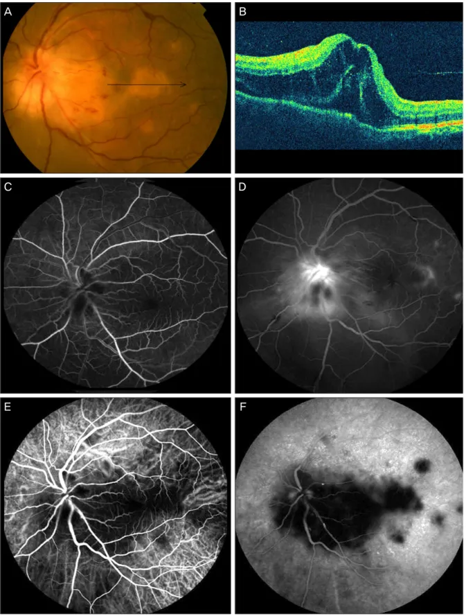
Figure 1. (A) Fundus photograph at the initial visit shows severe optic disc swelling and multiple yellowish chorioretinal
lesions. The black arrow indicates optical coherence tomography (OCT) scanning line. (B) OCT shows macular edema and subretinal fluid in the left eye (central subfield thickness: 820 μm). (C, D) Fluorescence angiography of the left eye.Hypofluorescence in the early phase. (C) hyperfluorescence in the late phase (D) are shown at the initial visit. (E, F) Indocyanine green angiography shows hypofluorescence during the early (E) and late phases (F) at the initial visit.
A B
C D
Figure 2. (A) High-resolution chest computerized tomography shows small clustered micronodules in the left lung apex
(black arrow). (B) Two weeks later, fundus photograph shows deceased optic disc swelling and diffuse choroiditis with amoeboid pattern in the posterior pole. The black arrow indicates optical coherence tomography (OCT) scanning line. (C) Fluorescence angiography shows hypofluorescence in the lesions and hyperfluorescence in the borders of lesions. (D) OCT shows an irregularly elevated retinal pigment epithelium line and fovea atrophy (central subfield thickness: 169 μm).였다(Fig. 1B). 형광안저혈관조영술에서는 노란 맥락망막 병변은 초기 저형광에서 후기로 가면서 과형광을 보였으며 (Fig. 1C, D) 인도시아닌그린혈관조영술에서 상기 병변은 초기 및 후기 모두 저형광을 보였다(Fig. 1E, F). 적혈구 침 강속도(28 mm/hr)를 제외한 기본적인 혈액검사 및 혈청검 사에서 모두 음성이었으며 시행한 뇌 자기 공명영상 및 흉 부 X선 촬영에서 정상 소견이었다. 하지만 결핵 피부 반응검 사에서 23 ×23 mm로 강양성을 보여 시행한 인터페론감마 분비 검사(QuantiFERON-TBTMGold; cellestis, Victoria, Australia) 상 양성결과(6.70 IU/mL; cut-off value 0.35 IU/mL)를 보였다. 고해상 흉부 전산화 단층촬영에서 좌측
폐 첨부에 무리를 이룬 작은 결절들이 보였다(Fig. 2A). 상 기 소견들을 바탕으로 결핵에 의한 뱀모양 맥락막염으로 진단하고 항결핵제와 하루 40 mg의 스테로이드 경구투여 로 치료를 시작했다. 치료 2주 후 좌안 최대교정시력은 안 전수지 10 cm였으나 시신경 유두 부종은 감소되었으며 망 막 및 맥락막의 침윤도 호전됐다(Fig. 2B). 형광안저혈관 조영술에서 병변 내 저형광이 나타나며 병변 경계부에서는 과형광이 나타났다(Fig. 2C). 빛간섭단층촬영으로 측정한 황반 중심 두께는 169 μm로 중심와의 위축을 보였다(Fig.
2D). 치료 12주 후 좌안의 최대교정시력은 안전수지 10 cm였으며 황반의 나타난 병변은 색소 침착과 함께 위축된
A B
Figure 3. (A) Twelve weeks later, fundus photograph shows chorioretinal atrophy with pigmentation in the macula. The
black arrow indicates OCT scanning line. (B) Twelve weeks later, OCT shows an irregularly elevated retinal pigment epi- thelium line and fovea atrophy (central subfield thickness: 162 μm).소견을 보였다(Fig. 3A). 빛간섭단층촬영에서 불규칙하게 올라간 망막색소상피층이 보였으며 내절/외절층의 파괴와 황반 중심 두께가 162 µm로 중심와 위축이 보였다(Fig.
3B). 치료 6개월 후 최대교정시력 및 황반 중심 두께는 이 전과 변화가 없었다.
고 찰
뱀모양 맥락막염은 망막색소상피, 맥락막과 맥락막 모세 혈관에 만성적으로 재발하며 진행하는 염증을 말한다. 이 뱀모양 맥락막염은 특징적으로 시신경 유두주위에서 병변 이 시작되어 황반부와 망막 주변부로 퍼져서 나가며 양안 에서 발생한다.7여러 연구가 진행되지만 원인은 아직 밝혀 지지 않았으나 스테로이드와 면역억제제를 이용한 치료가 병의 재발과 진행을 억제한다고 보고된다.7 Gupta et al8은 뱀모양 맥락막염으로 진단하고 면역억제제와 스테로이드 병합요법으로 치료하였지만 병이 진행하는 환자를 대상으 로 연구한 결과 공통적으로 결핵에 대한 감염이 있다는 사 실을 통해 결핵에 의한 뱀모양 맥락막염으로 진단하였다.
이러한 환자군에 항결핵제와 스테로이드 병합요법을 통하 여 좋은 예후를 보였다고 보고했다. 이 두 질환을 명확히 구분하여 진단하기는 어렵지만 뱀모양 맥락막염과 달리 결 핵에 의한 뱀모양 맥락막염은 결핵피부 반응 검사상 양성 소견을 보이고 대부분 단안이며, 약한 유리체염 소견을 볼 수 있다. 그리고 결핵에 의한 맥락막의 결절이나 망막 혈관 염, 시신경 유두 부종 등의 다른 안구내 조직의 침범 소견 을 볼 수 있다.1,7-10하지만 두 질환 모두 다수의 불규칙한 주변부와 후극부를 포함한 망막에 뱀모양의 병변을 보이며
BCG 접종이 의무화된 한국과 일본에서 결핵 피부 반응 검 사(PPD)는 큰 의미를 가지지 못하는 현실을 고려했을 때 진단은 더욱 어렵다. 상기 증례와 같이 위 질환은 일반적으 로 형광안저혈관조영술에서 병변이 초기 저형광에서 후기 로 가면서 과형광을 보이며, 인도시아닌그린혈관조영술에 서 병변은 초기 및 후기 모두 저형광을 보인다. 특히 각각 의 병변에서 뱀모양 맥락막염의 특징적인 소견인 중앙의 회복된 병변과 변연부의 활성화 병변을 확인하였다. 하지만 위 환자는 가족력 외 흉부 X선 촬영 및 호흡기 등의 증상이 결핵을 의심할 만한 소견은 없었지만 결핵 피부 반응 검사 상 강양성을 보였으며 고해상 흉부 단층촬영에서 결핵으로 의심되는 병변이 보이고 인터페론감마 분비 검사 상 양성 소견으로 결핵에 의한 뱀모양 맥락막염으로 진단을 내릴 수 있었다. 특히 인터페론감마 분비 검사는 결핵 피부 반응 검사 보다 높은 특이도(100%)와 좋은 민감도(93%)를 보 이는 검사이다. 검사 후 72시간 후에 결과가 나오는 결핵 피부 반응검사와 다르게 24시간내에 결과가 나오며 BCG 접종이 결과에 영향을 주지 않는 객관적인 검사이므로 앞 으로 눈 결핵이 의심되는 환자에게 유용한 검사로 생각된 다.11,12
Gupta et al8은 대부분의 결핵에 의한 뱀모양 맥락막염환 자의 경우 항결핵제와 스테로이드 경구 투여로 병의 관해 와 마지막 최대 교정시력이 20/30 이상으로 좋은 예후를 보였다고 보고하였다. Bansal et al10은 86.52%의 환자에서 황반부를 침범했으나 그 중 76%의 환자는 중심와를 보존 되었고 시력예후는 좋았다고 보고했다. 하지만 이번 증례는 처음부터 직접적으로 중심와를 침범했으며 항결핵제 및 스 테로이드 병합요법에도 불구하고 중심와의 위축이 발병 2
주내에 빠르게 진행하였다. 그리고 최대교정시력은 안전수 지 10 cm로 불량한 시력예후를 보였다. 이러한 불량한 시 력예후는 직접적인 중심와의 침범으로 인한 중심와의 위축 으로 생각되며 중심와 침범여부가 병의 좋은 예후인자가 될 것으로 생각한다.
저자들은 일반적인 결핵에 의한 뱀모양 맥락막염의 경우 는 황반부를 침범하더라도 대부분 중심와를 보존하여 좋은 시력예후를 보이지만10,13-15이번 증례와 같이 초기에 중심 와를 침범한 경우는 치료에도 불구하고 빠르게 진행하여 중심와 위축을 일으켜 좋지 않은 시력예후를 보인 1예를 경 험하였기에 문헌고찰과 함께 보고하는 바이다.
REFERENCES
1) Tabbara KF. Tuberculosis. Curr Opin Ophthalmol 2007;18:493- 501.
2) Sheu SJ, Shyu JS, Chen LM, et al. Ocular manifestations of tuberculosis. Ophthalmology 2001;108:1580-5.
3) Gupta A, Bansal R, Gupta V, et al. Ocular signs predictive of tu- bercular uveitis. Am J Ophthalmol 2010;149:562-70.
4) Sarvananthan N, Wiselka M, Bibby K. Intraocular tuberculosis without detectable systemic infection. Arch Ophthalmol 1998;116:
1386-8.
5) Hong JW, Park YC, Choi GJ. A case of tuberculous neuroretinitis.
J Korean Opthalmol Soc 2005;46:1582-5.
6) Song JS, Lee TW. Ocular manifestations of systemic tuberculosis :
report of 3 cases. J Korean Opthalmol Soc 1996;37:1561-9.
7) Lim WK, Buggage RR, Nussenblatt RB. Serpiginous choroiditis.
Surv Ophthalmol 2005;50:231-44.
8) Gupta V, Gupta A, Arora S, et al. Presumed tubercular serpiginous like choroiditis: clinical presentations and management. Opthalmology 2003;110:1744-9.
9) Vasconcelos-Santos DV, Rao PK, Davies JB, et al. Clinical features of tuberculous serpiginouslike choroiditis in contrast to classic ser- piginous choroiditis. Arch Ophthalmol 2010;128:853-8.
10) Bansal R, Gupta A, Gupta V, et al. Tubercular serpiginous-like choroi- ditis presenting as multifocal serpiginoid choroiditis. Ophthalmology 2012;119:2334-42.
11) Mackensen F, Becker MD, Wiehler U, et al. QuantiFERON TB- Gold--a new test strengthening long suspected tuberculous in- volvement in serpiginous-like choroiditis. Am J Ophthalmol 2008;
146:761-6.
12) Kang YA, Lee HW, Yoon HI, et al. Discrepancy between the tuber- culin skin test and the whole blood interferon gamma assay for the diagnosis of latent tuberculosis infection in an intermediate tuber- culosis-burden country. JAMA 2005;293:2756-61.
13) Suzuki J, Oh-I K, Kezuka T, et al. Comparison of patients with ocu- lar tuberculosis in the1990s and the 2000s. Jpn J Ophthalmol 2010;
54:19-23.
14) Gupta V, Gupta A, Sachdeva N, et al. Successful management of tubercular subretinal granulomas. Ocul Immunol Inflamm 2006;
14:35-40.
15) Gupta V, Bansal R, Gupta A. Continuous progression of tubercular serpiginous-like choroiditis after initiating antituberculosis treatment.
Am J Ophthalmol 2011;152:857-63.e2.
=ABSTRACT=
Rapidly Progressing Foveal Atrophy with Tuberculous
Serpiginous-Like Choroiditis Despite Combined Anti-Tuberculosis and Steroid Treatment
Jin Young Lee, MD, Kun Wook Kang, MD, Jae Pil Shin, MD, PhD, In Taek Kim, MD, PhD, Dong Ho Park, MD, PhD
Department of Ophthalmology, Kyungpook National University School of Medicine, Daegu, Korea
Purpose: To report a case of rapidly progressing foveal atrophy with tuberculous serpiginous-like choroiditis.
Case summary: A 54-year-old female patient had decreased vision of hand motions (os) for 3 days. Fundus examination showed optic disc swelling and yellowish chorioretinal lesions in the posterior pole (os). Optical coherence tomography (OCT) showed intraretinal edema and subretinal fluid in the left macula. Routine laboratory tests, serologic tests, and mag- netic resonance imaging results were normal except for erythrocyte sedimentation rate (28 mm/hr). Fluorescein angiog- raphy showed the chorioretinal lesions appeared to be early hypofluorescence followed by late hyperfluorescence.
Indocyanine green angiography showed hypofluorescence during early and late phases and the result of interferon-gamma release assay was positive. Under diagnosis of tuberculous serpiginous-like choroiditis, anti-tuberculous therapy com- bined with systemic corticosteroid was started. Despite decreased optic disc swelling, OCT showed a rapid progression of foveal atrophy within 2 weeks. Twelve weeks later, visual acuity was finger count at 10 cm. Six months later, best-cor- rected visual acuity and foveal atrophy were no interval change.
Conclusions: Tuberculous serpiginous-like choroiditis with foveal involvement can show rapidly progressive foveal atrophy and poor visual prognosis.
J Korean Ophthalmol Soc 2013;54(8):1287-1292
Key Words: Anti-tuberculous therapy, Foveal atrophy, Indocyanine green angiography, Interferon-gamma release assay, Tuberculous serpiginous-like choroiditis
Address reprint requests to Dong Ho Park, MD, PhD
Department of Ophthalmology, Kyungpook National University Hospital
#130 Dongdeok-ro, Jung-gu, Daegu 700-721, Korea
Tel: 82-53-420-5813, Fax: 82-53-426-6552, E-mail: [email protected]