https://doi.org/10.3341/jkos.2017.58.11.1282
Case Report
덱사메타손 임플란트 주입술 치료 후 재발된 점상내측맥락막병증 1예
Recurrence of Punctate Inner Choroidopathy with an Intravitreal Dexamethasone Implant
신민호1⋅강현지1⋅정인영1,2
Min Ho Shin, MD1, Hyun Ji Kang, MD1, In Young Chung, MD, PhD1,2
경상대학교 의학전문대학원 안과학교실1, 경상대학교 건강과학연구원2
Department of Ophthalmology, Gyeongsang National University School of Medicine1, Jinju, Korea Gyeongsang Institute of Health Science, Gyeongsang National University2, Jinju, Korea
Purpose: To report a case of punctate inner choroidopathy (PIC) treated with an intravitreal dexamethasone implant due to side effects of systemic steroid treatment.
Case Summary: A 23-year-old highly myopic female who presented with PIC in her right eye was treated with an intravitreal dex- amethasone implant due to side effects of systemic steroid treatment including facial edema and sleep disturbances. Three months after the implant she complained of severe acute visual disturbances in her right eye. Her visual acuity was classified as hand movement. Choroidal neovascularization (CNV) was observed on optical coherence tomography and indocyanine green angiography revealed more multiple hypofluorescent lesions compared to the initial visit. Six months after the systemic steroid and intravitreal bevacizumab injection treatments, visual acuity in right eye improved and the CNV disappeared.
Conclusions: This report describes a case of PIC after, treatment with an intravitreal dexamethasone implant due to the side ef- fects of systemic steroid treatment, which recurred with complications.
J Korean Ophthalmol Soc 2017;58(11):1282-1288
Keywords: Intravitreal bevacizumab, Intravitreal dexamethasone implant, Punctate inner choroidopathy, Systemic steroid
■Received: 2017. 8. 10. ■ Revised: 2017. 9. 19.
■Accepted: 2017. 10. 26.
■Address reprint requests to In Young Chung, MD, PhD Department of Ophthalmology, Gyeongsang National University Hospital, #79 Gangnam-ro, Jinju 52727, Korea Tel: 82-55-750-8171, Fax: 82-55-758-4158
E-mail: [email protected]
*Conflicts of Interest: The authors have no conflicts to disclose.
ⓒ2017 The Korean Ophthalmological Society
This is an Open Access article distributed under the terms of the Creative Commons Attribution Non-Commercial License (http://creativecommons.org/licenses/by-nc/3.0/) which permits unrestricted non-commercial use, distribution, and reproduction in any medium, provided the original work is properly cited.
점상내측맥락막병증은 젊은 근시 여성에서 발생하는 드 문 특발성 염증성 다초점 맥락망막병증으로서 임상적 양상, 다양한 영상학적 검사 양상, 유전적 양상 등이 전체포도막 염과 동반된 다초점맥락막염과 많은 부분이 겹쳐져서 동일 한 질환의 다른 표현형이라고 생각된다.1-3 다초점맥락막염 은 전방 염증과 유리체염이 흔히 동반되며 재발이 잦고 만
성적인 경과를 보임과 달리, 점상내측맥락막병증은 경미하 게 발생하는 경우 자연적으로 호전되며 예후가 좋다고 알 려져 있으나, 망막 중심오목 근처에 발생한 염증이나 신생 혈관은 심각한 시력저하를 일으킬 수 있어 질병의 경과와 합병증의 발생에 따라 전신적 면역억제제 투여 및 유리체 강 내 항혈관생성인자 주사, 레이저 치료, 광역학 치료, 황 반하 수술 등 다양한 치료가 시행되고 있다.3 최근 덱사메 타손 유리체강 내 임플란트 주입술이 점상내측맥락막병증 을 포함한 안구 후부 포도막염 치료에 긍정적인 결과를 보 였다는 여러 보고들이 있다.4-6 본 증례는 점상내측맥락막병 증에서 전신 스테로이드에 부작용을 보여 전신 스테로이드 중단 후 덱사메타손 유리체강 내 임플란트 주입술을 시행 하였으나 3개월만에 염증 재발과 함께 맥락막신생혈관이 발생하여 치료하였기에 관련 문헌고찰과 함께 이를 보고하
C D
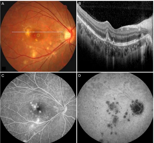
Figure 1. Fundus photograph, optical coherence tomography (OCT), fluorescein angiography and indocyanine green angiography at
initial visit. (A) Fundus photograph shows multiple, yellow, round lesions on posterior pole (dotted line is OCT section line). (B) Optical coherence tomography shows multiple irregular elevation of retinal pigment epithelium and photoreceptor. (C, D) Fluorescein angiography shows multiple hyperfluorescent lesions on posterior pole and indocyanine green angiography shows multi- ple hypofluorescent lesions on posterior pole (more prominent lesions on indocyanine green angiography).고자 한다.
증례보고
23세 여자가 약 1주일 전부터 발생한 우안의 시력저하를 주소로 내원하였다. 내원 시 최대교정 시력은 우안 20/25, 좌안 20/20이었으며 굴절률 검사상 우안 -8.0디옵터, 좌안 -8.50디옵터의 근시를 보였다. 세극등현미경 검사상 양안 모두 전방과 유리체의 염증 소견은 관찰되지 않았다. 안저 검사에서 우안의 후극부에 다발성의 노란색 원형의 병변들 이 관찰되었고(Fig. 1), 빛간섭단층촬영(optical coherence tomography, OCT) 검사에서 안저에서 관찰된 병변부위에
해당하는 곳의 망막상피세포와 시세포층의 불규칙한 융기 가 관찰되었다(Fig. 1).
형광안저혈관조영(fluorescein angiography, FAG)과 인도 시아닌그린안저혈관조영(indocyanine green angiography, ICGA) 결과 후기에서 형광조영보다 인도시아닌그린조영 에서 저명하게 드러나는 다수의 저형광병변들이 후극부에 관찰되었다(Fig. 1). 기타 염증 관련 후부 포도막염과 전신 질환과의 연관성을 감별하기 위한 혈액검사와 흉부단순촬 영에서 이상 소견은 관찰되지 않았다. 상기 결과들을 참고 하여 단안의 점상내측맥락막병증으로 진단하였고 경구 스테 로이드를 40 mg/day로 투여 시작하였고 1주일에 5 mg씩 감 량을 시행하였다. 스테로이드 치료 시작 1개월 후 우안 최
A B
C
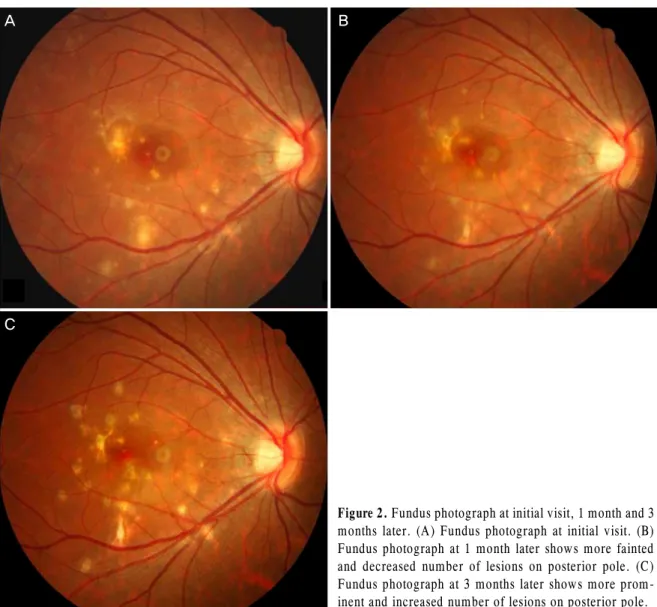
Figure 2. Fundus photograph at initial visit, 1 month and 3
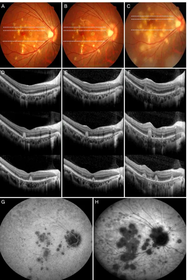
months later. (A) Fundus photograph at initial visit. (B) Fundus photograph at 1 month later shows more fainted and decreased number of lesions on posterior pole. (C) Fundus photograph at 3 months later shows more prom- inent and increased number of lesions on posterior pole.대교정시력은 20/20으로 호전되었고, 안저 검사에서 후극 부의 노란색의 다발성 병변들이 희미해지면서 호전되었다 (Fig. 2). 하지만, 환자가 얼굴이 붓고 잠을 잘 못 잔다는 스 테로이드 부작용을 호소하여 스테로이드를 10 mg/day로 감 량하여 유지하였다. 3개월 후 우안 최대교정시력 20/32로 감소하였고 안저 검사에서 후극부의 병변들이 증가하며 반 흔성 변화를 보여(Fig. 2), 경구 스테로이드 증량을 권유하 였으나, 경구 스테로이드는 부작용으로 복용을 거부하여 우안 덱사메타손 임플란트(Ozurdex®, Allergan, Irvine, CA, USA) 유리체강 내 주입술에 대해 설명하고 동의하에 이를 시행하였다. 우안 덱사메타손 임플란트 주입술 2개월 후 우 안 최대교정시력 20/20으로 호전되었고 안저 검사 및 빛간 섭단층촬영에서 안정되는 양상을 보였다(Fig. 3). 덱사메타 손 임플란트 주입술 3개월째, 우안 최대교정시력 안전수동 으로 악화되었고, 안저 검사 및 인도시아닌그린안저혈관조 영 검사에서 후극부의 다발성 황색병변들이 서로 합쳐지는
양상으로 병변의 크기 및 수가 증가하였으며(Fig. 3), 빛간 섭단층촬영에서 망막하액을 동반한 맥락막신생혈관이 관 찰되었다(Fig. 3). 환자에게 전신 스테로이드 사용이 필요하 며 부작용 재발 가능성에 대하여 설명하고 동의를 얻은 후 내과와 협진하에 경구 스테로이드 치료를 40 mg/day로 다 시 시작하여 서서히 감량하였고, 동시에 유리체강 내 베바 시주맙(Avastin, Roche Pharma. Ltd., Reinach, Switzerland) 1.25 mg (0.05 mL)을 1달 간격으로 2회 유리체강 내 주사 하였다. 마지막 베바시주맙 주사 6개월 후 우안 최대교정시 력은 20/32로 호전되었고 안저 검사 및 빛간섭단층촬영에서 망막하액 및 삼출물이 소실되었고 맥락막신생혈관도 소실되 었다(Fig. 4).
고 찰
점상내측맥락막병증은 질병 자체의 다양한 경과와 함께
G H
Figure 3. Fundus photograph, optical coherence tomography (OCT) and indocyanine green angiography at dexamethasone implant
injection, 2 months and 3 months after implant injection. (A-C) Fundus photograph at dexamethasone implant injection, 2 months and 3 months after implant injection. (D-F) OCT at dexamethasone implant injection, 2 months and 3 months after implant injection.(G, H) Indocyanine green angiography shows more confluent, increased number and enlarged lesions at 3 months after implant in- jection than initial visit.
A B
C D
Figure 4. Fundus photograph and optical coherence tomography (OCT) at 3 months and 6 months after implant injection. (A, B)
Fundus photograph and OCT at 3 months after implant injection (dotted line is OCT section line). (C) Fundus photograph at 6 months after bevacizumab injection shows decreased activity of inflammation and subretinal fibrosis (dotted line is OCT section line). (D) Optical coherence tomography at 6 months after bevacizumab injection shows absorbed subretinal fluid and decreased choroidal neovascularization.아주 드문 질환으로서 대규모의 잘 제어가 된 전향적인 연 구보다는 제어가 제대로 이루어지지 않은 연구, 전문가의 의견, 여러 증례보고 등을 치료 방법의 근거로 하고 있어 명확한 치료 방법이 현재까지도 확립되지 않고 있다.3 현재 까지 널리 의견의 일치가 이루어진 치료가 필요한 상태는 첫 번째, 새로이 발생했거나 활동성 염증성 병변이 특히 중 심오목근처에 있을 때, 두 번째, 점상내측맥락막병증에 이 차적으로 맥락막신생혈관이 발생하였을 경우이다.7
점상내측맥락막병증 치료에 있어서스테로이드의 2가지 역할은 첫 번째, 면역과 염증반응 경로를 신속히 차단하여 주로 급성기의 염증 반응을 억제하게 되며, 두 번째, 다른 치료와 동반하지 않더라도 점상내측맥락막병증에 이차적 으로 발생한 맥락막신생혈관을 쇠퇴시키는 항혈관생성 작 용을 하게 된다.8-10 점상내측맥락막병증에서 스테로이드는 전신적으로 혹은 국소주사(안구주위 주사, 유리체강 내 주 사)로 투여될 수 있는데, 이에 대한 비교연구가 불충분하여
어떤 투여 방법이 우위에 있다고 명확히 말하기 힘들다. Gerstenblith et al11의 조사에 의하면 환자들 중 60%는 전신 스테로이드, 22%는 안구 내 스테로이드, 10%는 안구 주위 스테로이드를 투여 받았다고 한다. Brueggeman et al8에 의 하면 점상내측맥락막병증에서 경구 스테로이드 치료는 병 변의 숫자를 줄이는 데는 효과적이었으나 중심오목아래 반 흔 형성은 막지 못하여 시력 호전에는 효과적이지 않다고 보고하였으며, Essex et al12도 점상내측맥락막병증에서 경 구 스테로이드 치료는 정상 반대안의 높은 맥락막신생혈관 발생과 연관이 있다고 보고하였다.
반면에, 점상내측맥락막병증에 특정되지 않지만 안구 후부 포도막염 치료에서 유리체강 내 덱사메타손 임플란 트 주입술이 효과적이라는 여러 보고들이 있다. Chronic Uveitis evaluation of the Intravitreal Dexamethasone Implant (HURON) study4에 의하면 1회의 덱사메타손 임플란트 유 리체강 내 주입술 치료 후 6개월의 경과관찰 동안 후부 포도
로 이루어진 환자군을 후향적으로 분석하였고 1회의 덱사 메타손 임플란트 주입술 후 6개월까지는 유의한 중심황반 두께감소 및 염증의 감소와 시력호전을 이루었으며, 추가 로 이루어진 덱사메타손 임플란트 주입술도 각각의 주입술 후 6개월까지는 1번째 주입술 이후와 비슷하게 좋은 효과 를 보였으며, 여러 차례의 주입술을 통하여 시력호전과 중 심황반두께의 감소를 유지하여 여러 차례의 주사가 시력과 중심황반두께에 유의한 축적된 효과를 가져온다고 한다.
하지만 이러한 연구들에서 후부 포도막염을 대상으로 치료 결과를 보고하고 있기에 점상내측맥락막병증이 포함된 증 례 수를 명확히 확인하기는 어려운 한계가 있다. 본 증례에 서는 경구 스테로이드를 첫 치료로 선택하였고 염증성 병 변의 감소와 시력 호전을 보였으나 환자가 수면장애, 안면 부종, 체중 증가 등 부작용을 호소하며 복용을 거부하여 부 득이하게 경구 스테로이드를 중단하였고 유리체강 내 덱사 메타손 임플란트 주입술을 시행하였다. 주입술 후 2개월까 지는 염증성 병변이 안정되며 시력의 호전이 유지되었으나 3개월 후 맥락막신생혈관이 동반된 염증성 병변의 급격한 악화로 심각한 시력손실을 가져왔다.
이는 이전의 여러 연구들4-6에서 덱사메타손 임플란트 주 입술 시행 시점에 환자들이 비교적 저용량의 스테로이드 및 면역억제제를 복용하고 있던 것과는 다르게 본 증례에 서는 경구 스테로이드를 중단하면서 염증의 억제 효과가 줄어들어 발생한 것으로 추측된다.
점상내측맥락막병증 환자 대부분이 단안의 증상을 호소 하여도 병변은 비대칭적으로 양안을 침범한다고 알려져 있 으나,1 본 증례는 오로지 단안에만 병변이 발생한 비전형적 인 증례이다. 경구 스테로이드 중단 후 덱사메타손 치료하 였지만 안구 내 유효 농도가 감소되는 시기에 맞추어 눈 속 염증이 악화되면서 더불어 맥락막신생혈관이 발생하였을 것으로 생각된다.
덱사메타손 임플란트를 유리체강내 주사 시행 시 최고 효과가 2-3개월 유지되고 5개월까지 그 약효가 지속되는
REFERENCES
1) Watzke RC, Packer AJ, Folk JC, et al. Punctate inner choroidopathy.
Am J Ophthalmol 1984;98:572-84.
2) Brown J Jr, Folk JC, Reddy CV, Kimura AE. Visual prognosis of multifocal choroiditis, punctate inner choroidopathy, and the dif- fuse subretinal fibrosis syndrome. Ophthalmology 1996;103:
1100-5.
3) Ahnood D, Madhusudhan S, Tsaloumas MD, et al. Punctate inner choroidopathy: a review. Surv Ophthalmol 2017;62:113-26.
4) Lowder C, Belfort R Jr, Lightman S, et al. Dexamethasone intra- vitreal implant for noninfectious intermediate or posterior uveitis.
Arch Ophthalmol 2011;129:545-53.
5) Pleyer U, Klamann M, Laurent TJ, et al. Fast and successful man- agement of intraocular inflammation with a single intravitreal dex- amethasone implant. Ophthalmologica 2014;232:223-9.
6) Tomkins-Netzer O, Taylor SR, Bar A, et al. Treatment with repeat dexamethasone implants results in long-term disease control in eyes with noninfectious uveitis. Ophthalmology 2014;121:1649-54.
7) Amer R, Lois N. Punctate inner choroidopathy. Surv Ophthalmol 2011;56:36-53.
8) Brueggeman RM, Noffke AS, Jampol LM. Resolution of punctate inner choroidopathy lesions with oral prednisone therapy. Arch Ophthalmol 2002;120:996.
9) Levy J, Shneck M, Klemperer I, Lifshitz T. Punctate inner choroid- opathy: resolution after oral steroid treatment and review of the literature. Can J Ophthalmol 2005;40:605-8.
10) Matsuda S, Gomi F, Oshima Y et al. Vascular endothelial growth factor reduced and connective tissue growth factor induced by tri- amcinolone in ARPE19 cells under oxidative stress. Invest Ophthalmol Vis Sci 2005;46:1062-8.
11) Gerstenblith AT, Thorne JE, Sobrin L, et al. Punctate inner choroid- opathy: a survey analysis of 77 persons. Ophthalmology 2007;114:
1201-4.
12) Essex RW, Wong J, Fraser-Bell S, et al. Punctate inner choroidop- athy: clinical features and outcomes. Arch Ophthalmol 2010;128:
982-7.
13) Chang-Lin JE, Attar M, Acheampong AA, et al. Pharmacokinetics and pharmacodynamics of a sustained-release dexamethasone in- travitreal implant. Invest Ophthalmol Vis Sci 2011;52:80-6.
= 국문초록 =
덱사메타손 임플란트 주입술 치료 후 재발된 점상내측맥락막병증 1예
목적: 점상내측맥락막병증에서 전신 스테로이드 부작용으로 유리체강 내 덱사메타손 임플란트 주입술로 치료하고 이를 문헌고찰과 함께 보고하고자 한다.
증례요약: 양안 고도근시인 23세 여자 환자가 우안에 발생한 점상내측맥락막병증으로 전신 스테로이드 치료 중 호전되는 경과를 보였 으나 안면부종, 수면장애 등 부작용으로 전신 스테로이드를 중단하고 덱사메타손 임플란트 유리체강 내 주입술을 시행하였다. 주입술 3개월 후 우안의 급격한 시력저하를 호소하였다. 당시 우안 최대교정시력은 안전 수동이었고, 빛간섭단층촬영에서 우안 맥락막신생혈 관이 관찰되었으며 인도시아닌그린안저혈관조영에서 후기에 다발성의 저형광병변들이 첫 방문 때보다 현저히 증가하였다. 전신 스테 로이드 치료와 유리체강 내 베바시주맙 주입술을 시행하였다. 6개월 후 우안 시력이 회복되었고, 맥락막신생혈관도 호전되었다.
결론: 점상내측맥락막병증에서 전신 스테로이드 부작용으로 유리체강 내 덱사메타손 임플란트 주입술 치료를 시행하였으나 3개월만 에 합병증과 함께 재발하였기에 관련 치료 경험을 보고하고자 한다.
<대한안과학회지 2017;58(11):1282-1288>