ISSN 0378-6471 (Print)⋅ISSN 2092-9374 (Online)
http://dx.doi.org/10.3341/jkos.2016.57.12.1970
Case Report
간접외상 이후에 발생한 급성 황반 신경망막병증 1예
A Case of Acute Macular Neuroretinopathy after Non-ocular Trauma
김세은⋅이시은⋅김윤영
Se Eun Kim, MD, Si Eun Lee, MD, Yun-young Kim, MD, PhD
대구가톨릭대학교 의과대학 안과학교실
Department of Ophthalmology, Catholic University of Daegu School of Medicine, Daegu, Korea
Purpose: In the present study, an unusual case of traumatic retinopathy presenting as acute macular neuroretinopathy was reported.
Case summary: A 69-year-old male was involved in a car accident and experienced a left 5th rib fracture. There was no direct oc- ular trauma. However, after the accident he noticed paracentral scotoma and loss of vision in his left eye. At initial examination 4 days after the trauma, central visual acuity was hand motion and visual field test revealed central scotoma in the left eye.
Spectral domain optical coherence tomography showed hyper-reflectivity of the outer nuclear layer and disruption of the ellipsoid zone. Fluorescein angiography did not show any leakage or vascular damage but near-infrared autofluorescence imaging showed a dark lesion in the macular area. Visual acuity was improved to 0.2 at 2 weeks after trauma and 0.6 at 6 months after trauma while mild ellipsoid zone defect and visual field defect persisted.
Conclusions: Traumatic retinopathy presenting as acute macular neuroretinopathy is an uncommon disease causing para- central scotomas after non-ocular trauma, and to the best of our knowledge, this is the first reported case in Korea.
J Korean Ophthalmol Soc 2016;57(12):1970-1975
Keywords: Acute macular neuroretinopathy, Central scotoma, Traumatic retinopathy
■Received: 2016. 8. 18. ■ Revised: 2016. 9. 20.
■Accepted: 2016. 11. 10.
■Address reprint requests to Yun-young Kim, MD, PhD Department of Ophthalmology, Daegu Catholic University Medical Center, #33 Duryugongwon-ro 17-gil, Nam-gu, Daegu 42472, Korea
Tel: 82-53-650-4728, Fax: 82-53-627-0133 E-mail: [email protected]
ⓒ2016 The Korean Ophthalmological Society
This is an Open Access article distributed under the terms of the Creative Commons Attribution Non-Commercial License (http://creativecommons.org/licenses/by-nc/3.0/) which permits unrestricted non-commercial use, distribution, and reproduction in any medium, provided the original work is properly cited.
급성 황반 신경망막병증(acute macular neuroretinopathy, AMN)은 1975년 Bos and Deutman1에 의해 처음 보고된 매 우 드문 질환이다. 안저 소견상 쐐기 혹은 꽃잎 모양의 갈색 병변이 관찰되고 병변에 상응하는 부위에 중심부근의 암점 이 증상으로 나타나는 것이 특징적이다. 초기에는 내망막층 의 손상이라고 생각되었으나, 빛간섭단층촬영(optical coher- ence tomography, OCT) 등 다양한 영상 기법의 발달로 망막 외층의 손상임을 확인하였다.2 OCT 상 외망상층(outer plexi-
form layer, OPL)과 외과립층(outer nuclear layer, ONL)의 고 반사(hyper-reflectivity)를 관찰할 수 있고 광수용체의 내외 층경계부(Inner segment/outer segment of photoreceptor junc- tion, ellipsoid zone)가 고반사 또는 결손으로 나타나기도 한 다. 급성 황반 신경망막병증은 무적색광 안저사진(red-free fundus photography)과 근적외안저자가형광(near-infrared au- tofluorescence)에서 병변부위가 좀 더 명확한 저형광으로 관 찰이 되는 경우가 많다.2 급성 황반 신경망막병증을 일으키 는 원인은 다양하게 보고되고 있다. 주로 비특이적인 독감 이후에 발생하고 피임약이나 에피네프린 등의 약제 복용 이후 발생한다는 보고도 있다.3 그러나 외상 이후의 급성 황 반 신경망막병증은 매우 드물며, 저자들은 국내에서 처음으 로 간접외상 이후 단안의 급성 황반 신경망막병증이 발생 한 증례가 있어 이에 대한 고찰과 함께 보고하는 바이다.
A B
C
Figure 1. Images of the left eye four days after car accident. Color fundus photography shows wedge-shaped brown discoloration
(arrow) (A). The lesion (arrow) shows more clearly on red-free fundus photography (B). Spectral domain optical coherence tomog- raphy revealed hyper-reflectivity of the outer plexiform layer and outer nuclear layer and disruption of ellipsoid zone and inter- digitation zone (arrows) (C).증례보고
과거력이 없는 69세 남자 환자가 교통사고로 좌측 5번 늑 골골절을 진단 받았으며 직접적인 안구외상은 없었다. 사고 직후 좌안의 중심부가 가려 보이는 증상이 있어 사고 후 4 일 뒤 안과에 의뢰되었다. 복시나 통증 등의 다른 증상은 호 소하지 않았다. 내원 당시 좌안의 중심 시력은 안전수동이 었고 굴절 이상은 +1.75디옵터로 교정되지 않았다. 안압은 정상범위 이내이고 동공검사 및 색각검사에서 특이소견은 없었다. 세극등현미경검사에서 양안의 전안부에 이상소견 은 관찰되지 않았다. 안저소견상 혈관 및 주변부 망막소견 은 정상이나 황반 주변부에 쐐기 모양의 갈색 병변이 관찰 되었고 무적색광 안저사진에서 병변부위가 더 잘 관찰되었
다(Fig. 1A, B). 스펙트럼영역 빛간섭단층촬영(spectral do- main optical coherence tomography) 상 황반 및 코쪽 망막에 OPL과 ONL의 반사도가 증가하였고 광수용체 내외층경계 부(ellipsoid zone)의 과반사 및 원뿔세포 외절끝층(cone out- er segment tips line line, interdigitation zone)의 결손을 확인 할 수 있었다(Fig. 1C). 형광안저혈관조영술에서 특이소견 은 없었으나 근적외안저자가형광에서는 OCT에서 나타나 는 병변과 상응하는 곳에 부분적으로 저형광이 관찰되었다 (Fig. 2). 시신경 이상은 관찰되지 않았으나 시야검사에서 중심부근암점이 관찰되었다(Fig. 3A). 특별한 치료 없이 경 과 관찰하였고 좌안의 시력은 외상 후 2주 뒤 0.2, 2달 뒤 0.4로 호전되었다. 외상 후 2달 뒤 OCT 상 반사도가 증가되 었던 부분은 사라졌으나 외망막층이 균일하지 않았고, 시야
A B
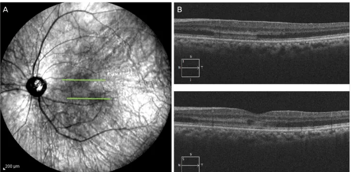
Figure 2. Near-infrared autofluorescence image of the patient. Near-infrared imaging presents multiple hyporeflective darker lesions in
left eye (A). The hyporeflective lesions (green lines) correlate to hyper-reflectivity in the outer nuclear layer and outer plexiform layer on spectral domain optical coherence tomography (B).A B
Figure 3. Humphrey visual field images of the patient. A Humphrey visual field study confirms paracentral scotoma in the left eye
(A). There is no definite visual field defect 2 months after trauma (B).검사소견은 호전되었다(Fig. 3B, Fig. 4). 경과관찰 후 6개월 이 지난 현재, 교정시력은 0.6이며 환자는 아직 약간의 중심 암점을 호소하고 있으며 광수용체 내외층경계부와 원뿔세 포 외절끝층에는 결손이 남아 있다. 또 OCT 상 광수용체
내외층경계부의 결손이 남아 있는 부분인 망막의 코쪽 부 위에 ONL이 위축된 소견을 관찰할 수 있었다(Fig. 4).
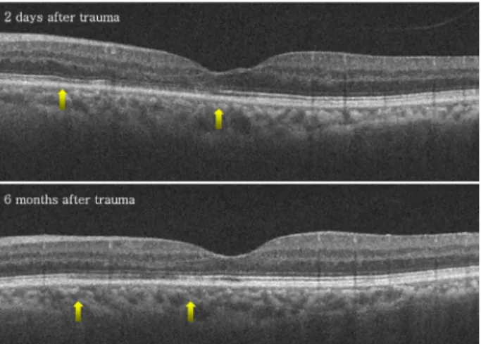
Figure 4. Spectral domain optical coherence tomography im-
ages of the left eye 2 days and 6 months after trauma. The im- ages display gradual improvement of the hyper-reflectivity within the outer plexiform layer and outer nuclear layer (ONL). But there are thinning of ONL and disruption of ellip- soid zone and interdigitation zone (between arrows).고 찰
급성 황반 신경망막병증(AMN)은 안저 소견상 쐐기 혹은 꽃잎 모양의 갈색 병변이 관찰되고 병변에 상응하는 부위 의 중심부근 암점이 특징적이다. 최근 다양한 영상기법의 발달로 정확한 병변의 위치를 관찰할 수 있게 되었는데, 일 반적인 안저사진보다 무적색광 안저 소견에서 쐐기 모양의 병변이 좀 더 명확히 관찰된다. 특히 OCT 상에서 광수용체 내외층경계부(ellipsoid zone)와 원뿔세포 외절끝층(interdigi- tation zone)의 손상이 나타나는 부위와 근적외안저자가형광 에서 저형광을 나타내는 부분이 일치하는 소견이 보이는데, 이를 통해 AMN에서는 망막색소상피내의 멜라닌이 소실되 거나 파괴되는 것이라고 생각된다.2 OCT가 발달하면서 최 근에는 초기 OCT에서 고반사가 나타나는 위치에 따라서 AMN을 두 부류로 분류한다. 전형적인 AMN (classic, type 2)의 경우, OCT 소견상 OPL과 그 외층인 ONL의 고반사가 관찰되는 것이 특징인데 이것은 망막의 심층모세혈관총 (deep capillary plexus)과 맥락막 모세혈관(choriocapillary) 사이의 분수계역(watershed zone)이 허혈에 취약하여 발생 하는 것으로 생각된다.4 또 다른 분류인 paracentral acute middle maculopathy (PAMM, type 1 AMN)는 Sarraf et al5이 처음 보고하였는데, AMN의 아형으로 전형적인 AMN보다 내층인 내과립층(inner nuclear layer, INL)과 외망막층(OPL) 에 고반사를 보인다.5-7 두 분류 모두 원인이 정확하게 밝혀 지지 않았으나 심부망막모세혈관총(deep retinal capillary plexus)의 허혈이나 손상으로 인한 망막의 징후로 생각된 다.2 보통 특별한 치료 없이 경과관찰을 하며 시간이 지나면
시력 및 시야장애가 호전되지만 시야결손이나 시력저하의 증상이 남기도 한다. 후기 AMN의 경과는 OCT에서도 잘 관찰되는데, 전형적인 AMN (classic, type 2)의 경우에는 ONL의 위축이 관찰되고, PAMM (type 1)은 INL의 위축이 동반되기도 한다.3,4 Bhavsar et al3은 1975년부터 보고된 101 건의 AMN 증례를 분석하였는데, 발병 전 관련된 요인으로 는 비특이적인 독감증상이 47.5%로 가장 많았으며 경구 피 임약이 35.6%, 에피네프린에 노출된 경우가 7.9%로 보고되 었다. 드물지만 5.9%인 6예에서 외상에 의해 발생한 경우를 보고하였으며 모두 직접적인 안구외상은 없었다.
간접 외상 이후 나타나는 황반의 변화로는 발살바망막병 증(valsalva retinopathy), 지방색전증후군(fat embolism) 등 혈 관의 직간접적인 손상이 관찰되는 경우가 대부분이다. 1997 년 Gillies et al8은 간접외상 이후 AMN과 유사한 증상을 보 이는 4명의 환자를 처음 보고하였다. 그러나 OCT가 발달하 지 않아 정확한 병변의 위치를 확인하지 못하였고 중심부근 암점과 안저소견상 황반부에 적갈색의 쐐기 모양의 병변으 로 진단하였다. 이후 Nentwich et al9은 간접 외상 이후 AMN 과 유사한 망막소견을 가진 5예를 보고하였고 Chinskey et al10도 5예를 보고하였다. OCT에서 망막외층, 특히 OPL 및 ONL의 고반사가 모두 관찰되었고 증례에 따라 광수용체 내 외층경계부 및 망막색소상피층(retinal pigment epithelium)의 결손이 보이는 경우도 있었다. Gillies et al8은 외상으로 인한 AMN의 발생기전을 제시하였는데, 갑자기 흉강의 압력이 높아지면 내측혈액망막장벽(inner-blood retinal barrier)이 파 괴되고, 그 결과 생긴 혈관외액(extravascular fluid)이 신속히 흡수되어 그 부위 망막의 손상으로 AMN이 발생한다는 것 이다. 그러나 OCT 등 발달된 영상기법으로 병변을 관찰한 결과, 급성기의 AMN에서 망막내 부종이나 혈관외액이 관 찰되지 않았고, 망막의 부종이 조직의 위축을 일으키지 않 으므로 가설과 증례의 소견이 맞지 않다. 그 후 많은 저자들 은 AMN의 발생기전에 대한 가설을 제시하였다. 그중 하나 가 염증으로 인한 것으로 Feigl and Haas11는 바이러스를 감 염원으로 한 자가면역 반응에 의해 망막에 염증물질이 축적 되어 망막외층의 고반사가 일어날 것이라고 생각하였고 Yeh et al12은 맥락막의 내층에 국소적인 염증이 축적되어 망 막외층의 허혈을 일으킬 것이라고 하였다. 그러나 AMN에 서 혈관조영술을 시행하였을 때 염증으로 인한 누출이 관찰 되지 않으며 유리체나 전안부에도 염증 소견이 관찰되지 않 는 점을 보았을 때 적절한 가설로 생각되지 않는다. 에피네 프린 등의 혈관수축제나 쇼크 등의 혈관질환이 선행하여 AMN이 관찰된 여러 증례를 통하여 최근에는 심부망막혈관 총의 국소적인 허혈로 인해 발생하였다는 가설이 많이 받아 들여지고 있으며, 실제 검사상 고배율 형광안저촬영(high
magnification fundus angiography)에서 국소적인 허혈이 관 찰되기도 한다.6 본 증례와 같이 교통사고 등의 큰 외상 이 후에 AMN이 발생하는 경우 또한 에피네프린이나 노르에피 네프린과 같은 카테콜라민이 분비되어13 혈관이 수축하거나 외상성 쇼크로 일시적인 저혈압이 발생하여 망막과 맥락막 의 분수계역에 허혈이 일어날 수 있다. 이처럼 여러 가지 혈 역학적인 원인에 의한 황반의 외망막층의 허혈성 손상이 AMN의 주요 기전으로 생각된다.10
국내에서 외상으로 인한 AMN은 본 증례가 첫 보고이며, 중심부 시야 장애 및 갈색 쐐기모양의 망막병변, OCT 상 망막외층의 고반사도와 광수용체 내외층경계부의 결손이 모두 관찰되었다. 본 증례환자를 6개월 동안 경과관찰한 결 과 시력은 호전되었으나 환자는 중심부 시야장애를 호소하 고 있으며 OCT에서 ONL이 위축된 것을 관찰할 수 있다.
전형적인 AMN에서는 시야 결손 부위 및 망막의 병변과 같 은 위치에 다초점망막전위도 검사에서 진폭 감소가 관찰되 는데 본 증례에서는 검사가 누락되어 확인하지 못하였다.
최근 다양한 원인에 의한 AMN이 보고되고 있다. 앞으로 빛간섭단층혈관조영술(optical coherence tomography angiog- raphy)이나 망막 산소측정법(retinal oximetry) 등 발전하고 있는 다양한 장비를 통하여 AMN의 기전에 대해 정확한 분 석이 필요할 것이다.
REFERENCES
1) Bos PJ, Deutman AF. Acute macular neuroretinopathy. Am J Ophthalmol 1975;80:573-84.
2) Fawzi AA, Pappuru RR, Sarraf D, et al. Acute macular neuro-
retinopathy: long-term insights revealed by multimodal imaging.
Retina 2012;32:1500-13.
3) Bhavsar KV, Lin S, Rahimy E, et al. Acute macular neuroretinopathy:
a comprehensive review of the literature. Surv Ophthalmol 2016;
61:538-65.
4) Yu S, Wang F, Pang CE, et al. Multimodal imaging findings in reti- nal deep capillary ischemia. Retina 2014;34:636-46.
5) Sarraf D, Rahimy E, Fawzi AA, et al. Paracentral acute middle maculopathy: a new variant of acute macular neuroretinopathy as- sociated with retinal capillary ischemia. JAMA Ophthalmol 2013;
131:1275-87.
6) Rahimy E, Sarraf D. Paracentral acute middle maculopathy spec- tral-domain optical coherence tomography feature of deep capil- lary ischemia. Curr Opin Ophthalmol 2014;25:207-12.
7) Chen X, Rahimy E, Sergott RC, et al. Spectrum of retinal vascular diseases associated with paracentral acute middle maculopathy.
Am J Ophthalmol 2015;160:26-34.e1.
8) Gillies M, Sarks J, Dunlop C, Mitchell P. Traumatic retinopathy re- sembling acute macular neuroretinopathy. Aust N Z J Ophthalmol 1997;25:207-10.
9) Nentwich MM, Leys A, Cramer A, Ulbig MW. Traumatic retinop- athy presenting as acute macular neuroretinopathy. Br J Ophthal- mol 2013;97:1268-72.
10) Chinskey ND, Rahimy E, Johnson MW. Acute macular neuro- retinopathy following non-ocular trauma: a hypothesis regarding pathophysiologic mechanism. Ophthalmic Surg Lasers Imaging Retina 2015;46:1013-20.
11) Feigl B, Haas A. Optical coherence tomography (OCT) in acute macular neuroretinopathy. Acta Ophthalmol Scand 2000;78:714-6.
12) Yeh S, Hwang TS, Weleber RG, et al. Acute macular outer retinop- athy (AMOR): a reappraisal of acute macular neuroretinopathy using multimodality diagnostic testing. Arch Ophthalmol 2011;129:365-8.
13) Davies CL, Newman RJ, Molyneux SG, Grahame-Smith DG. The relationship between plasma catecholamines and severity of injury in man. J Trauma 1984;24:99-105.
= 국문초록 =
간접외상 이후에 발생한 급성 황반 신경망막병증 1예
목적: 늑골골절 직후 발생한 중심부근암점을 증상으로 내원한 환자에서 급성 황반 신경망막병증의 소견이 관찰된 1예가 있어 이를 보고하고자 한다.
증례요약: 과거력이 없는 69세 남자 환자가 교통사고로 좌측 늑골골절을 진단 받았으며 직접적인 안구외상은 없었다. 사고 직후 좌안 이 가려보이는 증상을 주소로 본과에 의뢰되었다. 좌안의 중심부 시력은 안전수동이었고 시야검사상 중심부근암점이 관찰되었다. 빛 간섭단층촬영상 외망상층과 외과립층의 반사도가 증가하였고 광수용체내외절경계부의 결손을 확인할 수 있었다. 형광안저혈관조영 술상 특이소견은 관찰되지 않았지만 근적외안저자가형광에서 부분적으로 저형광을 보였으며 빛간섭단층촬영에서의 병변부와 상응하 는 위치에 있었다. 특별한 치료 없이 경과관찰한 결과 좌안의 시력은 2주 뒤 0.2, 6달 뒤 0.6으로 호전되었고 빛간섭단층촬영상 광수 용체내외절경계부의 결손부위가 관찰되고 시야검사상 미세한 중심부근 암점이 남아 있는 상태이다.
결론: 급성 황반 신경망막병증은 갑자기 발생하는 중심부근암점이 특징인 드문 질환으로, 아직 발병기전에 대해 명확히 밝혀지진 않 았지만 최근 빛간섭단층촬영의 발전으로 많이 보고되고 있다. 그러나 간접 외상 이후 급성 황반 신경망막병증의 소견을 보이는 경우 는 매우 드물며 국내에 처음 보고되는 증례이다.
<대한안과학회지 2016;57(12):1970-1975>