Endocrinol Metab 2013;28:320-325
http://dx.doi.org/10.3803/EnM.2013.28.4.320 pISSN 2093-596X · eISSN 2093-5978
Original Article
A Novel Mutation in the Von Hippel-Lindau Tumor Suppressor Gene Identified in a Patient Presenting with Gestational Diabetes Mellitus
Yun Hyi Ku1,2, Chang Ho Ahn2, Chan-Hyeon Jung2, Jie Eun Lee2, Lee-Kyung Kim2, Soo Heon Kwak2, Hye Seung Jung2, Kyong Soo Park2, Young Min Cho2
1Department of Internal Medicine, Korea Cancer Center Hospital; 2Department of Internal Medicine, Seoul National University College of Medicine, Seoul, Korea
Background: Von Hippel-Lindau (VHL) disease is an autosomal dominantly inherited, multisystemic tumor syndrome caused by mutations in the VHL gene. To date, more than 1,000 germline and somatic mutations of the VHL gene have been reported. We present a novel mutation in the VHL tumor suppressor gene that presented with gestational diabetes mellitus.
Methods: A 30-year-old woman presented with gestational diabetes mellitus. She sequentially showed multiple pancreatic cysts, spinal cord hemangioblastoma, cerebellar hemangioblastoma, and clear cell type renal cell carcinomas. Also, her father and brother had brain hemangioblastomas. Each of the three exons of the VHL gene was individually amplified by polymerase chain reaction and direct sequencing was performed using an ABI 3730 DNA analyzer.
Results: DNA sequence analysis to determine the presence of VHL mutation in her family revealed del291C, a novel frameshift mutation.
Conclusion: We found a novel mutation in the VHL tumor suppressor gene that presented with gestational diabetes mellitus.
Keywords: von Hippel-Lindau disease; VHL gene; Diabetes, gestational
INTRODUCTION
Von Hippel-Lindau (VHL) disease is a hereditary autosomal dominant, multiorgan tumor syndrome caused by a germline mutation in the VHL tumor suppressor gene [1]. Germline mu- tations in the VHL gene lead to the development of several be- nign or malignant tumors and cysts in many organ systems [1].
The most common tumors are hemangioblastomas of the reti- na and central nervous system, clear cell renal cell carcinoma (RCC) and pheochromocytoma [2]. Multiple cysts, serous mi-
crocystic adenomas and neuroendocrine tumors of the pancre- as have also been reported in patients with VHL disease [3].
More than 50% of subjects with VHL disease have pancreatic lesions including serous cystadenomas, multiple cysts, neuro- endocrine tumors, or combined lesions, but pancreatic lesions are generally asymptomatic or associated with only mild symp- toms [4]. Diabetes or pancreatitis seems to be relatively rare [3,4]. Clinically, types of VHL disease have been classified on the basis of the risk of pheochromocytoma and RCC [5,6].
VHL type 1 is characterized by a low risk of pheochromocyto-
Received: 19 July 2013, Accepted: 3 September 2013 Corresponding author: Young Min Cho
Department of Internal Medicine, Seoul National University College of Medicine, 101 Daehak-ro, Jongno-gu, Seoul 110-744, Korea
Tel: +82-2-2072-1965, Fax: +82-2-762-9662, E-mail: [email protected]
Copyright © 2013 Korean Endocrine Society
This is an Open Access article distributed under the terms of the Creative Com- mons Attribution Non-Commercial License (http://creativecommons.org/
licenses/by-nc/3.0/) which permits unrestricted non-commercial use, distribu- tion, and reproduction in any medium, provided the original work is properly cited.
ma, while VHL type 2 is characterized by a high risk of pheo- chromocytoma. Type 2 is subdivided into type 2A, which has a low risk of RCC; type 2B, which has a high risk of renal car- cinoma; and type 2C, which has a risk of pheochromocytoma but not the other manifestations of VHL disease [5,6]. The VHL gene was mapped by linkage analysis to the short arm of chromosome 3 in 1988 [7]. Several hundred germline muta- tions in the VHL gene have been reported since VHL gene was isolated in 1993 [8]. The VHL gene is a tumor suppressor ubiquitously expressed in adult tissues. Individuals with VHL disease carry one wild-type VHL allele and one inactivated VHL allele, and tumor or cyst development in VHL disease is linked to somatic inactivation or loss of the remaining wild- type VHL allele. Approximately 15% to 20% of VHL patients have large germline deletions, 27% have missense mutations, and 27% have nonsense or frameshift mutation [9]. In general, VHL mutations are extremely heterogeneous and are distribut- ed throughout the coding sequence, although intragenic mis- sense mutations are rarely seen with the first 50 codons. In to- tal, more than 150 different germline VHL mutations linked to VHL disease have been reported. We hereby report a patient with VHL disease caused by a novel frameshift mutation in the VHL gene and who initially presented with gestational dia- betes mellitus (GDM).
METHODS
A 30-year-old woman was referred to Seoul National Univer- sity Hospital diabetes clinic for glycemic control in July 2006.
Her height was 159 cm and her body weight was 52 kg, physi- cal examinations including vital signs were within normal limits, and her routine biochemical test results were normal except for the blood glucose level. She was diagnosed with GDM at gestational age 27 weeks of her first pregnancy (in January 2006). She used insulin for glycemic control during the pregnancy. The delivery occurred at full-term without any event, and the baby had no perinatal complications. After de- livery, she did not take any antidiabetes medications. At 3 months postpartum, fasting plasma glucose level was 252 mg/
dL, 2-hour postprandial glucose level was 572 mg/dL during a 75 g oral glucose tolerance test and her hemoglobin A1c level was 11.0%. Her antiglutamic acid decarboxylase antibody was negative and fasting C-peptide level was 0.2 ng/mL. She needed no more than 10 to 12 units of insulin a day to keep her blood glucose level under control, which was not main- tained with oral antidiabetes drugs in the place of insulin. An
abdominal computed tomography scan taken in October 2006 revealed that her pancreas and both kidneys were covered with numerous cysts (Fig. 1A); thereby, she was suspected to have VHL disease. Her family members had some features that ap- peared to be related to VHL disease. Her grandmother was troubled with severe headache during her lifetime, though the cause of death was unclear. Her father, who had diabetes mel- litus requiring insulin therapy, underwent brain surgery twice and was diagnosed with brain hemangioblastoma, which was his cause of death. In addition, her younger brother underwent brain surgery in his teens due to brain hemangioblastoma. He had not been diagnosed with diabetes mellitus.
In February 2007, she presented with paresthesia and weak- ness in both legs and voiding difficulty for 1 month prior to hospital admission. Magnetic resonance imaging (MRI) of the lumbosacral spine showed an intradural, brightly enhancing le- sion at the approximated level of the L2 to L3 disc space (Fig.
1B). She underwent complete resection of the tumor. Histo- pathological examination demonstrated hemangioblastoma. To screen the intracranial lesion, we performed brain MRI. Multi- ple cerebellar hemangioblastomas were detected on MRI (Fig.
1C), and she underwent gamma knife surgery. Ophthalmologic exam revealed no retinal hemangioblastoma. Two years later Fig. 1. Radiologic studies revealing the clinical features of the pa- tient. (A) Abdominal computed tomography (CT) scan shows multiple cysts in the pancreas and kidneys. (B) A hemangioblasto- ma is seen in the spinal cord (arrow). (C) Two hemangioblastomas are seen in the cerebellum (arrows). (D) A follow-up abdominal CT scan shows multiple renal cell carcinomas (arrow).
A B
C D
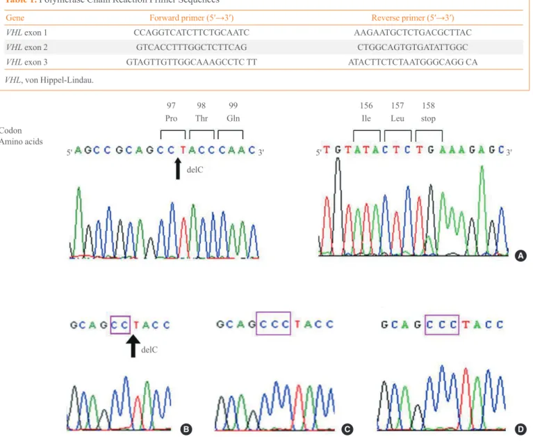
Codon Amino acids
5' 3' 5' 3'
97 98 99
Pro Thr Gln
delC
delC
Ile Leu stop 157
156 158
A
B C D
Fig. 2. The VHL gene mutation of the patient and her family members. (A) Sequencing analysis reveals a 291C deletion mutation, which produces a frameshift and an early stop codon. The subject’s brother (B) showed the same mutation, (C) whereas her clinically unaffect- ed sister and (D) second baby did not.
(in March 2011), she was diagnosed with RCC and underwent partial nephrectomy on both kidneys (Fig. 1D). Pathology re- vealed clear cell type RCC.
Genetic analysis of the VHL gene
Informed consent was obtained from the patient and her sib- lings for analysis of the VHL gene. Peripheral blood samples from the patient, her brother and her sister were collected.
The cord blood of the second baby of the patient was obtained at delivery with consent of the patient. A total 50 μL of ge- nomic DNA was extracted using Gentra Puregene Blood Kit
(Qiagen, Valencia, CA, USA) according to the manufacturer’s instructions. To screen the VHL gene for mutations, we per- formed direct sequencing of the coding region. Each of the three exons of the VHL gene was individually amplified by polymerase chain reaction (PCR), using primer pairs shown in Table 1. After purifying the PCR products by QIAquick PCR Purification Kit (Qiagen) according to the manufactur- er’s instructions, direct sequencing was performed using an ABI 3730 DNA analyzer (Applied Biosystems, Foster City, CA, USA).
Table 1. Polymerase Chain Reaction Primer Sequences
Gene Forward primer (5′→3′) Reverse primer (5′→3′)
VHL exon 1 CCAGGTCATCTTCTGCAATC AAGAATGCTCTGACGCTTAC
VHL exon 2 GTCACCTTTGGCTCTTCAG CTGGCAGTGTGATATTGGC
VHL exon 3 GTAGTTGTTGGCAAAGCCTC TT ATACTTCTCTAATGGGCAGG CA
VHL, von Hippel-Lindau.
Consent
Genetic analysis was approved by the Ethical Committee of Seoul National University Hospital, conforming to the Decla- ration of Helsinki. The patient and her siblings gave written informed consent to participate in this study and for publica- tion of this case report.
RESULTS
In DNA analysis, we found a 291C deletion mutation in the first exon of the subject’s VHL gene (Fig. 2A). This mutation switches the 97th codon from CCC to CCT, which codes for the same amino acid, proline. However, a newly organized 98th codon ACC from TAC produced an amino acid change from tyrosine to threonine (Tyr98Thr). From this point, a frame shift occurred, resulting in the 158th codon acting as an early stop codon (the human VHL consists of 213 amino ac- ids) (Fig. 2A). The human VHL gene is a 12 kb region located at 3p25.3 [5,10]. The gene consists of three exons; the first exon spans 1-113 codons, exon 2 spans 114-154 codons, and exon 3 spans 155-213 codons. Del291C mutation is located in exon 1, and an early stop was introduced in exon 3. Subse- quent genetic analysis of family members revealed the same mutation in her brother but not in her clinically unaffected sis- ter or baby (Fig. 2B-D). The assumed genogram based on family history and DNA analysis is shown in Fig. 3.
DISCUSSION
Pancreatic lesions including cysts or neuroendocrine tumors occur in approximately 70% of VHL patients [4,11]. About 90% of pancreatic lesions are single or multiple cysts [12,13].
Serous microcystic adenomas and neuroendocrine tumors of the pancreas have also been reported in patients afflicted with VHL disease. Most pancreatic lesions are asymptomatic and are discovered during screening of family members with VHL disease. However, mass effect of pancreatic lesions can result in acute pancreatitis [4,14], biliary obstruction [4,15] and gas- trointestinal obstruction [4,16]. In addition, pancreatic endo- crine insufficiency has been reported [4,17,18]. Hammel et al.
[4] reported diabetes mellitus to be present in three of 158 VHL patients, and the risk of diabetes was regarded to be rela- tively low [3,4]. Mukhopadhyay et al. [3] reported that exten- sive serous microcystic adenomas were associated with diabe- tes mellitus in patients with VHL disease. Intriguingly, in re- cent animal studies, loss of VHL tumor suppressor gene in β-cells lead to impaired glucose tolerance in mice [19,20], which indicates that impairment of β-cell function in a patient with VHL disease could be related to an inactivated VHL gene and change of VHL/HIF oxygen-sensing mechanisms.
VHL syndrome is rare in Korea. However, some mutations in the VHL gene have been identified in association with RCC [21], spinal cord hemangioblastoma [22], and pheochromocy- toma [23]. Although there have been no large-scale multi- center studies examining the genotype-phenotype correlation, a case series reported that the pancreatic involvement is as high as 89% (16/18) in Korean patients with VHL syndrome [24].
We found a novel mutation (del291C) in the VHL tumor suppressor gene of a patient presenting with GDM. This report suggests that VHL disease can be manifested as GDM as an initial presentation of the disease.
CONFLICTS OF INTEREST
No potential conflict of interest relevant to this article was re- ported.
ACKNOWLEDGMENTS
We thank Min Kim Ph.D. and Sang Gyu Park Ph.D. from Clinical Research Institute, Seoul National University Hospi- tal, Seoul, Korea for genetic analysis.
Fig. 3. Genogramof the family. The genogram was drawn based on the results of genetic analysis and probable symptoms and signs of von Hippel-Lindau (VHL) syndrome.
I-1
II-1 II-2 II-3 II-4 II-5
III-1 III-2
IV-2 IV-1
III-3 III-4
I-2 VHL syndrome
R/O brain tummor
REFERENCES
1. Kim WY, Kaelin WG. Role of VHL gene mutation in hu- man cancer. J Clin Oncol 2004;22:4991-5004.
2. Woodward ER, Maher ER. Von Hippel-Lindau disease and endocrine tumour susceptibility. Endocr Relat Cancer 2006;
13:415-25.
3. Mukhopadhyay B, Sahdev A, Monson JP, Besser GM, Reznek RH, Chew SL. Pancreatic lesions in von Hippel- Lindau disease. Clin Endocrinol (Oxf) 2002;57:603-8.
4. Hammel PR, Vilgrain V, Terris B, Penfornis A, Sauvanet A, Correas JM, Chauveau D, Balian A, Beigelman C, O’Toole D, Bernades P, Ruszniewski P, Richard S. Pancreatic involve ment in von Hippel-Lindau disease: the Groupe Francophone d’Etude de la Maladie de von Hippel-Lindau. Gastroenterol- ogy 2000;119:1087-95.
5. Richard S, David P, Marsot-Dupuch K, Giraud S, Beroud C, Resche F. Central nervous system hemangioblastomas, en- dolymphatic sac tumors, and von Hippel-Lindau disease.
Neurosurg Rev 2000;23:1-22.
6. Zbar B, Kishida T, Chen F, Schmidt L, Maher ER, Richards FM, Crossey PA, Webster AR, Affara NA, Ferguson-Smith MA, Brauch H, Glavac D, Neumann HP, Tisherman S, Mul- vihill JJ, Gross DJ, Shuin T, Whaley J, Seizinger B, Kley N, Olschwang S, Boisson C, Richard S, Lips CH, Linehan WM, Lerman M. Germline mutations in the Von Hippel-Lindau disease (VHL) gene in families from North America, Europe, and Japan. Hum Mutat 1996;8:348-57.
7. Seizinger BR, Rouleau GA, Ozelius LJ, Lane AH, Farmer GE, Lamiell JM, Haines J, Yuen JW, Collins D, Majoor-Krak auer D, Bonner T, Mathew C, Rubenstein A, Halperin J, Mc- Conkie-Rosell A, Green JS, Trofatter JA, Ponder BA, Eier- man L, Bowmer MI, Schimke R, Oostra B, Aronin N, Smith DI, Drabkin H, Waziri MH, Hobbs WJ, Martuza RL, Con- neally PM, Hsia YE, Gusella JF. Von Hippel-Lindau disease maps to the region of chromosome 3 associated with renal cell carcinoma. Nature 1988;332:268-9.
8. Latif F, Tory K, Gnarra J, Yao M, Duh FM, Orcutt ML, Stackhouse T, Kuzmin I, Modi W, Geil L, Schmidt L, Zhou F, Li H, Wei MH, Chen F, Glenn G, Choyke P, Walther MM, Weng Y, Duan DS, Dean M, Glavac D, Richards FM, Crossey PA, Ferguson-Smith MA, Le Paslier D, Chumakov I, Cohen D, Chinault AC, Maher ER, Linehan WM, Zbar B, Lerman MI. Identification of the von Hippel-Lindau disease tumor suppressor gene. Science 1993;260:1317-20.
9. Maher ER, Kaelin WG Jr. von Hippel-Lindau disease.
Medicine (Baltimore) 1997;76:381-91.
10. Nordstrom-O’Brien M, van der Luijt RB, van Rooijen E, van den Ouweland AM, Majoor-Krakauer DF, Lolkema MP, van Brussel A, Voest EE, Giles RH. Genetic analysis of von Hippel-Lindau disease. Hum Mutat 2010;31:521-37.
11. Horton WA, Wong V, Eldridge R. Von Hippel-Lindau dis- ease: clinical and pathological manifestations in nine fami- lies with 50 affected members. Arch Intern Med 1976;
136:769-77.
12. Lubensky IA, Pack S, Ault D, Vortmeyer AO, Libutti SK, Choyke PL, Walther MM, Linehan WM, Zhuang Z. Multi- ple neuroendocrine tumors of the pancreas in von Hippel- Lindau disease patients: histopathological and molecular genetic analysis. Am J Pathol 1998;153:223-31.
13. Libutti SK, Choyke PL, Bartlett DL, Vargas H, Walther M, Lubensky I, Glenn G, Linehan WM, Alexander HR. Pan- creatic neuroendocrine tumors associated with von Hippel Lindau disease: diagnostic and management recommenda- tions. Surgery 1998;124:1153-9.
14. Tenner S, Roston A, Lichtenstein D, Sica G, Carr-Locke D, Banks PA. Von Hippel-Lindau disease complicated by acute pancreatitis and Evan’s syndrome. Int J Pancreatol 1995;18:271-5.
15. Beerman MH, Fromkes JJ, Carey LC, Thomas FB. Pancre- atic cystadenoma in Von Hippel-Lindau disease: an unusu- al cause of pancreatic and common bile duct obstruction. J Clin Gastroenterol 1982;4:537-40.
16. Kunzli BM, Shrikhande SV, Buchler MW, Friess H. Pan- creatic lesions in von Hippel-Lindau syndrome: report of a case. Surg Today 2004;34:626-9.
17. Lamiell JM, Salazar FG, Hsia YE. von Hippel-Lindau dis- ease affecting 43 members of a single kindred. Medicine (Baltimore) 1989;68:1-29.
18. Thompson RK, Peters JI, Sirinek KR, Levine BA. von Hip- pel-Lindau syndrome presenting as pancreatic endocrine insufficiency: a case report. Surgery 1989;105:598-604.
19. Cantley J, Selman C, Shukla D, Abramov AY, Forstreuter F, Esteban MA, Claret M, Lingard SJ, Clements M, Harten SK, Asare-Anane H, Batterham RL, Herrera PL, Persaud SJ, Duchen MR, Maxwell PH, Withers DJ. Deletion of the von Hippel-Lindau gene in pancreatic beta cells impairs glucose homeostasis in mice. J Clin Invest 2009;119:125-35.
20. Puri S, Cano DA, Hebrok M. A role for von Hippel-Lindau protein in pancreatic beta-cell function. Diabetes 2009;58:
433-41.
21. Kim WT, Ham WS, Ju HJ, Lee JS, Lee JS, Choi YD. Clini-
cal characteristics of renal cell carcinoma in Korean pa- tients with von Hippel-Lindau disease compared to spo- radic bilateral or multifocal renal cell carcinoma. J Korean Med Sci 2009;24:1145-9.
22. Na JH, Kim HS, Eoh W, Kim JH, Kim JS, Kim ES. Spinal cord hemangioblastoma: diagnosis and clinical outcome after surgical treatment. J Korean Neurosurg Soc 2007;42:
436-40.
23. Kang HC, Kim IJ, Park JH, Shin Y, Jang SG, Ahn SA,
Park HW, Lim SK, Oh SK, Kim DJ, Lee KW, Choi YS, Park YJ, Lee MR, Kim DW, Park JG. Three novel VHL germline mutations in Korean patients with von Hippel- Lindau disease and pheochromocytomas. Oncol Rep 2005;
14:879-83.
24. Lee KH, Lee JS, Kim BJ, Lee JK, Kim SH, Kim SH, Lee KT. Pancreatic involvement in Korean patients with von Hippel-Lindau disease. J Gastroenterol 2009;44:447-52.