대 한 방 사 선 의 학 회 지 1992 ; 28 ( 1) : 1 04~ 1 Oï Journal of Korean Radiological Society, January, 1992
Multiple Coronary Arteriovenous Fistulae Combined with Ventricular Septal Defect*: A Case Report
Kun Sik Jung, M.D., Seok Kil Zeon, M.D., Ki Sik Kim, M.D. **, Yeon Hee Oh, M.D. ***
Department o[ Radiology, Keimyung University. School o[ Medicine
INTRODUCTION
treatment
During a nine month period. symptoms developed and she was medicated with seldatives and antacids.
Congenital coronary arteriovenous fistulas are all but her condition was gradually aggravated. She was of those anomalies and malformations that result in then transfered to our hospital and a heart murmur a direct communication of a coronary artery and/or was discovered on physical exam.
any of its branches with a cardiac chamber or ex- She had no remarkable past medical history or tracardiac vessel resulting in a shunt ofvarying pro- family history. Physical examination revealed a well portions. The majority of these fistulas form an A-V developed and well nourished woman in no acute connection between a coronary artery and the right distress. Her pulse rate was 82/min and regular. and heart chambers. and rarely their is communication her blood pressure was 100/80mmHg.
with the left heart chambers. A grade 4/6 pansystolic murmur was best heard In most reported cases. the abnormal fistula in- at the cardiac apex. There is no p외pable thri1l noted.
volves only one coronary artery which developed An electrocardiogram revealed normal sinus rhythm
alone. with a primary A-V block and left ventricular hyper-
This report describes a unique case in which multi- trophy. The chest X-ray was within normal limits.
ple coronary fistulas associated with VSD were en- Cardiac catheterization revealed oxygen step-up(9%)
countered. at the right ventricular level, and the Qp/Qs was 1.8.
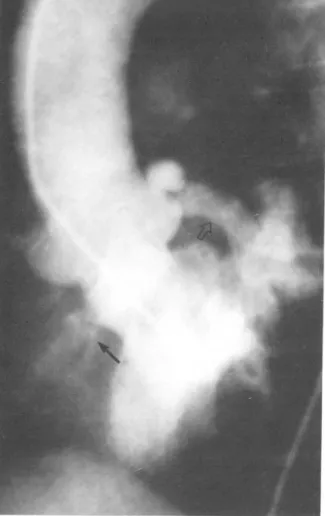
Left vetricular cineangiography in the left anterior CASE REPORT oblique pr이 ection demonstrated an extremely dilated and tortous vessel that corresponded to the left cir- A 53 year old woman was admitted because of ex- cumf1ex coronary artery. A small VSD and opacifica- ertional dyspnea. intermittent palpitation. easy tion of the right ventricle(Fig. 1) was also seen fatigability and mild chest discomfort. She experienc- A selective left coronary arteriogram satisfactori- ed frequent episodes of palpitation for 10 years. but Iy opacified this vessel. The coronary sinus. right had managed to live a relatively normallife without atrium. right ventricle and pulmonary artery(Fig. 2)
Index Words: Fistula. coronary 54.18 Ventricular septal defect 52.14 Coronary Angiography 54.12 Angiocardiography 52.12
were also visible.
A right coronary arteriogram demonstrated an enlarged vessel of the terminal branch of the posterior descending artery which terminated in a small fistulous stoma emptying into the left ventricle (Fig.
*01 논문은 1991년 계명대학교 을종연구비 및 동산의료원 조사연구비로 이루어졌음
**계명대학교 의과대학 내과학교실
**Department of Internal Medicine. Keimyung Uiversity. SchooJ of Medicine
***동국대학교 포항병원 방사선과학교실
***Department of RadioJogy. Pohang HospitaJ. Dongguk University
이 논문은 1991년 5 월 9 일 접수하여 1991 년 11 월 14 일에 채택되었음
Received May 9. Accepted November 14. 1991
- 104 -
Kun Sik Jung. et al Multiple Coronary Arteriovenous Fistulae Combined with Ventricular Septal Defect
Fig. 1. LAO view of left cineventriculogram reveals an extremely dilated tortous left coronary artery(open ar.
row) and a small ventricular septal defect(arrow).
a
3).
The left coronary arterial fistulous tract was ligated by transfixion sutures and the VSD was repaired by direct closure
However. the right coronary arterial fist비a was not repaired, because the 02 step.up at the ventricular level was mainly due to the left coronary arterial fistula and VSD. The patient did well postoperative- ly, and the murmur completely disappeared.
DISCUSSION
Coronary arterial fistulas may be formed by a well defined vessel emptying into a cardiac chamber or by a plexus of multiple fine vessels. Myocardial vessels in the adult heart are derived embryological- ly from both endothelial protrusions into the myocar- dium and coronary arteries and veins. This endothelial protrusion into the myocardium extends to the epicardial surface, forming an intertrabecular network. The outermost intertrabecular network becomes almost completely obliterated and forms a capillary network as the myocardium grows, where as the innermost intertrabecular vessels retain their communication with chambers ofthe heart, forming Thebesian vessels of the adult heart.
Investigators reporting earlier cases, in which sinusoidal communication existed between involved coronary arteries and the heart chamber, ascribed the
b
Fig.2. a. b. Selective left coronary arteriogram reveals tortuous aneurysmally dilated left circumflex coronary artery communicating with the dilated coronary sinus(arrowj(A:AP B:LAO)
Journal of Korean Radiological Society 1992 ; 28( 1) : 1 04~ 1 07
a
Fig.3. a.b. Selective right coronary arteriogram reveals an enlarged vessel of its terminal branch of the posterior desoending artery which terminated in a small fistulous stoma emptying into the left ventricle(arrow)(A:AP B:LAO}‘
anomaly to persistence of the outermost inter- The majority of patients in the younger age group trabecular space. there by permitting communication with this anomality are asymptomatic and may reach between the coronary artery and the heart chamber the age of 30 to 40 years without difficuIty. However through the Thebesian vessels(3). most of them eventually will become symptomatic The coronary artery involved is usually extreme- later in life as a resuIt of the long.standing ly dilated and tortous. The dilatation starts from the hemodynamic abnormality. complications of the origin of the involved vessel and extends to the point disease. the development of coronary arteriosclerosis.
of communication. However. the size ofthe orifice of or hypertension(6)
the communicating vessel is relatively small in com- If symptoms occur. most frequent symptoms are parison with the dilated coronary artery. The bran- chest pain. dyspnea. palpitations and easy fatigabili- ches distal to the point of communication are ty. Chest pain is probably due to myocardial ischemia relatively small. secondary to the shunt. that is. a large amount of ox-
The recipient coronary sinus or vein. which also ygenated blood that normally would pass through the becomes aneurysmally dilated and thin-walled. myocardial capillaries is shunted instead through the serves as a site of blood c10t formation. fistula(6).
Congenital coronary arterial fistulas usually arise Pulmonary hypertension. congestive heart failure from a single artery. most frequently the right cor- and subacute bacterial endocarditis are the most onary artery. and usually communicate with the right common complication(7). Because ofthese complica- ventricle. right atrium or pulmonary artery(4). the tions. surgical correction ofthe anomalies is the treat- fistula between the right coronary artery and left ven- ment of choice.
tricle is quite rare. One case was reported in Korea. In this case. palpitations developed at age 43. and which revealed a marked dilatation of the right cor- surgical intervention was chosen because of the onary artery(diameter 3cm) and a large fistulous significant left to right shunt through the coronary stoma(diameter 1.5cm} into the left ventricle(5}. arterial fistula and VSD.
One of the two fistulas in this case was the right In most cases. the congenital coronary arterial coronary artery which communcated the left ventri- fistula develops alone. But infrequently several com-
cle. The stoma ofthe fistula opened into the left ven- bined anomalies are reported; Patent ductus
tricle but was small enough to be closed during the arteriosus. Tetralogy of Fallot‘ Mitral stenosis systolic phase by ventricular contraction after which etc(8-1O).
mild dilatation of the right coronary artery was noted. To the best of our knowledge. this is the first case
1 (、/、
Kun Sik Jung, et al Multiple Coronary Arteriovenous Fistulae Combined with Ventricular Septal Defect
of a congenital coronary arterial fistula combined with an interventricular septal defect. written in the English Iiterature. We propose that. in a congenital coronary arterial fistula. a complete cardiac catheterization and a cardiac angiogram should be performed for further evaluation of combined cardiac anomalies.
REFERENCES
fistula. Surgery 1969;65:59.69
5. Lee BH. Yu SJ. Moon ES. Kim SH. Choi YH: A case report of congenital coronary artery fistula to the left ventricle. The journal of the Korean radiological society 1987;23:420.423
6. Kimbiris 0‘ Kasparian H‘ Knibbe P. Brest AN: Cor onary artery.coronary sinus fistula. Am J Cardiology 1970‘26:532-539
7. Steinberg I. Baldwin JS‘ Dotter CT: Coronary arteriovenous fistula. Circulation 1958: 1 7‘372-390 1. Yu YJ‘ Han MC‘ Park JH: Congenital coronary 8. Schaffer AB. St Ville J. Mackler SA‘ Coronary arteriovenous fistulae. The journal of the Korean arteriovenous fistula with patent ductus. Am Heart
radiological society 1982;18:744-750 J 1963:65:758
2. Oh YH. Kim H. Zeon SK. Suh SJ: Congenital cor- 9. Busch Vw‘ ct al: Late deferioration in tetralogy of onary artery fistula. The journal of the Korean Fallot. Arch Internal Med 1978‘138‘1423
radiological society 1986;22:1083-1086 10. King SB. Schonmaker FW: Coronary artery to left 3. Reddy K. Gupta M. Hamby RI: Multiple coronary atrial fistula in association with severe arteriosystemic fistulas. Am J Cardiology 1974: atherosclerosis and mitral stenosis. Chest 1975:
33:304-306 67:361
4. McNamara JJ. Gross RE: Congenital coronary artery
〈국문 요약〉
심실 중격 결손증과 동반된 다발성 관상 동정맥루 |예 보고
*계명대학교 의과대학 방사선과학교실, 내과학교실** 동국대학교 부속포항병원 방사선과학교실***
정 건 식 • 전 석 길 • 김 기 식* • 오 연 희**
선천성 관상동맥루는 매우 드문 심질환으로서, 국내에서도 몇 예가 보고된 바가 있으나 0.2) 다발성 관상 동맥루는 보고 된 바가 없으며 세계적으로도 희귀한 증례이다.
최근 저자들은 53세의 여자에서 우측 관상동맥이 와심실과, 그리고 화측 만곡 관상동맥이 우심실과 연결된 다발성 관상 동맥루와 동반된 심실중격 결손증 l예를 경험 하였기에 보고하는 바이다.