721 Korean Circulation J 2004;34(7):721-722 IMAGE IN CARDIOVASCULAR MEDICINE
Asymptomatic Giant Coronary Aneurysm Presented as Cardiac Murmur
Eun-Ju Cho, MD, Chong-Jin Kim, MD, Jin-Man Cho, MD and Jae-Hyung Kim, MD Division of Cardiology, Department of Internal Medicine, College of Medicine, The Catholic University of Korea, Seoul, Korea
Case
A 27-year-old man presented with cardiac murmur detected on routine examination. He has no history of the illness consistent with Kawasaki disease on his childhood.
On admission, his blood pressure was 120/80 mmHg and heart rate was 78 beats per minute. On the chest aus- cultation, heart beat was regular, and early systolic murmur and thrill was detected on left side of mid sternal border.
The intensity of murmur aggravated on expiration and attenuated on inspiration. The electrocardiogram and the plain chest X-ray revealed no definite abnormal findings.
Transthoracic and transesophageal echocardiogram show- ed enlarged left main coronary artery (maximal diameter of 54 mm) with enlarged left anterior descending coron- ary artery and left circumflex artery. The patient under- went left and right heart catheterization with coronary angiography. The coronary angiography revealed diffuse ectatic change on three coronary arteries with sluggish antegrade flow. Thrill was aggravated with forced hand injection of dye to the left coronary artery. On the exercise treadmill test, there was no evidence of myocardial is- chemia, arrhythmia or symptoms even at the stage 4 by Bruce protocol. The patient discharged with the medica- tion of antiplatelet and anticoagulant agent. He got along without any complications and remained free of symp- toms for the entire 3-year follow-up period.
Received:January 15, 2004 Accepted:March 10, 2004
Correspondence:Chong-Jin Kim, MD,Division of Cardiology, Department of Internal Medicine, 620-56 Jeon Nong-dong, Dong Dae Moon-gu, St. Paul’s Hospital, College of Medicine, The Catholic University of Korea, Seoul 130-708, Korea Tel:82-2-958-2388, Fax:82-2-968-72506 E-mail:[email protected]
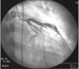
Figure 2. Angiographic view of the left coronary artery at the anteroposterior projection with 20 degrees of cra- nial angulation shows very tortuous dilated left anterior descending coronary artery (maximal diameter of 7.4 mm) with sluggish antegrade flow.
Figure 1. Upper esophageal horizontal image of transe- sophageal echocardiogram shows markedly dilated left main coronary artery and proximal portion of left anterior descending coronary artery.
Giant Coronary Aneurysm
Korean Circulation J 2004;34(7):721-722 722
Figure 3. Angiographic view at the 30-degree right an- terior oblique projection with 25 degrees of caudal an- gulation shows proximal to mid portion of left circumflex artery dilatation (maximal diameter of 6.3 mm) with slight sluggish flow.
Figure 4. 60-degree left anterior oblique view with 25 de- grees cranial angulation of right coronary artery shows markedly dilated (maximal diameter of 10.0 mm) mid and distal portion and very sluggish antegrade flow.