submit.radiology.or.kr J Korean Soc Radiol 2012;66(4):333-335
333 INTRODUCTION
Coronary-to-bronchial artery fistula (CBF) is a rare anomaly of the coronary artery. Most CBFs are clinically silent and are generally diagnosed incidentally during coronary angiography.
In the largest coronary cineangiography series conducted to date by Matsunaga et al. (1), the CBF were observed in 16 of the 2922 consecutive patients who underwent selective coronary cinean- giography during a 10 year at one institution. Although cardiac catheterization is considered the best method for identifying these types of vessel communications, the recently development of multi-detector computed tomography (MDCT) systems could be used to detect CBF (2, 3). In this report, we present a case of CBF in a 64-year-old man with clinical manifestation of aortic dissection that was detected by aortic CT angiography and diag- nosed by coronary CT angiography.
CASE REPORT
A 64-year-old man presented with acute onset epigastric
pain. On examination, his blood pressure was 180/100 mm Hg with a heart rate of 95 beats/min. Blood test findings, including cardiac enzymes, such as, creatine kinase MB (CK-MB), tropo- nin I, and d-dimer, were all within the normal range. Aortic CT angiography (CTA; 320-row MDCT; Aquilion ONE, Toshiba Medical Systems, Otawara, Japan) was performed to rule out aortic dissection. We injected intravenously 100 mL of nonion- ic contrast material (Xenetix®; iobitridol 350 mgI/mL; Guerbet, Roissy, France) at 4 mL/s into patients and a CT scan was start- ed using automatic bolus triggering in the ascending aorta (the triggering level was 300 HU) in the craniocaudal direction.
Aortic CTA revealed a diffusely dilated aorta with intimal dis- section from the just distal portion of the left subclavian arteri- al origin to both common iliac arteries (Fig. 1A). In addition, tortuous vascular structures were identified along the pericar- dial reflections (transverse sinus, oblique sinus, and atrioven- tricular groove) and left bronchovascular bundles (Fig. 1B), and seemed to be a continuation of the coronary arteries. Fur- thermore, in the lung window setting chest CT, cystic bronchi- ectasis was well demonstrated in the left lower lobe (Fig. 1C).
Case Report
pISSN 1738-2637
J Korean Soc Radiol 2012;66(4):333-335
Received December 26, 2011;
Accepted February 20, 2012
Corresponding author: Ki-Nam Lee, MD
Department of Radiology, Dong-A University Hospital, Dong-A University College of Medicine,
26 Daesingongwon-ro, Seo-gu, Busan 602-715, Korea.
Tel. 82-51-240-5367 Fax. 82-51-253-4931 E-mail: [email protected]
Copyrights © 2012 The Korean Society of Radiology
We report the case of a 64-year-old man with coronary-to-bronchial artery fistula (CBF) that was detected incidentally; the patient presented with the clinical mani- festations of aortic dissection. Coronary CT angiography demonstrated tortuous vessels originating from the left circumflex coronary artery that were in communi- cation with the bronchial arteries. CBF is a rare coronary artery anomaly and CBFs are asymptomatic in most cases. Familiarity with CT findings of CBF may help diag- nose the incidental finding of CBF through the use of CT angiography undertaken for a different diagnostic purpose.
Index terms
Coronary-to-Bronchial Artery Fistula Cardiac
CT Angiography
Diagnosis of Incidental Coronary-to-Bronchial Artery Fistula with CT Angiography: A Case Report
CT 혈관조영술에서 우연히 발견된 관상동맥기관지 동맥루의 진단: 1예 보고
Eun-Ju Kang, MD, Ki-Nam Lee, MD, Woo-Jeong Kim, MD, Jin-Hwa Lee, MD, Jong Young Oh, MD
Department of Radiology, Dong-A University Hospital, Dong-A University College of Medicine, Busan, Korea
Coronary-to-Bronchial Artery Fistula
submit.radiology.or.kr
J Korean Soc Radiol 2012;66(4):333-335
334
DISCUSSION
The prevalence of coronary artery anomalies has been estimat- ed to be 1.3% in patients that undergo coronary arteriography, and coronary fistulas account for 13% of these anomalies (4).
However, CBF is encountered very rarely, and most reports have been of isolated cases (2, 5). In a recent study, the incidence of CBF was estimated to be 0.61% among those undergoing CCTA (5). CBFs are believed to be congenital anastomosis between the coronary arteries and bronchial arteries. Some underlying pul- monary diseases, such as bronchiectasis or chronic pulmonary inflammation can cause anastomoses to dilate (2). It has been suggested that CBFs are present in all patients from birth and normally remain closed, but that a sizable CBF may result from considerable and persistent disturbance of the pressure equilibri- um (3, 6). CBFs usually originate from the left circumflex artery via a left atrial branch (in 75% to 81%) (1, 3). In our case, we be- lieve that the CBF was caused by bronchiectasis with parenchy- mal destruction of the left lung. Although cardiac catheterization Subsequent coronary CT angiography (CCTA) was per-
formed to determine the origins of the anomalous tortuous vessels using the same CT scanner. Nonionic contrast material (70 mL) was injected at 4 mL/sec followed by 30 mL of con- trast-saline mixture (2 : 8 diluted), and a CT scan was started using automatic bolus triggering in the ascending aorta at a triggering level of 100 HU with a 5 sec delay. CCTA images were obtained by prospective data acquisition at 70-80% of the R-R interval using the following scan parameters: tube voltage 100 kV, tube current 520 mA, and gantry rotation time 350 msec. CCTA demonstrated that the dilated anomalous vessels originated from the left circumflex coronary artery and coursed along the left side of the left atrium, which communicated with the bronchial circulation (Fig. 1D-G). Since there were no symptoms associated with coronary artery steal phenomenon (i.e. ischemic symptoms, such as chest pain or tightness) and no evidence of hemorrhage, the patient did not undergo con- ventional coronary angiography for treatment. No clinical events occurred during 3 months of follow-up.
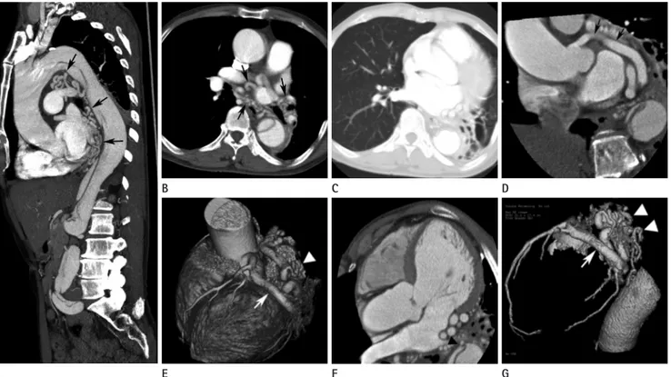
Fig. 1. A 64-year-old man with a coronary-to-bronchial artery fistula and dissecting aortic aneurysm.
A-C. Aorta CT angiography shows a diffusely dilated aorta with intimal dissection from just the distal portion of left subclavian arterial origin to both common iliac arteries (A). Tortuous vascular structures are identified along the pericardial reflections and bronchovascular bundles (arrows in A, B). In the lung window setting, cystic bronchiectasis with markedly decreased volume is observed in the left lower lobe (C).
D-G. Coronary CT angiography shows that the tortuously dilated anomalous vessels originate from the left circumflex coronary artery (arrows) and course along the left side of the left atrium to communicate with the bronchial circulation and the thoracic aorta (arrowheads).
A E
B
F C
G D
Eun-Ju Kang, et al
submit.radiology.or.kr J Korean Soc Radiol 2012;66(4):333-335
335
studies. Radiology 1993;186:877-882
2. Lee CM, Leung TK, Wang HJ, Lee WH, Shen LK, Chen YY.
Identification of a coronary-to-bronchial-artery commu- nication with MDCT shows the diagnostic potential of this new technology: case report and review. J Thorac Imaging 2007;22:274-276
3. Lee ST, Kim SY, Hur G, Hwang YJ, Kim YH, Seo JW, et al.
Coronary-to-bronchial artery fistula: demonstration by 64-multidetector computed tomography with retrospec- tive electrocardiogram-gated reconstructions. J Comput Assist Tomogr 2008;32:444-447
4. Yamanaka O, Hobbs RE. Coronary artery anomalies in 126,595 patients undergoing coronary arteriography. Cathet Cardiovasc Diagn 1990;21:28-40
5. Kim SK, Kim HW, Kim PJ, Chun HJ. Concomitant left and right coronary to bronchial artery fistulae resulting in myo- cardial ischaemia. Eur J Cardiothorac Surg 2011;39:278 6. Moberg A. Anastomoses between extracardiac vessels and
coronary arteries. Acta Med Scand Suppl 1968;485:5-26 7. Yoon W, Kim JK, Kim YH, Chung TW, Kang HK. Bronchial
and nonbronchial systemic artery embolization for life- threatening hemoptysis: a comprehensive review. Radio- graphics 2002;22:1395-1409
8. Yoon YC, Lee KS, Jeong YJ, Shin SW, Chung MJ, Kwon OJ.
Hemoptysis: bronchial and nonbronchial systemic arteries at 16-detector row CT. Radiology 2005;234:292-298 is widely regarded to be the best diagnostic method for identify-
ing such communications, it is invasive and has a high degree of risk. On the other hand, CCTA is reliable in identifying and de- fining anomalous coronary arteries and their courses (2, 3). Fur- thermore, in our case described, conventional angiography could not have been performed because of extensive aortic dissection.
Most patients with CBF are asymptomatic, but cardiovascular symptoms and signs, such as, continuous machinery murmur, angina due to coronary steal phenomenon, congestive heart fail- ure, infective endocarditis, and rupture of an aneurysmal fistula, may develop (3-5). CBFs can also cause hemoptysis (7, 8). Stent grafting and coil embolization are regarded as the best treatment methods for coronary steal phenomenon (5). In cases of severe coronary artery disease, bypass surgery with surgical ligation of the CBF can be an appropriate treatment, and in some cases of recurrent hemoptysis, embolization of the coronary artery may be considered to stop bleeding (7, 8). Our patient did not exhibit any cardiovascular symptoms or hemoptysis and his chief symp- toms were associated with aortic dissection. Therefore, our pa- tient did not undergo endovascular treatment.
REFERENCES
1. Matsunaga N, Hayashi K, Sakamoto I, Ogawa Y, Matsuoka Y, Imamura T, et al. Coronary-to-pulmonary artery shunts via the bronchial artery: analysis of cineangiographic
CT 혈관조영술에서 우연히 발견된 관상동맥기관지 동맥루의 진단:
1예 보고
강은주 · 이기남 · 김우정 · 이진화 · 오종영
저자들은 대동맥류 박리를 주소로 내원한 64세 남자에서 대동맥 CT상 우연히 발견된 관상동맥기관지 동맥루를 보고하고 자 한다. 관상동맥 CT에 좌회전 관상동맥에서 기시하여 기관지 혈관과 연결되는 확장된 혈관들이 확인되었다. 관상동맥기 관지 동맥루는 드문 관상동맥 기형이며 대부분 무증상이기 때문에 다른 목적으로 촬영한 CT에서 우연히 발견될 수 있다.
동아대학교 의과대학 동아대학교병원 영상의학과학교실