INTRODUCTION
Metaplastic ossification has been described in mucosal polyps in the gastrointestinal tract.1-3) However, meta- plastic ossification is a rare event in nasal polyps, which has been reported only ten cases in the English language literature.1-5) Herein, we report a case of huge nasal polyp with metaplastic ossification, which obstructed left nasal cavity and nasopharynx, and provoked both nasal obstruc- tion and sleep apnea.
CASE
A 60-year-old male was referred with a 30 years history of both nasal obstruction and sleep apnea. The patient has been treated for colon cancer with liver and lung metasta-
sis. Thirty years earlier, the patient was underwent nasal mass removal at local hospital. However, the patient did not recognize the operation site and biopsy result of the nasal mass. On endoscopic examination, nasal polyp was noted in the left nasal cavity (Fig. 1). The polyp filled the left nasal cavity and extended into the nasopharynx. The right nasal cavity appeared clear, however right posterior nasal cavity was occupied by nasal polyp extending from J Rhinol 20(2), 2013
- 136 -
www.ksrhino.or.kr
Nasopharynx Obstruction by Huge Nasal Polyp with Metaplastic Ossification
Dong Hoon Lee, MD, Hyun Seok Choi, MD, John Jae Woon Lee, MD and Sang Chul Lim, MD, PhD
Department of Otolaryngology-Head and Neck Surgery, Chonnam National University Medical School
& Chonnam National University Hwasun Hospital, Hwasun, South Korea.
ABSTRACT
We present a case of huge nasal polyp with metaplastic ossification, which obstructed left nasal cavity and nasopharynx, and provoked both nasal obstruction and sleep apnea. The patient had a history of previous sinus surgery at local hospital 30 years ago. Nasal polyp with metaplstic ossification was removed by endoscopic sinus surgery. This case highlights the importance of including metaplastic ossification of nasal polyp in the differential diagnosis of nasal cavity mass.
KEY WORDS : Nasal Polyp·Metaplasia·Ossification.
Address correspondence and reprint requests to Sang Chul Lim, MD, PhD, Chonnam National University Medical School and Hwasun Hospi- tal, 160 Ilsimri, Hwasun, Jeonnam, South Korea 519-809
Tel: +82-61-379-8190 · Fax: +82-61-379-7761 E-mail: [email protected]
Received for publication on April 30, 2013 Accepted for publicatoin on August 29, 2013
Fig. 1. On endoscopic exami- nation, nasal polyp fills the left nasal cavity.
Fig. 2. A CT scan shows a multi- lobulated soft tissue mass with calcification in the left nasal cavity, which extends to the nasopharynx.
Fig. 3. Axial T1-WI (A), T1-WI with enhancement (B), and T2-WI (B) scans show a multi-lobulated, soft tissue lesion with adjacent bony remodeling and diffuse irregular ossification in left nasal cavity. The mass has slightly hypointensity on T1-weighted MR images, and hyperintensity on T2-weighted MR images.
A B C
Lee et al : Nasal Polyp Ossification / 137
the left nasal cavity. A computed tomography (CT) scan showed multi-lobulated soft tissue lesion with adjacent bony remodeling and diffuse irregular ossification in left nasal cavity (Fig. 2). We performed the magnetic reso- nance imaging (MRI) for further evaluation of diffuse ir- regular ossification lesion on CT. On MRI, the soft tissue components of lesion demonstrated slightly hypointensity on T1-weighted MR images, and hyperintensity on T2- weighted MR images (Fig. 3).
We initially diagnosed left chronic sinusitis accompa- nied by calcification or metastasis from the colon cancer.
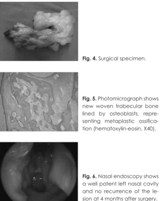
Endoscopic sinus surgery was performed under general anesthesia. After removal of the left nasal polyps, macro- scopic examination showed a lobulated gelatinous polyp with areas of calcification (Fig. 4). Histopathologic ex- amination showed a benign nasal polyp with extensive metaplastic bone formation in the stroma (Fig. 5). No ma- lignant features were seen in any of the tissue sections of the resected mass. According to the radiologic images and histopathologic examination, this case was diagnosed as huge nasal polyp with metaplastic ossification. The post- operative course was uneventful. Four months after en- doscopic sinus surgery, nasal endoscopy showed a well patent left nasal cavity and no recurrence of the lesion (Fig. 6). And the patient symptoms of nasal obstruction and sleep apnea were disappeared.
This report was approved by the Institutional Review Board of Chonnam National University Hwasun Hospi- tal.
DISCUSSION
Nasal polyps are the most common expansile lesions in the nasal cavity. There are a number of secondary changes with nasal polyps, such as infarction, surface ulceration, mucoid liquefaction, and metaplasia of the surface epithe- lium.3) However, metaplastic ossification of nasal polyp is very rare.1-5)
The pathogenesis of nasal polyp with metaplastic ossifi- cation is not fully understood, but two theories have been proposed. The first theory implies that pleuripotential cells present in the stroma of the polyp or dedifferentia- tion of cells present in the polyp into a pluripotential cell, which may then differentiate along an osteoblastic lineage leading to ossification.1-4) The second theory postulates that new bone formation might occur in the nasal pol- yps themselves possibly from bony remnants left behind during previous surgery.5) However, in nine of ten previ- ously reported cases, had no history of previous sinonasal surgery. Thus the second theory was thought to be less plausible. However, the patient, in our case, had history of previous nasal surgery. Thus we suggest that both first and second theory is possible for pathogenesis of nasal polyp with metaplastic ossification.
Typical symptom of nasal polyp with metaplastic ossi- fication is unilateral nasal obstruction.1) 2) 5) Occasionally, patients presented bilateral nasal obstruction, anosmia, and headache.2) 5) In our case, the patient complained of both nasal obstruction and sleep apnea, because nasophar- ynx was obstructed by nasal polyp with metaplastic os- sification.
For the preoperative diagnosis of nasal polyp with metaplastic ossification, radiologic examination is useful and essential.3) CT is useful for identifying not only cal- cifications, but also surrounding tissues patterns and ero- sions. A variety of pathologic conditions can mimic nasal polyp with metaplastic ossification on CT and MRI and include rhinolith, fungus ball, inverted papilloma, chond- rosarcoma, osteosarcoma, and fibroosseous lesions, such as fibrous dysplasia and ossifying firboma.3) MRI may be useful when fungus ball or inverted papilloma cannot be rule out on CT scan.3)
The treatment of nasal polyp with metaplastic ossifica- tion is surgical removal via endoscopic approach or open appraoch.1-5) Most of nasal polyps with metaplastic ossifi- cation can be removed endoscopically, as was done in our case. Surgical removal of the nasal polyp with metaplastic ossification resulted in a cure of the patient’s symptoms.1) 2)
There are some unique characteristics in this case. First, the nasal polyp with metaplastic ossification, in our case, extended from the vestibule to the nasopharynx, and thus this is the hugest nasal polyp with metaplastic ossifica-
Fig. 4. Surgical specimen.
Fig. 5. Photomicrograph shows new woven trabecular bone lined by osteoblasts, repre- senting metaplastic ossifica- tion (hematoxylin-eosin, X40).
Fig. 6. Nasal endoscopy shows a well patent left nasal cavity and no recurrence of the le- sion at 4 months after surgery.
138 / J Rhinol 20(2), 2013
tion among previously reported cases. Second, there may be other, similar, unreported cases in which surgeons removed nasal polyps but remained unaware that it con- tained metaplastic ossification. This case highlights the importance of including metaplastic ossification of nasal polyp in the differential diagnosis of nasal cavity mass.
저자역할(Author Contributions)
이동훈, 최현석, 이재운, 임상철은 본 연구에서 모든 자료에 접근할 수 있으며, 자료의 완전성과 자료 분석의 정확성에 책임을 지고 있습니 다. 연구 기획 : 이동훈, 최현석, 이재운, 임상철. 자료 해석 및 분석 : 이동훈, 최현석, 이재운, 임상철. 논문초안 : 이동훈. 논문수정 : 임상 철. 연구 총괄 : 임상철
REFERENCES
1) Ramachandran K, Thomas MA, Denholm RB. Osseous metaplasia of a nasal polyp. J Otolaryngol 2005;34:72-3.
2) Marquez Moyano JA, Navarro Cantero A, Garrido Iniesta FJ, Poy- ato Zamorano C, Margues Asin J. Metaplastic ossification in nasal polyp. Acta Otorrinolaringol Esp 2007;58:276-7.
3) Kim YK, Kim HJ, Kim J, Chung SK, Kim E, Ko YH, et al. Nasal polyps with metaplastic ossification: CT and MR imaging findings.
Neuroradiology 2010;52:1179-84.
4) Jacono AA, Sclafani AP, Van De Water T, McCormick S, Frenz D.
Metaplastic bone formation in nasal polyps with histologic pres- ence of transforming growth factor beta-1 (TGFbeta-1) and bone morphogenetic proteins (BMPs). Otolaryngol Head Neck Surg 2001;125:96-7.
5) de Vries N. New bone formation in nasal polyps. Rhinology 1988;26:217-9.