Brief Report
Vol. 29, No. 4, 2017 497
Received May 30, 2016, Revised July 25, 2016, Accepted for publication July 28, 2016
*These authors contributed equally to this work and should be considered co-first authors.
Corresponding author: Chun Wook Park, Department of Dermatology, Kangnam Sacred Heart Hospital, Hallym University College of Medicine, 1 Singil-ro, Yeongdeungpo-gu, Seoul 07441, Korea. Tel: 82-2-829-5221, Fax:
82-2-832-3237, E-mail: [email protected]
Hye One Kim, Department of Dermatology, Kangnam Sacred Heart Hospital, Hallym University College of Medicine, 1 Singil-ro, Yeong- deungpo-gu, Seoul 07441, Korea. Tel: 82-2-829-5221, Fax: 82-2-832-3237, E-mail: [email protected]
This is an Open Access article distributed under the terms of the Creative Commons Attribution Non-Commercial License (http://creativecommons.
org/licenses/by-nc/4.0) which permits unrestricted non-commercial use, distribution, and reproduction in any medium, provided the original work is properly cited.
Copyright © The Korean Dermatological Association and The Korean
Society for Investigative Dermatology Fig. 1. A solitary, tender, and reddish nodule on the right third toe.
with concomitant involvement of the lips. Ann Dermatol 2010;22:106-109.
2. Del Pozo J, Sacristán F, Martínez W, Paradela S, Fernández-Jorge B, Fonseca E. Neutrophilic dermatosis of the hands: presentation of eight cases and review of the literature. J Dermatol 2007;34:243-247.
3. Imaoka K, Kaneko S, Harada Y, Ota M, Furumura M, Morita E. Neutrophilic dermatosis of the palms. J Dermatol
2012;39:949-951.
4. Weenig RH, Bruce AJ, McEvoy MT, Gibson LE, Davis MD.
Neutrophilic dermatosis of the hands: four new cases and review of the literature. Int J Dermatol 2004;43:95-102.
5. Cohen PR, Kurzrock R. Sweet's syndrome: a neutrophilic dermatosis classically associated with acute onset and fever.
Clin Dermatol 2000;18:265-282.
https://doi.org/10.5021/ad.2017.29.4.497
Fibro-Osseous Pseudotumor of the Digit Presenting as an Enlarging Erythematous Subungual Nodule
Yong Se Cho*, Sook Young Park*, Yong Won Choi, Jee Hee Son, Yun Sun Byun, Bo Young Chung, Hee Jin Cho
1, Hye One Kim, Chun Wook Park
Department of Dermatology, Hallym University Kangnam Sacred Heart Hospital, Seoul, 1Department of Dermatology, Hallym University Chuncheon Sacred Heart Hospital, Chuncheon, Korea
Dear Editor:
A 27-year-old man presented with a tender nodule on the distal aspect of the right third toe, slowly growing in size over a 2-month period. He had a history of trauma in the right third toe during exercise two months prior to his visit. Initially, the nodule was soft in consistency but with time enlarged in size and became hard. An examination
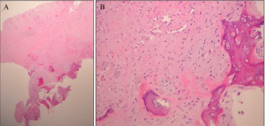
revealed an erythematous, eroded, hard mobile nodule measuring 0.5×0.5 cm in size (Fig. 1). The initial clinical suspicion was that it was a viral wart; thus, a punch biop- sy was done. Microscopic examination showed the lesion was multinodular with irregular margins in the dermis.
The nodules consisted of a mixture of fibroblasts, mixoid matrix, and focal deposits of osteoid with irregularly dis- tributed osteoblasts (Fig. 2A). The osseous trabeculae were
Brief Report
498 Ann Dermatol
Fig. 2. (A) The lesion had variable cellularity and consisted of a mixture of loosely or compactly arranged fibroblasts and trabeculae of the bone showing varying degrees of maturation (H&E, ×40). (B) Newly formed trabeculae of the osteoid and bone were distributed hapha- zardly without zoning phenomenon.
The trabeculae of the bone were rimmed by fibroblasts and osteo- blasts (H&E, ×200).
haphazardly distributed without peripheral zoning. The osseous trabeculae were rimmed by proliferated fibro- blasts and osteoblasts (Fig. 2B). The diagnosis was con- firmed by histopathologic findings as fibro-osseous pseu- dotomor of the digits (FOPD). The tumor was completely excised, and no connection with the underlying bone was observed. No recurrence was noted during a follow-up pe- riod of 6 months.
FOPD is a rare benign ossifying lesion proposed as a uni- fying term by Dupree and Enzinger1. FOPD may occur anywhere in the body but is most common in the finger, especially in the region of the proximal phalange1,2. In Korea, only 3 cases of FOPD of the finger and 1 case of FOPD of the toe have been reported. Typically, this lesion affects young adults, mainly women, and presents as a soft tissue mass growing over a period of weeks or a few months2. Pain, tenderness, and functional limitation may be present. The pathogenesis of FOPD is thought to be re- lated to repeated trauma to the area; however, a specific history of antecedent trauma was revealed in a small num- ber of cases1.
The essential histologic features include localization in the subcutaneous tissue without muscular involvement, a dis- orderly multinodular growth pattern with indistinct bor- ders, and a fibroblastic proliferation showing varying de- grees of cellular atypia and haphazardly arranged osseous trabeculae without the zoning phenomenon1,3. Main patho- logic differentials to this entity are myositis ossificans, ex- traskeletal osteosarcoma, and subungual exostosis. Mysositis ossificans usually occur after trauma, in the deeper aspect of proximal soft tissues and histopathologically show a typical zonation pattern3. Extraskeletal osteosarcoma should always be ruled out; however, it shows destructive stromal invasion, obvious cytologic atypia and immature osteoid directly formed by tumor cells4. Subungual exo- stosis can appear clinically and histopathologically very
similar to FOPD, except for the presence of a connection to the underlying phalangeal bone, the presence of bone marrow tissue, and the feature of an overlying fi- bro-cartilaginous cap5.
FOPD has an excellent prognosis following complete ex- cision with low risk of recurrence (0%∼14%)5. No cases of malignant transformation or metastases are on record1,5. We report herein a rare and typical case of FOPD of the toe and suggest that FOPD should be considered in the differential diagnosis of any digital mass.
ACKNOWLEDGMENT
This study was supported by grants of the National Research Foundation of Korea (NRF), funded by the Ministry of Science, ICT & Future Planning (NRF-2017 R1A2B4006252), Korea Healthcare technology R&D proj- ect, funded by Ministry of Health & Welfare, Republic of Korea (HI17C0597), and the Hallym University Research Fund (HURF-2017-35).
CONFLICTS OF INTEREST
The authors have nothing to disclose.
REFERENCES
1. Dupree WB, Enzinger FM. Fibro-osseous pseudotumor of the digits. Cancer 1986;58:2103-2109.
2. Nishio J, Iwasaki H, Soejima O, Naito M, Kikuchi M. Rapidly growing fibro-osseous pseudotumor of the digits mimicking extraskeletal osteosarcoma. J Orthop Sci 2002;7:410-413.
3. de Silva MV, Reid R. Myositis ossificans and fibroosseous pseudotumor of digits: a clinicopathological review of 64 cases with emphasis on diagnostic pitfalls. Int J Surg Pathol 2003;11:187-195.
Brief Report
Vol. 29, No. 4, 2017 499
Received March 11, 2016, Revised July 25, 2016, Accepted for publication August 4, 2016
Corresponding author: Hwa Jung Ryu, Department of Dermatology, Korea University Ansan Hospital, 123 Jeokgeum-ro, Danwon-gu, Ansan 15355, Korea. Tel: 82-31-412-5186, Fax: 82-31-412-4208, E-mail: [email protected] This is an Open Access article distributed under the terms of the Creative Commons Attribution Non-Commercial License (http://creativecommons.
org/licenses/by-nc/4.0) which permits unrestricted non-commercial use, distribution, and reproduction in any medium, provided the original work is properly cited.
Copyright © The Korean Dermatological Association and The Korean
Society for Investigative Dermatology Fig. 1. Erythematous patch, bullae, and ulceration on right T3 dermatome.
4. Park SG, Song JY, Song IG, Kim MS, Shin BS. Cutaneous extraskeletal osteosarcoma on the scar of a previous bone graft. Ann Dermatol 2011;23(Suppl 2):S160-S164.
5. Tan KB, Tan SH, Aw DC, Lee YS. Fibro-osseous pseudotumor of the digit: Presentation as an enlarging erythematous cuta- neous nodule. Dermatol Online J 2010;16:7.
https://doi.org/10.5021/ad.2017.29.4.499
A Case of Wolf’s Isotopic Response Presenting as Bullous Pemphigoid
Seung Hyun Chun, Bo Young Kim, Chang Min Kim, Jae Beom Park, Hwa Jung Ryu
Department of Dermatology, Korea University Ansan Hospital, College of Medicine, Korea University, Ansan, Korea
Dear Editor:
Wolf’s isotopic response refers to a phenomenon in which a new skin disorder develops at the site of another, un- related, and already healed skin disease. According to a report by Ruocco et al.1 in 2014, there are approximately 200 cases of such a condition. Majority of primary in- fections includes infection by varicella zoster virus. In contrast the secondary skin lesion includes various cuta- neous conditions such as granulomatous reaction, malig- nant tumor, dysimmune reaction, and morphea2. In this report the authors present a case of bullous pemphigoid (BP) with Wolf’s isotopic response in a Korean female patient.
An 80-year-old female patient presented with an itchy er- ythematous patch, bullae, and ulceration on the right T3 dermatome. (Fig. 1) The bullous lesions had persisted for the last 2 months, and the patient had suffered from her- pes zoster at the same site 6 months prior to this visit.
Incisional biopsy of the affected lesion revealed subepi- thelial vesicles with dermal infiltration of lymphocytes,
histiocytes, and eosinophils (Fig. 2A). Direct immuno- fluorescence with fluorescein isothiocyanate revealed line- ar deposition of immunoglobulin G in the basement mem- brane zone (Fig. 2B). With the final diagnosis of BP with Wolf’s isotopic response, the patient was started on sys- temic steroid therapy and she showed clinical improvement.
The patient has tapered oral methylprednisolone and is now free of new bulla with topical steroid and 1 gram of tetracycline and nicotinamide per day.
Recently Ruocco et al.1 analyzed the previously reported cases of Wolf’s isotopic response. Although numerous skin conditions have been identified as a result of Wolf’s isotopic response, bullous disorders such as pemphigus vulgaris and BP were seldom seen. Up until now only one