Esculetin의 caspase-3 활성을 통한 U937 인체 혈구암세포의 세포사멸 유도
박철1․현숙경1․신우진2․정경태1,3․최병태4․권현주1,5․황혜진1,6․김병우1,5․박동일2․이원호7․최영현1,8*
동의대학교 1블루바이오 소재개발센터, 한의과대학 2내과학교실 및 8생화학교실, 자연과학대학 3임상병리학과, 5생명응용학과 및
대학원 바이오물질제어학과, 6생활과학대학 식품영양학과, 부산대학교 4한의학전문대학원 해부학교실 및 7자연과학대학 생물학과
Received December 16, 2008 /Accepted January 30, 2009
Esculetin Induces Apoptosis through Caspase-3 Activation in Human Leukemia U937 Cells. Cheol Park
1, Sook Kyung Hyun
1, Woo-Jin Shin
2, Kyung Tae Chung
1,3, Byung Tae Choi
4, Hyun Ju Kwon
1,5, Hye Jin Hwang
1,6, Byung-Woo Kim
1,5, Dong Il Park
2, Won Ho Lee
7and Yung Hyun Choi
1,8*.
1Blue-Bio Industry RIC, Departments of
2Physiology and 8Biochemistry, College of Oriental Medicine, Departments of
3
Biomedical Laboratory Science and
5Life Science and Biotechnology, College of Natural Sciences and Department of Biomaterial Control (BK21 program), Graduate School,
6Department of Food and Nutrition, College of Human Ecology, Dong-Eui University, Busan 614-052, South Korea;
4Division of Meridian and Structural Medicine, School of Oriental Medicine,
7Department of Biology, College of Natural Sciences, Pusan National University, Busan 609-735, South Korea - Esculetin, a coumarin compound, has been known to inhibit proliferation and induce apoptosis in several types of human cancer cells. However, the molecular mechanisms in- volved in esculetin-induced apoptosis are still uncharacterized in human leukemia cells. In this study, we have investigated whether esculetin exerts anti-proliferative and apoptotic effects on human leuke- mia U937 cells. It was found that esculetin could inhibit cell viability in a time-dependent manner, which was associated with the induction of apoptotic cell death such as increased populations of apop- totic-sub G1 phase. Apoptosis of U937 cells by esculetin was associated with an inhibition of Bcl-2/Bax binding activity, formation of tBid, down-regulation of X-linked inhibitor of apoptotic protein (XIAP) expression, and up-regulation of death receptor 4 (DR4) and FasL expression. Esculetin treatment also induced the degradation of β-catenin and DNA fragmentation factor 45/inhibitor of caspase-activated DNase (DFF45/ICAD). Furthermore, a caspase-3 specific inhibitor, z-DEVD-fmk, significantly inhibited sub-G1 phase DNA content, morphological changes and degradation of β-catenin and DEE45/ICAD.
These results indicated that a key regulator in esculetin-induced apoptosis was caspase-3 in human leukemia U937 cells.
Key words : Esculetin, U937, apoptosis, caspase-3
*Corresponding author
*Tel:+82-51-850-8649, Fax:+82-51-853-4036
*E-mail : [email protected]
서 론
Apoptosis는 배아의 발생, 조직의 재배치 및 손상된 세포의 제거 등에 관련된 생리적인 세포의 죽음이며, apoptosis의 비 정상적인 조절은 암 및 자가면역질환 등과 같은 질병의 원인 이 될 수 있다[7,9,14]. Apoptosis는 세포의 수축, 염색질 응축, DNA 단편화 및 세포막의 수포화 현상 등과 같은 형태적인 특징을 가지며, 여러 가지 유전자들의 증가 및 감소와 같은 상호작용에 의하여 조절된다[10,13,22]. Apoptosis 과정은 외 재적 경로(extrinsic pathway) 및 내재적 경로(intrinsic path- way)에 의해 유발되는데, 먼저 외재적 경로는 Fas ligand (FasL/CD95L) 및 tumor necrosis factor-related apoptosis-in- ducing ligand (APO2L/TRAIL)와 같은 단백질 ligand들이 세 포 표면에 존재하는 Fas/CD95, 죽음의 수용체(death receptor 4, DR4) 및 DR5와 같은 세포막 수용체들에 결합함으로서 유발
된다[2,5]. 죽음의 배위자(death ligand)가 death receptor와 결 합하게 되면 세포막 내부에 death-inducing signaling com- plex (DISC)를 형성하게 되고, 이러한 pro-apoptotic 신호는 개시 caspases인 caspase-8 및 -10의 활성화를 일으켜 효과 caspases인 caspase-3, -6 및 -7의 활성화와 그에 따른 DNA의 단편화 현상을 유발시킨다[1,19]. 한편 내재적 경로는 미토콘 드리아 의존적인 apoptosis 과정으로서 약물, 방사선 및 활성 산소종(reactive oxygen species, ROS) 등과 같은 여러 가지 자극에 의하여 미토콘드리아 외막에 존재하는 Bcl-2 family의 발현에 영향을 주게 되면 미토콘드리아 막 전위(mitochondrial membrane potential, MMP)의 변화가 유발되어 cytochrome c가 세포질로 방출된다[4,18]. 이렇게 방출된 cytochrome c는 apoptotic protease activating factor-1 (Apaf-1) 및 pro-cas- pase-9과 결합하여 apoptosome을 형성하여 caspase-9 및 -3을 활성화시켜서 apoptosis를 유발시킨다[3,31].
매실, 레몬, 인진쑥 등을 포함한 다양한 식물에 많이 함유되
어 있는 coumarin 유도체인 esculetin (6,7-dihydroxycoumarin)
은 혈소판응집과 xanthine oxidase 활성 억제, N-meth-
yl-N-nitrosourea에 의하여 유도된 유방암 및 benzo[a]pyrene
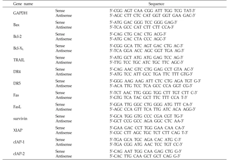
Table 1. Sequence of primers used for RT-PCR
Gene name Sequence
GAPDH Sense
Antisense
5'-CGG AGT CAA CGG ATT TGG TCG TAT-3' 5'-AGC CTT CTC CAT GGT GGT GAA GAC-3'
Bax Sense
Antisense
5'-ATG GAC GGG TCC GGG GAG-3' 5'-TCA GCC CAT CTT CTT CCA-3'
Bcl-2 Sense
Antisense
5'-CAG CTG CAC CTG ACG-3' 5'-ATG CAC CTA CCC AGC-3'
Bcl-XL Sense
Antisense
5'-CGG GCA TTC AGT GAC CTG AC-3' 5'-TCA GGA ACC AGC GGT TGA AG-3'
TRAIL Sense
Antisense
5'-ATG GCT ATG ATG GAG TCC AG-3' 5'-TTG TCC TGC ATC TGC TTC AGC-3'
DR4 Sense
Antisense
5'-CAG AAC GTC CTG GAG CCT GTA AC-3' 5'-ATG TCC ATT GCC TGA TTC TTT GTG-3'
DR5 Sense
Antisense
5'-GGG AAG AAG ATT CTC CTG AGA TGT G-3' 5'-ACA TTG TCC TCA GCC CCA GGT CG-3'
Fas Sense
Antisense
5'-TCT AAC TTG GGG TGG CTT TGT CTT C-3' 5'-GTG TCA TAC GCT TTC TTT CCA T-3'
FasL Sense
Antisense
5'-GGA TTG GGC CTG GGG ATG TTT CA-3' 5'-AGC CCA GTT TCA TTG ATC ACA AGG-3'
survivin Sense
Antisense
5'-GCA TGG GTG CCC CGA CGT TG-3' 5'-GCT CCG GCC AGA GGC CTC AA-3'
XIAP Sense
Antisense
5'-GAA GAC CCT TGG GAA CAA CA-3' 5'-CGC CTT AGC TGC TCT CTT CAG T-3'
cIAP-1 Sense
Antisense
5'-TGA GCA TGC AGA CAC ATG C-3' 5'-TGA CGG ATG AAC TCC TGT CC-3'
cIAP-2 Sense
Antisense
5'-CAG AAT TGG CAA GAG CTG G-3' 5'-CAC TTG CAA GCT GCT CAG G-3'
plus 4-(methylnitrosoamino)-1-(3-pyridyl)-1-butanone에 의
하여 유도된 폐암의 억제효과를 가지는 등 약리학 및 생화학 적 활성이 뛰어난 것으로 보고되어지고 있다[11,20,21]. 최근에 는 esculetin이 암세포의 apoptosis 유발 및 세포주기 억제와 같은 항암작용을 가지는 것으로 알려져 있으나 분자적 기작은 명확히 밝혀져 있지 않다[16,17,23,27].
본 연구에서는 인체 혈구암세포인 U937 세포에서 esculetin 이 유발하는 항암효과의 생화학적 기전의 해석을 위하여 암세 포의 생존율 및 형태에 미치는 esculetin의 영향을 조사하였고, apoptosis 과정 중 내재적 및 외재적 경로에 관여하는 몇 가지 중요한 유전자들의 발현 변화를 조사하여 유의적인 결과를 얻었기에 이를 보고하는 바이다.
재료 및 방법
실험 재료
본 실험에 사용된 esculetin은 Sigma-Aldrich (St. Louis, MO, USA)에서 구입하였다. mRNA 분석을 위하여 Bioneer (Taejeon, Korea)에서 구입한 primer는 Table 1에 나타내었고, immunoblotting을 위하여 사용된 항체들은 Santa Cruz
Biotechnology Inc. (Santa Cruz, CA, USA) 및 CalBiochem (San Diego, CA, USA)에서 구입하였으며, 2차 항체로 사용된 peroxidase-labeled donkey anti-rabbit 및 peroxidase-labeled sheep anti-mouse immunoglobulin은 Amersham Life Science Corp. (Arlington Heights, IL, USA)에서 구입하였다.
세포의 배양
실험에 사용한 인체 혈구암 U937 세포는 American Type Culture Collection (ATCC, Rockville, MD, USA)에서 분양 받았으며 90%의 RPMI-1640 배지(Gibco BRL, Grand Island, NY, USA)에 10% fetal bovine serum (FBS), 2 mM glutamine, 100 U/ml penicillin 및 100 μg/ml streptomycin (Gibco BRL) 이 포함된 성장배지를 사용하여 5% CO
2, 37
oC의 조건하에서 배양하였다.
MTT assay에 의한 세포 성장억제 조사
30 μg/ml의 esculetin을 일정시간 동안 처리하여 배양한 다음 배지를 제거하고 0.5 mg/ml 농도의 tetrazolium bro- mide salt (MTT, Amresco, Solon, OH, USA)를 분주하였다.
2시간 동안 CO
2배양기에서 배양시킨 다음 DMSO로 생성된
Fig. 1. Growth inhibition and apoptosis induction in U937 human leukemia cells by esculetin. (A) U937 cells were seeded at 1X10⁴/ml in a 6-well plate and treated with variable times of esculetin for 30 μg/ml. The growth inhibition was measured by the metabolic-bye-based MTT assay. Data are expressed as mean±SD of three independent experiments. (B) The cells treated with esculetin for the indicated times and then the cells were stained with PI for flow cytometry analysis. The percentages of cells with hypodiploid DNA (sub-G1 phase) contents represent the fractions undergoing apoptotic DNA degradation.
Data are expressed as mean±SD of three independent experiments. (C) The morphological changes of U937 cells after treatment with esculetin were visualized by an inverted microscopy. Magnification, X200.
formazin을 모두 녹인 후 ELISA reader (Molecular Devices, Sunnyvale, CA, USA)를 이용하여 540 nm에서 흡광도를 측 정하였다. 측정은 모두 세 번을 하였으며, 그에 대한 평균값과 표준 오차를 Microsoft EXCEL program을 사용하여 분석하 였다.
Flow cytometry 분석
일정시간 동안 esculetin을 처리한 세포를 모은 다음 CycleTEST PLUS DNA REAGENT Kit (Becton Dickinson, San Jose, CA, USA)를 이용하여 고정 및 염색을 하여 4
oC, 암실에서 30분 동안 반응을 시켰다. 반응시킨 세포를 35-mm mesh를 이용하여 단일세포로 분리한 후 FACSCalibur (Becton Dickinson)를 이용하여 형광반응에 따른 세포 내 DNA 함량을 CellQuest software 및 ModiFit LT (Becton Dickinson) 프로그램을 이용하여 분석하였다.
위상차 현미경을 이용한 세포의 성장과 형태의 관찰 세포 배양용 100 mm petri dishes에 세포를 1×10
5개/ml 정도로 분주하고 esculetin을 처리하여 일정시간 동안 배양 한 후, 위상차 현미경(inverted microscope, Carl Zeiss, Germany)을 이용하여 200배의 배율로 형태 변화를 관찰하 였다.
Reverse transcription-polymerase chain reaction (RT-PCR)에 의한 mRNA의 분석
상기와 동일한 조건에서 준비된 U937 세포에 TRIzol re- agent (Invitrogen Co., Carlsbad, CA, USA)를 4
oC에서 1시간 동안 처리하여 총 RNA를 분리하였다. 분리된 RNA를 정량한 후, 각각의 primer, DEPC water 그리고 ONE-STEP RT-PCR PreMix Kit (Intron, Korea)를 넣고 Mastercycler gradient
(Eppendorf, Hamburg, Germany)를 이용하여 증폭한 후, 1%
agarose gel을 이용하여 100 V에서 전기영동을 하였다. 전기영 동으로 DNA 분리가 끝난 gel을 ethidium bromide (EtBr)를 이용하여 염색한 후 관찰하였다.
Immunoprecipitation, gel electrophoresis 및 Western blot analysis
준비된 암세포에 lysis buffer [25 mM Tris-Cl (pH 7.5), 250 mM NaCl, 5 mM EDTA, 1% NP-40, 1 mM phenyme- thylsulfonyl fluoride, 5 mM dithiothreitol]를 첨가하여 단백 질을 분리한 다음 Bio-Rad 단백질 정량 시약(Bio-Rad, Hercules, CA, USA)을 이용하여 정량하였다. 면역침전을 위 해서 해당 항체가 들어있는 extraction buffer를 준비된 단백질 에 첨가한 후 4
oC에서 1시간 동안 반응시켜 만들어진 면역복 합체를 protein G/A-Sepharose bead를 이용하여 모았다. 면 역침전 및 총 단백질을 sodium dodecyl sulphate (SDS)- poly- acrylamide gel을 이용하여 전기영동으로 분리한 다음 nitro- cellulose membrane (Schleicher and Schuell, Keene, NH, USA)으로 electroblotting에 의해 전이시켰다. 준비된 mem- brane을 관찰대상 단백질에 해당되는 1차 및 2차 항체에 반응 시킨 다음 enhanced chemiluminoesence (ECL) 용액 (Amersham Life Science Corp., Arlington Heights, IL, USA) 을 처리하여 암실에서 X-ray 필름에 감광시켜 특정단백질의 양을 분석하였다.
결 과
U937 세포의 증식 및 형태에 미치는 esculetin의 영향
인체 혈구암세포인 U937 세포의 증식에 미치는 esculetin
의 영향을 MTT assay를 이용하여 조사한 결과, Fig. 1A에 나
타난 바와 같이 30 μg/ml의 esculetin 처리시간이 증가함에 따라 암세포의 증식이 감소하여 24시간 처리군의 경우에는 약 70% 정도의 증식억제 효과가 나타났다. 이러한 증식억제 에 따른 암세포의 형태 변화를 위상차 현미경을 이용하여 관 찰한 결과, Fig. 1C에서와 같이 esculetin 처리시간이 증가할 수록 전체적인 세포의 밀도가 감소하였고 심한 형태적 변형 이 관찰되었다.
Esculetin에 의한 apoptosis의 유발
Esculetin 처리에 의한 U937 세포의 증식억제 및 형태변화 가 apoptosis 유발과 연관이 있을 것으로 예상되어 apoptosis 유발의 정도를 flow cytometry를 이용하여 정량적인 분석을 한 결과는 Fig. 1B에 나타난 바와 같다. 정상 배지에서 자란 암세포에서는 apoptosis가 거의 유발되지 않은 것으로 나타났 지만 esculetin의 처리시간이 증가할수록 sub-G1의 비율이 증 가하여 24시간 처리군에서는 sub-G1의 비율이 70% 이상으로 나타났다. 이는 esculetin 처리에 의한 U937 세포의 증식 억제 및 형태변화가 apoptosis 유발과 밀접한 관련이 있음을 의미 한다.
Bcl-2 family의 발현 및 Bcl-2/Bax의 binding activity에 미치는 esculetin의 영향
Esculetin 처리에 의한 암세포의 apoptosis 유발에 관여하 는 유전자의 탐색을 위하여 먼저 Bcl-2 family에 속하는 몇 가지 유전자 발현의 정도에 미치는 esculetin의 영향을 RT-PCR 및 Western blot analysis 방법으로 조사하였다.
Fig. 2에 나타낸 바와 같이 esculetin 처리 시간 의존적으로 extrinsic pathway에 의하여 유발되는 Bid의 단편화 현상이 관찰되었다. 또한 Bcl-2/Bax의 결합력을 조사한 결과, Fig.
2C에 나타난 바와 같이 esculetin 처리에 의하여 Bcl-2 및 Bax의 mRNA 및 단백질 수준에서는 아무런 변화가 관찰되 지 않았지만 Bcl-2/Bax의 결합력은 현저하게 감소하는 것으 로 나타났다. 이는 esculetin 처리에 의한 apoptosis 유발에 있어서 외재적 및 내재적 경로가 모두 관여하여 apoptosis 유발 관련 효소들의 활성화가 이루어지고 있음을 시사하여 주는 것이다.
TRAIL, DR4, DR5, Fas 및 FasL의 발현에 미치는 esculetin의 영향
외재적 경로를 통한 apoptosis 과정에 있어서 중요한 역할 을 하는 죽음의 수용체에 속하는 유전자들의 발현 변화를 관 찰한 결과, Fig. 3에 나타난 바와 같이 DR4 및 FasL의 발현이 단백질 수준에서 현저하게 증가하는 것으로 나타났다. 이는 esculetin에 의한 apoptosis 유발에 있어서 죽음의 수용체들의 증가와 같은 외재적 경로의 활성화를 통한 Bid 단백질의 단편 화가 중요한 역할을 한다는 것을 보여주는 결과이다.
Fig. 2. Effects of esculetin treatment on the Bcl-2 family ex- pression in U937 human leukemia cells. (A) The cells treated with esculetin for the indicated times and total RNAs were isolated and reverse-transcribed. The result- ing cDNAs were subjected to PCR with indicated pri- mers and the reaction products were subjected to elec- trophoresis in 1% agarose gel and visualized by EtBr staining. Glyceraldehyde-3-phosphate dehydrogenase (GAPDH) was used as an internal control. (B) Whole cell lysates (0.5 mg of protein) from control cells and cells treated with esculetin were immunoprecipitated with anti-Bax antibody. Immuno-complexes were sepa- rated by 10% SDS-polyacrylamide gel electrophoresis, transferred to nitrocellulose membrane, and probed with anti-Bcl-2 antibody. Proteins were detected by ECL detection. (C) The cells were lysed and then cellular pro- teins were separated by SDS-polyacrylamide gels and transferred onto nitrocellulose membranes. The mem- branes were probed with the indicated antibodies.
Proteins were visualized using an ECL detection system.
Actin was used as an internal control.
IAP family의 발현에 미치는 esculetin의 영향
다음은 esculetin에 의한 apoptosis 유발에 있어서 caspases 의 활성을 억제하는 것으로 알려진 IAP family가 관여하는지 의 여부를 조사하였다. Fig. 4A 및 B에 나타난 바와 같이 조사 된 IAP family 중 XIAP가 단백질 수준에서 발현이 감소하는 것으로 나타나, esculetin이 XIAP의 발현을 감소시켜 caspases 의 활성을 억제하지 못하게 함으로서 apoptosis 유발을 촉진 시키는 것으로 생각되어 진다.
β-catenin, CAD 및 ICAD의 발현에 미치는 esculetin의 영향
Apoptosis 유발 시 활성화된 caspase-3은 여러 종류의 기
질 단백질의 분해를 유발하게 되는 것으로 알려져 있으므로
몇 가지 중요한 기질 단백질의 발현이 esculetin 처리에 의하
여 어떠한 변화가 유발되는 지를 조사하였다. Fig. 4B에서와
같이 세포내 골격 유지와 세포 유착에 관여하는 β-catenin 및
DNA의 단편화에 관여하는 것으로 알려진 DFF45/ICAD의
Fig. 5. Suppression of esculetin-induced apoptosis by caspase-3 inhibitor, z-DEVD-fmk, in U937 human leukemia cells. Cells were pretreated with 50 μM of z-DEVD-fmk for 1 hr, then treated with 30 μg/ml esculetin for 6 hr. (A) The cells were collected and stained with PI for flow cytometry analysis. The percentages of cells with hypodiploid DNA (sub-G1 phase) contents represent the fractions undergoing apoptotic DNA degradation. Data are expressed as mean±SD of three independent experiments. (B) Cell morphology was visualized by an inverted microscopy. Magnification, X200. (C) The cells were lysed and then cellular proteins were separated by SDS-polyacrylamide gels and transferred onto nitrocellulose membranes. The membranes were probed with the indicated antibodies. Proteins were visualized using an ECL detection system. Actin was used as an internal control.
Fig. 3. Effects of esculetin treatment on the TRAIL, DR4, DR5, Fas and FasL expression in U937 human leukemia cells.
(A) Total RNAs were isolated and reverse-transcribed.
The resulting cDNAs were subjected to PCR with in- dicated primers and the reaction products were sub- jected to electrophoresis in 1% agarose gel and visualized by EtBr staining. GAPDH was used as an internal control. (B) The cells were lysed and then cellular pro- teins were separated by SDS-polyacrylamide gels and transferred onto nitrocellulose membranes. The mem- branes were probed with the indicated antibodies.
Proteins were visualized using an ECL detection system.
Actin was used as an internal control.
발현의 감소 또는 단편화 현상이 관찰되었다. 즉 esculetin 처 리에 의한 apoptosis 유발은 caspase-3의 활성화와 이에 따른 기질 단백질들의 분해가 중요한 역할을 한다는 것을 알 수 있었다.
Caspase-3 inhibitor에 의한 apoptosis의 억제
이상의 결과들을 살펴볼 때 esculetin에 의한 apoptosis 유 발에 있어서 caspase-3의 활성이 중요한 역할을 하는 것으로 생각되어 caspase-3 저해제가 esculetin에 의한 apoptosis에 어
Fig. 4. Effects of esculetin on the IAP family, β-catenin, CAD/
DFF40 and ICAD/DFF45 in U937 human leukemia cells.
(A) Total RNAs were isolated and reverse-transcribed.
The resulting cDNAs were subjected to PCR with in- dicated primers and the reaction products were sub- jected to electrophoresis in 1% agarose gel and visualized by EtBr staining. GAPDH was used as an internal control. (B) The cells were lysed and then cellular pro- teins were separated by SDS-polyacrylamide gels and transferred onto nitrocellulose membranes. The mem- branes were probed with the indicated antibodies.
Proteins were visualized using an ECL detection system.
Actin was used as an internal control.
떠한 영향을 주는 지를 조사하여 보았다. Fig. 5A에서 나타난
바와 같이 esculetin 처리에 의하여 유도되는 apoptosis가 cas-
pase-3 저해제인 z-DEVD-fmk 선 처리에 의하여 거의 완벽하
게 억제되는 것으로 나타났다. 또한 esculetin에 의한 형태 변
화 및 caspase-3의 기질 단백질들의 발현 변화에서도 동일한
결과가 나타났다(Fig. 5B 및 C). 따라서 esculetin에 의하여 유
발되는 apoptosis에 있어서 caspase-3이 중요한 조절자로서 역
할을 하는 것으로 생각된다.
고 찰
본 연구에서는 인체 혈구암 U937 세포를 대상으로 escule- tin의 항암기전을 조사하였다. 이를 위하여 먼저 정상배지 및 esculetin이 함유된 배지에서 자란 암세포의 증식의 정도 및 형태 변화를 비교하였다. Fig. 1A 및 C에서 알 수 있듯이 escu- letin의 처리 시간 의존적으로 암세포의 증식이 감소되었으며, 심한 형태적 변형을 유발하였다. 이러한 증식의 억제 및 형태 변화가 apoptosis 유발과 밀접한 연관성이 있을 것으로 기대 되어 flow cytometry를 이용하여 정량적으로 분석한 결과, Fig. 1B에서와 같이 apoptosis 유발 세포군에 해당하는 sub-G1 기에 속하는 세포들의 빈도가 esculetin 처리 시간 의존적으로 증가되어 esculetin 처리에 따른 U937 세포의 증식 억제 및 형태 변화는 apoptosis 유발과 직접적인 연관이 있음을 알 수 있었다.
한편 미토콘드리아 외막에 존재하는 Bcl-2 family에 속하는 단백질들은 네 가지의 Bcl homology (BH) domains (BH1–
BH4) 중 최소한 한 개의 domain을 포함하고 있으며, 미토콘드 리아 보존과 미토콘드리아에 의해 유도되는 apoptosis를 조절 하는 중요한 조절자이다. 이들 중 대표적인 단백질로는 pro-apoptotic 유전자인 Bax와 anti-apoptotic 유전자인 Bcl-2 가 존재하는데 이들은 서로 dimer 형태로 결합하여 존재하지 만 여러 가지 자극에 의하여 발현 수준에 변화가 유발되면 apoptosis가 일어나는 것으로 알려져 있다[6,25]. 따라서 escu- letin이 Bcl-2 family의 발현에 어떠한 영향을 미치는 지를 조 사한 결과, Fig. 2에서와 같이 Bid의 단편화 및 Bcl-2/Bax 결합 력의 감소를 유발되어 esculetin에 의한 암세포의 apoptosis 유발에 있어서 Bcl-2 family에 속하는 유전자들의 dimer 형성 균형의 교란이 중요한 역할을 하는 것으로 생각되어진다.
외재적 경로를 통한 apoptosis에 있어서 죽음의 수용체에 속하는 유전자들 중, TRAIL은 DR4 및 DR5와 결합하고 FasL 는 Fas와 결합함으로서 apoptosis를 유발하는 것으로 알려져 있다[15,24]. 이러한 죽음의 수용체에 속하는 단백질들이 escu- letin에 의하여 어떠한 변화가 유발되는지를 확인한 결과, Fig.
3에서와 같이 DR4 및 FasL의 발현이 증가하는 것으로 나타나 esculetin에 의한 apoptosis 유발은 외재적 경로가 어느 정도 관여하는 것으로 생각되어진다. Apoptosis 유발에 있어서 ef- fector caspase인 caspase-3은 apoptosis의 최종 단계에서 중요 한 역할을 하는 것으로 알려져 있는데, 전사 조절인자인 nu- clear factor kappa B (NF-κB)에 의해 조절 받는 것으로 밝혀진 IAP family에 속하는 유전자들 중 일부는 caspase-3과 직접적 인 결합을 통하여 이들의 활성을 억제하는 것으로 알려져 있 다[12,26]. 또한 caspase-3이 활성화되면 세포의 생존에 필요한 여러 기질 단백질들을 분해시킴으로서 apoptosis가 유발되는
데, 대표적인 기질 단백질인 β-catenin은 세포내 골격의 유지 와 다양한 부착성 세포의 전사 조절에 중요한 역할을 하며 정상 세포의 경우 β-catenin은 92 kDa의 분자량을 가지나 apoptosis가 일어나면 62-72 kDa로 단편화가 일어난다[8,30].
또 다른 기질 단백질로는 DNA 단편화에 관여하는 DFF40/
CAD와 DFF45/ICAD가 존재하는데 이들은 서로 복합체를 형 성하고 있으며, apoptosis가 유발되면 caspase-3에 의하여 ICAD의 두 군데 Asp 잔기에서 단편화가 일어나는 것으로 알 려져 있다[28,29]. 따라서 esculetin이 IAP family 및 caspase-3 기질 단백질의 발현에 어떠한 영향을 미치는지를 조사한 결 과, Fig. 4에 나타난 바와 같이 XIAP의 발현이 esculetin 처리 시간 의존적으로 감소하였고, caspase-3 기질 단백질인 β- catenin 및 DFF45/ICAD의 단편화 현상이 유발되어, esculetin 에 의한 apoptosis에 있어서 caspase-3의 활성 증가가 중요한 역할을 하는 것으로 나타났다. 따라서 caspase-3의 활성을 인 위적으로 억제하였을 경우 esculetin에 의한 apoptosis가 억제 되는지를 조사한 결과, esculetin에 의해 증가된 sub-G1의 비 율이 caspase-3 inhibitor인 z-DEVD-fmk 선 처리에 의하여 현 저하게 억제되었으며, 이러한 결과는 형태 변화 및 caspase-3 기질 단백질들의 발현 변화도 억제하는 것으로 나타나(Fig.
5), esculetin에 의해 유발되는 apoptosis는 caspase-3이 중요한 조절자로서 작용한다는 것을 알 수 있었다.
이상의 결과에서 esculetin에 의한 인체 혈구암세포인 U937 세포에서의 증식억제 및 형태 변화는 apoptosis 유발과 밀접 한 연관이 있었으며, 이러한 apoptosis 유발에는 죽음의 수용 체, Bcl-2 family 및 IAP family의 발현 변화와 그에 따른 cas- pase-3의 활성화에 의한 기질 단백질들의 단편화 현상과 직접 적인 연관성이 있음을 알 수 있었다. 본 연구의 결과는 escule- tin의 생화학적 항암기전과 연관된 실험의 기초 자료로서 그 가치가 매우 높을 것으로 생각된다.
감사의 글
이 연구는 지식경제부·부산광역시 지원 지역혁신센터사업 (RIC) 동의대학교 블루바이오 소재 개발 및 실용화 지원 센터 지원에 의하여 이루어진 결과입니다.
References
1. Algeciras-Schimnich, A., E. M. Pietras, B. C. Barnhart, P.
Legembre, S. Vijayan, S. L. Holbeck, and M. E. Peter. 2003.
Two CD95 tumor classes with different sensitivities to anti- tumor drugs. Proc. Natl. Acad. Sci. USA 100, 11445-11450.
2. Ashkenazi, A. 2002. Targeting death and decoy receptors of the tumour-necrosis factor superfamily. Nat. Rev. Cancer
2, 420-430.
3. Bratton, S. B., G. Walker, S. M. Srinivasula, X. M. Sun, M.
Butterworth, E. S. Alnemri, and G. M. Cohen. 2001.
Recruitment, activation and retention of caspases-9 and -3 by Apaf-1 apoptosome and associated XIAP complexes.
EMBO J. 20, 998-1009.
4. Coultas, L. and A. Strasser. 2003. The role of the Bcl-2 pro- tein family in cancer. Semin. Cancer Biol. 13, 115-123.
5. Debatin, K. M. and P. H. Krammer. 2004. Death receptors in chemotherapy and cancer. Oncogene 23, 2950-2966.
6. Donovan, M. and T. G. Cotter. 2004. Control of mitochon- drial integrity by Bcl-2 family members and caspase-in- dependent cell death. Biochim. Biophys. Acta. 1644, 133-147.
7. Evans, V. G. 1993. Multiple pathways to apoptosis. Cell Biol.
Int. 17, 461-476.
8. Fukuda, K. 1999. Apoptosis-associated cleavage of β-catenin in human colon cancer and rat hepatoma cells. Int. J.
Biochem. Cell Biol. 31, 519-529.
9. Fulda, S. and K. M. Debatin. 2006. Extrinsic versus intrinsic apoptosis pathways in anticancer chemotherapy. Oncogene
25, 4798-4811.
10. Han, S. I., Y. S. Kim, and T. H. Kim. 2008. Role of apoptotic and necrotic cell death under physiologic conditions. BMB
Rep. 41, 1-10.
11. Hecht, S. S., P. M. Kenney, M. Wang, N. Trushin, S.
Agarwal, A. V. Rao, and P. Upadhyaya. 1999. Evaluation of butylated hydroxyanisole, myo-inositol, curcumin, escu- letin, resveratrol andlycopene as inhibitors of benzo[a]pyr- ene plus 4-(methylnitrosamino) 1-(3-pyridyl)-1-butanone-in- duced lung tumorigenesis in A/J mice. Cancer Lett. 137, 123-130.
12. Holcik, M., H. Gibson, and R. G. Korneluk. 2001. XIAP:
apoptotic brake and promising therapeutic target. Apoptosis
6, 253-261
13. Jin, Z. and W. S. El-Deiry. 2005. Overview of cell death sig- naling pathways. Cancer Biol. Ther. 4, 139-163.
14. Kerr, J. F., A. H. Wyllie, and A. R. Currie. 1972. Apoptosis:
a basic biological phenomenon with wide-ranging im- plications in tissue kinetics. Br. J. Cancer 26, 239-257.
15. Kimberley, F. C. and G. R. Screaton. 2004. Following a TRAIL: update on a ligand and its five receptors. Cell Res.
14, 359-372.
16. Kuo, H. C., H. J. Lee, C. C. Hu, H. I. Shun, and T. H. Tseng.
2006. Enhancement of esculetin on Taxol-induced apoptosis in human hepatoma HepG2 cells. Toxicol. Appl. Pharmacol.
210, 55-62.
17. Lee, S. H., C. Park, C. Y. Jin, G. Y. Kim, S. K. Moon, J.
W. Hyun, W. H. Lee, B. T. Choi, T. K. Kwon, Y. H. Yoo, and Y. H. Choi. 2008. Involvement of extracellular signal-re- lated kinase signaling in esculetin induced G1 arrest of hu- man leukemia U937 cells. Biomed. Pharmacother. 62, 723-729.
18. Letai, A. 2005. Pharmacological manipulation of Bcl-2 fam- ily members to control cell death. J. Clin. Invest. 115,
2648-2655.
19. Li, H., H. Zhu, C. J. Xu, and J. Yuan. 1998. Cleavage of BID by caspase 8 mediates the mitochondrial damage in the Fas pathway of apoptosis. Cell 94, 491-501.
20. Matsunaga, K., N. Yoshimi, Y. Yamada, M. Shimizu, K.
Kawabata, Y. Ozawa, A. Hara, and H. Mori. 1998. Inhibitory effects of nabumetone, a cyclooxygenase-2 inhibitor, and es- culetin, a lipoxygenase inhibitor, on N-methyl-N-nitro- sourea-induced mammary carcinogenesis in rats. Jpn. J.
Cancer Res. 89, 496-501.
21. Okada, H. and T. W. Mak. 2004. Pathways of apoptotic and non-apoptotic death in tumour cells. Nat. Rev. Cancer 4, 592-603.
22. Okada, Y., N. Miyauchi, K. Suzuki, T. Kobayashi, C.
Tsutsui, K. Mayuzumi, S. Nishibe, and T. Okuyama. 1995.
Search for naturally occurring substances to prevent the complications of diabetes. II. Inhibitory effect of coumarin and flavonoid derivatives on bovine lens aldose reductase and rabbit platelet aggregation. Chem. Pharm. Bull. (Tokyo)
43, 1385-1387.
23. Park, C., C. Y. Jin, G. Y. Kim, I. W. Choi, T. K. Kwon, B.
T. Choi, S. J. Lee, W. H. Lee, and Y. H. Choi. 2008. Induction of apoptosis by esculetin in human leukemia U937 cells through activation of JNK and ERK. Toxicol. Appl.
Pharmacol. 227, 219-228.
24. Peták, I. and J. A. Houghton. 2001. Shared pathways: death receptors and cytotoxic drugs in cancer therapy. Pathol.
Oncol. Res. 7, 95-106.
25. Rosse, T., R. Olivier, L. Monney, M. Rager, S. Conus, I.
Fellay, B. Jansen, and C. Borner. 1998. Bcl-2 prolongs cell survival after Bax-induced release of cytochrome c. Nature
391, 496-499.
26. Salvesen, G. S. and C. S. Duckett. 2002. IAP proteins: block- ing the road to death's door. Nat. Rev. Mol. Cell Biol. 3, 401-410.
27. Wang, C. J., Y. J. Hsieh, C. Y. Chu, Y. L. Lin, and T. H.
Tseng. 2002. Inhibition of cell cycle progression in human leukemia HL-60 cells by esculetin. Cancer Lett. 183, 163-168.
28. Widlak, P, and W. T. Garrard. 2005. Discovery, regulation, and action of the major apoptotic nucleases DFF40/CAD and endonuclease G. J. Cell Biochem. 94, 1078-1087.
29. Widlak, P. 2000. The DFF40/CAD endonuclease and its role in apoptosis. Acta. Biochim. Pol. 47, 1037-1044.
30. Wijnhoven, B. P., W. N. Dinjens, and M. Pignatelli. 2000.
E-cadherin-catenin cell-cell adhesion complex and human cancer. Br. J. Surg. 87, 992-1005.
31. Zou, H., Y. Li, X. Liu, and X. Wang. 1999. An APAF-1.cytochrome c multimeric complex is a functional apoptosome that activates procaspase-9. J. Biol. Chem. 274, 11549-11556.