225 서 론
Apoptosis에 의한 세포의 죽음은 개체의 발생단계나
DNA 손상, 바이러스 감염 등에 의한 유전적 조절 하에 서 일어나는 정교한 개체의 생존을 위한 방어기전이란 점에서 necrossis와 구별된다.1) 즉 apoptosis는 개체 보존수 준에서 손상된 세포들의 제거를 위한 중요한 수단이며,
인체 위암 및 흑색종세포에서 Genistein의 Caspaseꠂ3 활성화를 통한 Apoptosis 유도
동의대학교 한의과대학 1생화학교실, 2해부학교실,
3대학원 바이오물질제어학과(BK21 Program), 4경상대학교 농과대학 식품공학과, 5부산대학교 생물학과
김정임1․박 철1․최병태2․류충호4․이원호5․최영현1,3
Induction of Apoptosis by Genistein through Caspase-3 Activation in Human Gastric Carcinoma and Melanoma Cells
Jung Im Kim1, Cheol Park1, Byung Tae Choi2, Chung-Ho Rhu4, Won Ho Lee5 and Yung Hyun Choi1,3 Departments of 1Biochemistry and 2Anatomy, Dongeui University College of Oriental Medicine,
3Department of Biomaterial Control (BK21 Program), Dongeui University Graduate School, Busan 614-052,
4Division of Applied Life Science, Gyeongsang National University, Jinju 660-701,
5Department of Biology, Busan National University, Busan 609-735, Korea
Genistein, an isoflavone in soybean products, has been reported to influence many biological processes, of which suppression of cell proliferation and stimulation of apoptosis are considered to be the major pathways underlying its inhibition of tumorigenesis. In this study, we investigated the effect of genistein on apoptosis of AGS human gastric carcinoma and SK-MEL-2 melanoma cells. Genistein treatment resulted in the inhibition of cell proliferation in a concentration-dependent manner. The anti-proliferative effect of genistein was associated with formation of apoptotic bodies, and flow cytometry analysis con- firmed that genistein treatment increased populations of apoptotic-sub G1 phase. Apoptotic cell death by genistein was connected with a down-regulation of anti-apoptotic Bcl-2 expression and an up-regulation of pro-apoptotic Bax expression. Genistein induced the proteolytic activation of caspase-3 and down-re- gulation of inhibition of apoptosis protein (IAP) family expression, and a concomitant degradation of poly (ADP-ribose) polymerase (PARP) and phospholipas C-γ1 (PLCγ1). Pretreatment of AGS and SK-MEL-2 cells with the selective caspase-3 inhibitor z-DEVD-fmk significantly blocked the genistein- induced apoptosis. These data confirmed that genistein-induced apoptosis is mediated through activation of caspase- 3-dependent pathway. Taken together, these findings partially provide novel insights into the possible molecular mechanism of the anti-cancer activity of genistein. (Cancer Prev Res 11, 225-234, 2006) ꠏꠏꠏꠏꠏꠏꠏꠏꠏꠏꠏꠏꠏꠏꠏꠏꠏꠏꠏꠏꠏꠏꠏꠏꠏꠏꠏꠏꠏꠏꠏꠏꠏꠏꠏꠏꠏꠏꠏꠏꠏꠏꠏꠏꠏꠏꠏꠏꠏꠏꠏꠏꠏꠏꠏꠏꠏꠏꠏꠏꠏꠏꠏꠏꠏꠏꠏꠏꠏꠏꠏꠏꠏꠏꠏꠏꠏꠏꠏꠏꠏꠏꠏꠏꠏꠏꠏꠏꠏꠏꠏꠏꠏꠏꠏꠏꠏꠏꠏꠏꠏꠏ Key Words: Genistein, AGS, SK-MEL-2, Apoptosis, Caspase-3
책임저자:최영현, 614-051, 부산시 부산진구 양정동 산45번지 동의대학교 한의과대학 생화학교실
Tel: 051-850-7413, Fax: 051-853-4036 E-mail: [email protected]
접수일:2006년 8월 9일, 게재승인일:2006년 9월 5일
Correspondence to:Yung Hyun Choi
Department of Biochemistry, Dongeui University College of Oriental Medicine, San 45, Yangjung-dong, Busanjin-gu, Busan 614-052, Korea Tel: +82-51-850-7413, Fax: +82-51-853-4036
E-mail: [email protected]
정상적인 세포주기의 이탈이나 cyclin-dependent kinases (Cdks) 활성의 변화가 apoptotic cell death의 주원인이 될 수 있다.2) 특히 apoptosis에는 세포질 및 염색질 응축, 세 포막 수포화 현상, DNA 단편화 등이 수반되는데 이러한 현상은 세포내부의 정교한 신호전달에 의해 조절된다.3) Apoptosis의 유발에 종양억제 유전자 p53이나, Bcl-2 fam- ily에 속하는 Bcl-2 및 Bax와 같은 유전자가 관여한다는 사실이 알려지면서 apoptosis와 연관된 분자적 기전이 최 근 많이 밝혀지고 있는데, Bcl-2는 apoptosis를 억제하는 반면, Bax는 과발현되었을 때 apoptosis를 유도한다.4) 두 단백질은 서로 dimer를 형성하면서 cytochrome c와 같은 apoptosis 유발에 관여하는 인자들의 조절에 관여하며 caspase의 활성을 조절하고, 활성화된 caspase는 직․간접 적으로 세포 내 존재하는 많은 표적 단백질의 분해에 관 여한다. 또한 caspase의 활성은 inhibitor of apoptosis pro- teins (IAPs), 전사 조절인자인 nuclear factor kappa B (NF- κB) 등에 의해서도 직접 또는 간접적으로도 조절될 수 있다.5)
Flavonoids는 과일과 야채를 포함한 많은 식물에 함유 되어 있으며, genistein은 flavonoid의 한 group인 isoflavones 에 속하는 색소의 일종으로 phenol계 화합물이다. Genis- tein은 protein tyrosine kinase (PTK)의 작용을 특이적으로 억제한다고 알려졌는데,6) PTK는 tumorigenesis 과정에 중 요한 역할을 하며, 성장 조절 수용체들과도 밀접하게 연 관되어 있는 것으로 알려져 있다.7,8) Genistein은 또한 DNA와 topoisomerase II 사이의 공유 결합을 촉진함으로 써 topoisomerase II의 작용을 억제하고,9∼11) 세포분화를 유도하거나 DNA 나선의 절단 효과12) 등도 있다고 보고 된 바 있다. 특히 genistein은 여러 종류의 암세포들에서 apoptosis를 유발하며13∼15) 혈관형성을 억제할 수 있다고 알려져 있다.16) 그리고 생쥐의 배 발생단계의 성장 및 세 포증식을 억제하거나, 세포분화를 유도하며,17) 여러 종류 의 인체 암세포에서 세포분열을 억제시키는18∼21) 등의 효 과가 있는 것으로 보고되고 있다. 최근 이러한 genistein 및 그와 유사한 flavonoid계 화합물에 많은 관심을 갖는 이유 는 대두와 같은 콩류를 많이 섭취하는 한국을 포함한 아 시아계 여성들에서의 유방암뿐만 아니라 남성들에서의 전립선 및 췌장암의 빈도가 서양인들에 비하여 상대적으 로 매우 낮다는 통계자료에 기초를 두고 있다.22∼24) 최근 genistein의 항암작용 가능성이 대두되면서 인체 전립선암세포, 대장암세포, 췌장암세포, 유방암세포, 폐암세포, 흑색종세포 등을 포함한 몇 가지 in vitro 세포 주에서 genistein에 의한 암세포 증식의 억제기전의 연 구가 수행된 결과에 의하면 genistein의 항암작용이 ap-
optosis 유발과 밀접한 관련이 있는 것으로 보고되고 있
다.13∼15,18∼21,25∼30) 본 연구에서는 선행연구들에 의해 관
찰된 genistein의 항암작용에 관한 추가적인 작용 기전을 조사하기 위하여 genistein에 의한 AGS 인체위암세포 및 SK-MEL-2 흑색종세포의 생존율 저하와 연관된 apoptosis 유발에 관여하는 몇 가지 유전자 산물의 발현 변화를 조 사하였다.
재료 및 방법 1. 실험재료
본 실험에 사용된 genistein은 Sigma Chemical Co. (St. Louis, MO, USA)에서 구입하였고 이를 dimethyl sulfoxide (DMSO) 에 녹여 100 mM의 stock 용액으로 제조한 뒤 -20oC에 보 관하였고 적정 농도로 배지에 희석하여 처리하였다.
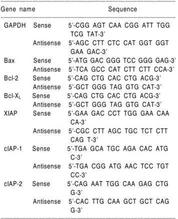
mRNA 분석을 위하여 Bioneer (Taejeon, Korea)에서 구입한 primer는 Table 1에 나타내었고 단백질 분석을 위하여 사 용된 Bcl-2, Bcl-XL, XIAP, cIAP-1, cIAP-2, caspase-3, ca- spase-8, caspase-9, poly(ADP-ribose) polymerase (PARP) 및 phospholipas C (PLC)-γ1 항체는 Santa Cruz Biotechnology Inc. (Santa Cruz, CA, USA) 및 CalBiochem (San Diego, CA,
Table 1. Sequence of primers used for RT-PCR
ꠏꠏꠏꠏꠏꠏꠏꠏꠏꠏꠏꠏꠏꠏꠏꠏꠏꠏꠏꠏꠏꠏꠏꠏꠏꠏꠏꠏꠏꠏꠏꠏꠏꠏꠏꠏꠏꠏꠏꠏꠏꠏꠏꠏꠏꠏꠏꠏꠏꠏꠏꠏꠏꠏꠏ
Gene name Sequence
ꠏꠏꠏꠏꠏꠏꠏꠏꠏꠏꠏꠏꠏꠏꠏꠏꠏꠏꠏꠏꠏꠏꠏꠏꠏꠏꠏꠏꠏꠏꠏꠏꠏꠏꠏꠏꠏꠏꠏꠏꠏꠏꠏꠏꠏꠏꠏꠏꠏꠏꠏꠏꠏꠏꠏ GAPDH Sense 5’-CGG AGT CAA CGG ATT TGG
TCG TAT-3’
Antisense 5’-AGC CTT CTC CAT GGT GGT GAA GAC-3’
Bax Sense 5’-ATG GAC GGG TCC GGG GAG-3’
Antisense 5’-TCA GCC CAT CTT CTT CCA-3’
Bcl-2 Sense 5’-CAG CTG CAC CTG ACG-3’
Antisense 5’-GCT GGG TAG GTG CAT-3’
Bcl-XL Sense 5’-CAG CTG CAC CTG ACG-3’
Antisense 5’-GCT GGG TAG GTG CAT-3’
XIAP Sense 5’-GAA GAC CCT TGG GAA CAA CA-3’
Antisense 5’-CGC CTT AGC TGC TCT CTT CAG T-3’
cIAP-1 Sense 5’-TGA GCA TGC AGA CAC ATG C-3’
Antisense 5’-TGA CGG ATG AAC TCC TGT CC-3’
cIAP-2 Sense 5’-CAG AAT TGG CAA GAG CTG G-3’
Antisense 5’-CAC TTG CAA GCT GCT CAG G-3’
ꠏꠏꠏꠏꠏꠏꠏꠏꠏꠏꠏꠏꠏꠏꠏꠏꠏꠏꠏꠏꠏꠏꠏꠏꠏꠏꠏꠏꠏꠏꠏꠏꠏꠏꠏꠏꠏꠏꠏꠏꠏꠏꠏꠏꠏꠏꠏꠏꠏꠏꠏꠏꠏꠏꠏ
USA)에서 구입하였다. Immunoblotting을 위해 2차 항체 로 사용된 peroxidase-labeled donkey anti-rabbit 및 peroxi- dase-labeled sheep anti-mouse immunoglobulin은 Amersham Life Science Corp. (Arlington Heights, IL, USA)에서 구입하 였다. Caspases의 in vitro 활성 측정을 위한 colorimetric as- say kits는 R&D Systems (Minneapolis, MN, USA)에서 구입 하였으며, caspase-3 inhibitor (z-DEVD-FMK)는 CalBiochem (Darmstadt, Germany)에서 구입하였다.
2. 암세포의 배양
실험에 사용한 AGS 인체위암세포 및 SK-MEL-2 흑색 종세포는 생명공학연구소(KRIBB, Taejeon, Korea)에서 분 양 받았으며 90%의 RPMI-1640 배지(Gibco BRL, Grand Island, NY, USA), 10% fetal bovine serum (FBS, Gibco BRL) 에 1%의 penicillin 및 streptomycin (Gibco BRL)이 포함된 성장배지를 사용하여 5% CO2, 37oC의 조건하에서 배양 하였다.
3. Hemocytometer를 이용한 세포 생존율의 측정
세포 배양용 6 well plate에 암세포를 5×10⁴개/ml의 개 수로 well당 2 ml씩 분주하고 24시간 동안 안정화시킨 다 음 genistein을 적정농도로 처리하여 배양하였다. 48시간 후 배지를 제거하고 0.05% trypsin-EDTA를 처리하여 세 포를 부유시켜서 phosphate-buffered saline (PBS)를 각 well 당 적정량을 첨가하여 세포를 모은 다음, 2,000 rpm으로 5분간 원심분리하였다. 상층액을 제거하고 세포만 남긴 다음 다시 PBS를 1 ml 첨가하여 잘 섞은 후 세포 부유액 과 0.5% trypan blue (Gibco BRL)를 동량으로 섞은 후 2분 간 처리한 후 위상차 현미경(Carl Zeiss, Germany)을 이용 하여 살아있는 세포를 계수하였다. 이에 따른 결과를 Microsoft EXCEL program을 사용하여 분석하였다.
4. DAPI staining에 의한 세포핵의 형태 관찰
Genistein의 처리에 의한 암세포의 apoptosis 유발 여부 를 확인하기 위한 핵의 형태변화 관찰을 위하여 genistein 이 처리된 세포들을 PBS로 세척하고 3.7% paraformal- dehyde로 상온에서 10분간 고정시킨 후 형광 염색물질인 4,6-diamidino-2-phenylindole (DAPI, Sigma) 용액을 이용하 여 10분간 염색하였다. 이들 세포를 다시 PBS로 2회 수 세한 후 형광현미경을 이용하여 핵의 형태 변화를 정상 군과 비교하였다.
5. Flow cytometry 분석
정상 및 genistein이 함유된 배지에서 자란 암세포들을
PBS로 두세 번 씻어내고, 고정액(70% ethyl alcohol, 0.5%
Tween 20)을 첨가하여 4oC에서 고정시킨 후, 핵산에 특이 적으로 결합하는 형광물질인 DNA intercalating dye propidium iodide (PI, concentration, 50μg/ml; Sigma)와 10 kunit의 RNase (Sigma)를 처리하고 4oC에서 1시간 동안 염 색하였다. 이를 다시 PBS로 두 번 씻어낸 후 DNA flow cytometry (Becton Dickinson, San Jose, CA, USA)에 적용시 켜 형광반응에 따른 histogram을 ModiFit LT (Becton Dick- inson) 프로그램으로 분석하였다.
6. Reverse transcription-polymerase chain reaction (RT-PCR) 분석
동일한 조건에서 준비된 암세포를 대상으로 TRIzol reagent (Invitrogen Co., Carlsbad, CA, USA)를 이용하여 total RNA를 분리하였다. 분리된 RNA를 정량한 후 primer, DEPC water, ONE-STEP RT-PCR PreMix Kit (Intron, Korea) 를 넣고 Mastercycler gradient (Eppendorf, Hamburg, Ger- many)를 이용하여 증폭하였다. 이때 housekeeping 유전자 인 glyceraldehyde-3-phosphate dehydrogenase (GAPDH)를 internal control로 사용하였다. 각 PCR 산물들을 1% aga- rose gel을 이용하여 전기영동하고 ethidium bromide (EtBr, Sigma)로 염색한 후 UV하에서 확인하였다.
7. 단백질의 분리, 전기영동 및 Western blotting
정상 및 genistein이 처리된 배지에서 자란 세포들을 lysis buffer로 용해한 후, 고속원심분리로 세포 내 잔사물 을 분리시킨 후 동량의 단백질을 SDS-polyacrylamide gel 전기영동으로 분리하였다. 분리된 단백질을 nitrocellulose membrane (Schleicher and Schuell, Keene, NH, USA)으로 전 이시킨 후, 특정 단백질에 대한 항체와 그에 대한 이차 항체 반응을 실시한 후 enhanced chemiluminoesence (ECL) 용액 (Amersham Life Science)을 적용시킨 다음 X-ray film 에 감광시켜 특정 단백질의 양을 분석하였다.
8. In vitro caspase-3, -8 및 -9의 activity 측정
정상 및 genistein이 처리된 배지에서 48시간 배양된 세 포를 모은 뒤 단백질을 추출하고 정량하여 각각 100μg 의 단백질을 fluorogenic peptide 기질 100 uM이 함유된 extraction buffer [40 mM HEPES (pH 7.4), 20% glycerol (v/v), 1 mM EDTA, 0.2% NP-40 and 10 mM DL-DTT] 50 μl에 혼합하였으며, microtiter plate에 다시 extraction buffer에 희석하여 각 sample 당 총 volume이 100μl가 되게 하였다.
실험에 사용된 기질은 caspase-3의 경우에는 Asp-Glu-Val- Asp (DEVD)-p-nitroaniline (pNA)이었고 caspase-8의 경우에
는 Ile-Glu-Thr-Asp (IETD)-pNA이었으며, caspase-9은 Leu- Glu-His-Asp (LEHD)-pNA였다. 준비된 plate를 37oC에서 3 시간 동안 incubation 시킨 후 ELISA reader를 이용하여 405 nm의 흡광도를 이용하여 반응의 정도를 측정하였다.
결 과
1. 암세포의 생존에 미치는 genistein의 영향
Genistein을 적정농도로 희석하여 AGS 및 SK-MEL-2 세 포에 48시간 동안 처리한 후, hemocytometer로 살아있는 세포의 수를 계수하여 genistein이 처리되지 않은 대조군 과 비교한 결과는 Fig. 1에 나타낸 바와 같다. 결과에서 알 수 있듯이 처리된 genistein의 농도가 증가될수록 생존 율은 두 세포주 모두에서 농도 의존적으로 감소함을 알 수 있었으며, 특히 40μM 처리군에서 AGS 세포의 생존 율은 약 10% 정도로 나타난 반면 SK-MEL-2 세포의 생존 율은 약 40%로 나타나 AGS 세포가 genistein에 대해 민감 하게 반응하는 것을 알 수 있었으며, 이는 MTT assay에 의한 결과와 거의 유사하였다.30)
2. Genistein 처리에 의한 apoptosis 유도
Genistein의 처리에 따른 암세포의 증식억제에는 암세
포의 전체적인 모양에서 다양한 형태적 변형을 유발하 였으며(data not shown), 이러한 암세포의 성장억제와 형 태적 변형이 apoptosis 유발과 상관성이 있는지를 조사하
Fig. 2. Formation of apoptotic bodies by genistein treatment in AGS human gastric carcinoma cells. Cells were treated with genistein for 48 h and then stained with DAPI solution. After 10 min incubation at room temperature, the cells were washed with PBS and nuclear morphology was photographed with a fluorescent microscope using blue filter. Magnification, ×400.
0 10
20 30 40 Genistein ( M)µ
Fig. 1. Effects of genistein on the viability of AGS human gastric carcinoma and SK-MEL-2 melanoma cells. Cells were seeded at 5×10⁴/ml in a 6-well plate and incubated for 24 h. Cells were treated with variable concentrations of genistein for 48 h. The cells were trypsinized and the viable cells were scored by hemocytometer counts of trypan blue-excluding cells. The data shown are means±SD of three independent experiments.
Fig. 3. Induction of apoptosis by genistein in (A) AGS human gastric carcinoma cells and (B) SK-MEL-2 melanoma cells. Cells were exposed for 48 h with increasing concentration of genistein and then the cells were collected and stained with PI for flow cytometry analysis. The percentages of cells with hypodiploid DNA (sub-G1 phase) contents represent the fractions undergoing apoptotic DNA degradation. Data are means average of two separate experiments.
Fig. 4. Effects of genistein treatment on the Bcl-2 and IAP family expression in AGS human gastric carcinoma cells. (A) After 48 h incubation with genistein, total RNAs were isolated and reverse-transcribed. The resulting cDNAs were subjected to PCR with indicated primers and the reaction products were subjected to electrophoresis in 1% agarose gel and visualized by EtBr staining.
GAPDH was used as an internal control. (B) The cells were lysed and then cellular proteins were separated by SDS-polyacrylamide gels and transferred onto nitrocellulose membranes. The membranes were probed with the indicated antibodies. Proteins were visualized using an ECL detection system. Actin was used as an internal control.
Bax
Bcl-2
A
0 10 20 30 40
GAPDH Bcl-XL Genistein ( M)µ
XIAP
cIAP-1
0 10 20 30 40
GAPDH cIAP-2 Genistein ( M)µ
Bax
Bcl-2
B
0 10 20 30 40
Actin Bcl-XL Genistein ( M)µ
XIAP
cIAP-1
0 10 20 30 40
Actin cIAP-2 Genistein ( M)µ
기 위하여 DAPI staining을 실시하여 핵의 형태를 조사하 였다. Fig. 2에 나타낸 바와 같이 AGS 세포에서 genistein 이 처리되지 않은 정상 배지에서 자란 암세포에서 핵의 형태가 뚜렷하게 정상으로 염색이 되었으나 genistein이 처리된 암세포의 경우, 염색질 응축에 의한 apoptosis가 일어난 세포에서 전형적으로 관찰되는 apoptotic body가 농도 의존적으로 증가되었으며, 이는 SK-MEL-2 세포에 서도 유사하게 관찰된 바 있다.30)
이상의 형태적 분석에서 genistein에 의한 암세포의 증 식억제는 apoptosis 유발과 직접적인 관련이 있었기에 genistein 처리에 따른 apoptosis 유발의 정도를 flow cyto- metry를 이용하여 정량적인 분석을 한 결과는 Fig. 3과 같다. 즉 정상 배지에서 자란 암세포에서의 자연적 apoptosis 유발 빈도는 매우 낮았지만(2% 미만) genistein의 처리 농도가 증가될수록 apoptosis의 유발 정도가 증가하 여 40μM 농도에서 AGS 세포는 sub-G1의 비율이 약 32%
였고 SK-MEL-2 세포는 약 16%로 나타나 genistein의 처리 에 따른 sub-G1의 경우도 AGS 세포에서 높게 나타났음 을 알 수 있었다.
3. Bcl-2 및 IAP family의 발현에 미치는 genistein의 영향
다음은 genistein의 처리에 의한 apoptosis 유발에 관여하
는 기전을 조사하기 위하여 AGS 세포를 대상으로 Bcl-2 및 IAP family에 속하는 유전자의 발현 수준을 RT-PCR 및 Western blot analysis 방법으로 조사하였다. Fig. 4A 및 B에 나타낸 바와 같이 genistein 처리 농도 의존적으로 apoptosis 유도에 관여하는 Bax의 발현이 증가하였고 apoptosis 유발 억제에 관여하는 Bcl-2의 발현이 감소하였 음을 알 수 있었다. 하지만 SK-MEL-2 세포에서는 Bax 및 Bcl-2 모두 genistein 처리에 따른 변화가 거의 관찰되지 않았다.30) IAP family의 경우, Fig. 5에 나타난 바와 같이 AGS 세포에서 genistein의 처리에 의해 전사 및 번역수준 에서 IAP family의 선택적 감소 현상이 관찰되었다.
4. Caspases의 발현 및 활성에 미치는 genistein의 영향
지금까지 알려진 caspase 중 대부분의 apoptosis가 유발 된 세포에서 높은 활성도를 보여주는 caspase-3, -8 및 -9 의 발현에 미치는 genistein의 영향을 조사하였다. Fig. 5A 의 결과에서 볼 수 있듯이 AGS 세포에서 genistein의 처리 에 의하여 불활성형인 pro-caspase-3 단백질의 발현은 농 도 의존적으로 감소되었으나 뚜렷한 활성형의 단백질 발현 증가를 정확히 관찰하기는 어려웠다. 따라서 caspases의 활성 정도를 직접 분석한 결과, Fig. 5B에 나타 난 바와 같이 genistein이 처리된 암세포에서는 처리 농도
Fig. 5. Activation of caspase-3, and degradation of PARP and PLCγ1 by genistein in AGS human gastric carcinoma cells. (A) After 48 h incubation with genistein, the cells were lysed and then cellular proteins were separated by SDS-polyacrylamide gels and transferred onto nitrocellulose membranes.The membranes were probed with indicated antibodies. Proteins were visualized using an ECL detection system. Actin was used as an internal control. (B) After 48 h incubation with genistein, aliquots (150μg protein) were incubated with substrates, DEVD-pNA, IETD-pNA and LEHD-pNA, for in vitro caspase-3, -8 and -9 activity, respectively, at 37oC for 3 h. The released fluorescent products were measured. Data are means average of two separate experiments.
A
Pro-caspase-3 Pro-caspase-8
0 10 20 30 40
PARP Pro-caspase-9 Genistein ( M)µ
PLC- 1γ
Actin
B
Caspase 8 Caspase 3 Caspase 9
0 10 20 30 40
Caspase activity
Genistein ( M)µ 0
1.0 2.0 3.0 4.0 5.0
의존적으로 caspase-3의 활성이 모두 증가되었음을 알 수 있었다. 특히 genistein을 40μM 처리하였을 경우 caspase- 3의 활성이 약 4.7배 증가하였다.
5. PARP 및 PLC-γ1의 발현에 미치는 genistein의 영향
다음은 apoptosis 유발시 caspase에 의하여 특이하게 분 해가 일어나는 몇 가지 표적 단백질의 발현에 미치는 genistein의 영향을 Western blot analysis 방법으로 조사하 였다. Fig. 5A에서 볼 수 있듯이 AGS 세포에서 genistein의 처리 농도 증가에 따라 DNA repair와 genomic stability에 관여하는 PARP 단백질의 발현의 단편화가 나타났으며, 세포의 성장과 증식에 중요한 역할을 하는 PLC-γ1 단백 질의 발현 또한 genistein의 처리 농도 의존적으로 발현의
감소 또는 단편화현상이 나타났다.
6. Caspase-3 inhibitor에 의한 apoptosis의 억제
이상의 결과들을 살펴볼 때 caspase-3의 활성이 genis- tein에 의한 apoptosis에 중요한 역할을 하는 것으로 생각 되어 caspase-3 inhibitor가 genistein에 의한 apoptosis를 어떠 한 영향을 주는지 조사하여 보았다. Fig. 6에서 나타낸 바와 같이 AGS 위암세포 및 SK-MEL-2 흑색종세포 모두 에서 genistein의 처리에 의해 유도되는 apoptosis가 ca- spase-3 inhibitor인 z-DEVD-FMK 전 처리에 의해서 약 15% 정도 억제되는 것으로 나타났다. 따라서 genistein에 의해서 유도되는 apoptosis에는 caspase-3이 중요한 역할을 하는 것으로 생각된다.
Fig. 6. Suppression of genistein-induced apoptosis by caspase-3 inhibitors in AGS human gastric carcinoma and SK-MEL-2 melanoma cells. Cells were pretreated with 50μM of caspase-3 inhibitor (Z-DEVD-FMK) for 2 h, then treated with 40μM genistein for 48 h. (A) Effects of capase-3 inhibitor on morphological changes in genistein-treated cells. (B) The viable cell number was determined by hemocytometer counts of trypan blue-excluding cells. The data shown are means±SD of three independent experiments.
A
- + - +
Relative viability
0 0.2 0.4 0.6 0.8 1.0 1.2
AGS
SK-MEL-2
Control Genistein z-DEVD-FMK Genistein + z-DEVD-FMK
B
- - + +
AGS
- + - +
Relative viability
0 0.2 0.4 0.6 0.8 1.0 1.2
- - + +
SK-MEL-2
Genistein z-DEVD-FMK
고 찰
본 연구에서는 AGS 인체위암세포 및 SK-MEL-2 흑색 종세포의 성장에 미치는 genistein의 영향을 조사하기 위 하여 genistein이 함유된 배지에서 자란 암세포의 생존율 을 정상배지에서 자란 대조군과 비교하였다. Fig. 1에서 알 수 있듯이 두 세포주 모두에서 genistein의 첨가농도 의존적으로 암세포의 생존율은 감소되었으며, 특히 AGS 세포에서 genistein에 의한 생존율 감소가 두드러졌다. 이 러한 암세포 증식 억제 및 형태 변화의 관찰에서 geni- stein의 처리에 의한 암세포 증식의 억제는 apoptosis 유발 과 밀접한 연관성이 있을 것으로 기대되어 이에 대한 증 거를 제시하기 위하여 암세포의 핵 형태 변화를 관찰한 결과(Fig. 2), 대조군의 경우 모든 암세포에서 핵의 형태 가 뚜렷하게 정상으로 염색이 되었으나 genistein이 처리 된 암세포의 경우, genistein 처리 농도 의존적으로 염색 질 응축에 의한 apoptosis가 일어난 세포에서 전형적으로 관찰되는 apoptotic body의 증가 현상을 관찰할 수 있었 다.1,2) 즉 이는 nucleosome의 linker DNA 부분의 절단에 의 한 DNA 단편화의 결과이므로 genistein의 처리에 의한 암세포의 증식억제 및 형태적 변형이 암세포의 apoptosis 유발과 밀접한 관련이 있음을 시사하여 주는 것으로 사 료된다. 아울러 flow cytometry 분석에 의한 apoptosis 유발 세포군에 해당하는 sub-G1기에 속하는 세포들의 빈도 역시 genistein 처리 농도 의존적으로 증가되어 genistein 처리에 따른 암세포의 증식 억제는 apoptosis 유발과 직접 적인 연관이 있음을 알 수 있었다(Fig. 3). 특히 AGS 세포 에서 sub-G1기에 속하는 세포들의 빈도가 더욱 높게 나 타났으며, 이는 genistein의 처리에 의한 두 세포주에서의 생존율의 차이와도 부합되는 결과였다.
다음은 genistein의 처리에 의한 apoptosis 유발에 관여 하는 유전자의 탐색을 위하여 apoptosis와 연관성을 가지 는 가장 대표적인 유전자인 Bcl-2 family에 속하는 유전자 의 발현을 조사하였다. Bcl-2 family에 속하는 몇 가지 중 요한 인자들은 apoptosis 유발 조절에 가장 대표적인 유전 자로 알려져 있는데, 그중 Bcl-2는 anti-apoptotic 분자로서 apoptosis의 유발을 억제하는 기능을 가지며, Bax는 pro- apoptotic 분자로 Bax 단백질 발현의 증가는 apoptosis의 유 발과 관계가 있다.31) 이들 두 유전자는 세포 내 소기관 중 mitochondria로부터의 cytochrome c를 유리시켜 cysteine- related protease인 caspase, 종양억제 유전자인 p53, DNA의 단편화와 연관된 endonuclease 등의 활성을 조절하는 것 으로 알려져 있다. 이들은 서로 dimer의 형태로 존재하며
그들의 발현 수준에 변화가 초래되면 apoptosis가 유발된 다.31) Fig. 4는 다양한 농도의 genistein이 함유된 배지에서 배양된 암세포를 대상으로 전사 및 번역수준을 조사한 결과로서, AGS 세포의 경우 전사와 번역수준 모두에서 pro-apoptotic 유전자인 Bax의 증가 및 anti-apoptotic 유전 자인 Bcl-2의 감소가 관찰되었다. 즉 AGS 세포의 경우 genistein에 의한 apoptosis 유발에 Bcl-2 family에 속하는 유 전자의 발현의 차이가 중요한 역할을 하는 것으로 생각 된다.
한편 apoptosis에 관여하는 여러 인자들 중 최근 밝혀진 IAP family에 속하는 여러 단백질은 곤충세포에서 바이 러스 감염에 의한 apoptosis를 억제하기 위한 baculoviral 단백질 군들로서 외부 신호에 의한 세포 내 anti-apoptotic 활성을 지닌다.32,33) 그리고 이들 중 몇 가지는 caspase와 의 직접적인 결합을 통하여 그들의 apoptotic 활성을 억 제할 수 있을 것으로 밝혀졌다.32,33) Genistein 처리에 의한 암세포의 apoptosis 유발에 IAP family가 관여하는지의 여 부를 조사한 결과 Fig. 4에서 나타낸 바와 같이 SK-MEL-2 세포에서 보다는 다소 약하지만,30) genistein 처리 농도 의 존적으로 AGS 세포에서 IAP family의 감소가 관찰되었 다. 이는 SK-MEL-2 세포에서 genistein의 처리에 따른 apoptosis 유발에 Bcl-2 family의 변화 보다 IAP family의 발 현 감소가 중요한 역할을 하는 것으로 생각할 수 있다.
Caspase protease라는 효소 역시 apoptosis 유발에 중요한 조절인자로서 작용한다. 이들 family에 속하는 단백질들 은 세포에서 핵과 mitochondria의 외막에 불활성 상태로 존재하며, Bcl-2/Bax family 발현의 변화에 따라 이들의 활 성도가 조절될 수 있다.34,35) 이들은 proenzyme 형태로 존 재하다가 apoptosis 유도를 활성화시키는 신호에 의해 활 성화된 protease로 전환되어 직접 또는 간접적으로 세포 내에 존재하는 많은 표적 단백질의 분해에 관여한다. 따 라서 caspase의 활성화는 apoptosis의 유발에 대한 또 다른 증거가 될 수 있으며 많은 선행연구 등에서 검증되어 왔 다. 따라서 genistein이 caspases에 미치는 영향을 조사한 결과 Fig. 5A에 나타난 바와 같이 caspase-3의 경우 AGS 세포에서 pro-caspase-3 단백질의 발현이 감소하였으나 활성형 단백질은 관찰할 수 없었다. 따라서 caspases의 활 성 정도를 직접 관찰한 결과 Fig. 5B에서와 같이 caspase-8 및 caspase-9의 할성은 크게 변하지 않은 반면 caspase-3의 경우는 처리 농도 의존적으로 활성이 증가하는 것으로 나타났으며, 이는 SK-MEL-2 세포에서도 유사하였다.30) 또한 caspase-3 inhibitor인 z-DEVD-FMK를 이용하여 geni- stein에 의한 apoptosis가 억제되는지를 알아본 결과 Fig.
6에서와 같이 두 세포주 모두에서 15% 정도의 억제효과
가 나타났다. 이상의 결과를 살펴볼 때 genistein에 의한 두 세포주에서의 apoptosis 유발에는 caspase-3이 중요한 역할을 하는 것으로 나타났다.
한편 apoptosis가 유발되었을 경우 특이하게 분해가 일 어나는 몇 가지 표적 단백질 중 DNA repair나 genomic stability의 유지에 매우 중요한 역할을 하는 PARP 단백질 은 정상적인 세포의 경우 116 KDa의 분자량을 가지지만 활성화된 caspase에 의하여 분해가 일어나면 85 kDa 크기 의 단편이 관찰되거나 주 band의 발현이 감소되어 정상 적인 DNA repair 과정이 억제된다.36,37) 또한 최근 연구에 의하면 epidermal growth factor, transforming growth factor, insulin-like growth factor-1 및 cytokine 등과 같은 세포 성장 을 촉진하는 인자들이 apoptosis를 억제할 수 있는 것으로 보고되고 있다. 이들 성장인자의 수용체들은 phospha- tidylinositol 3-kinase (PI3K), G-protein 및 phospholipase C-γ (PLC-γ1) 등과 같은 주요 신호전달 분자들을 활성화시 킬 수 있다. PI3K에 의해 생산되는 phosphatidylinositol 3, 4, 5-trisphosphate (PIP3)는 protein kinase B (PKB)/Akt 및 PLC-γ1을 활성화시킨다. 특히 PLC-γ1은 phosphatidy- linositol 4,5-bisphosphate를 hydrolyze시켜 protein kinase C (PKC) activator인 diacylglycerol 및 세포 내 Ca2+ 조절에 중 요한 역할을 하는 inositol 1,4,5-trisphosphate (IP3)를 생산 한다. 따라서 PI3K나 Ras와 같은 세포성장 신호분자와 같이 PLC-γ1 역시 세포의 증식에 중심적인 역할을 하는 것으로 알려져 있다. 그러나 apoptosis가 유발될 경우 활 성화된 caspase 효소에 의하여 PLC-γ1 단백질은 분해될 수 있기 때문에 apoptosis 유발의 생화학적 표식자로 사용 이 가능하다.38) 따라서 genistein 처리에 따른 PARP 및 PLC-γ1 단백질의 발현 변화를 살펴본 결과 Fig. 5에서 볼 수 있듯이 genistein 처리 농도 의존적으로 발현의 감 소 또는 단편화현상이 나타났다.30) 이는 genistein 처리에 의한 암세포에서의 apoptosis 유발은 caspases의 활성화와 연관된 caspase 기질 단백질의 분해에 의해서 발생한다는 것을 의미하는 결과로서 genistein의 apoptosis 유발 경로 가 전형적인 mitochondria 경로를 통하는 유전자들의 발 현에 전반적으로 영향을 미쳤음을 의미하는 것으로 추 정된다.
이상의 결과에서 genistein에 의한 AGS 인체위암세포 및 SK-MEL-2 흑색종세포의 성장억제는 apoptosis 유발과 밀접한 관련이 있었다. 이러한 apoptosis 유발에는 Bcl-2 family 및 IAP family의 발현 변화와 그에 따른 caspase의 활성화에 의한 표적 단백질들의 발현 저하와 직접적인 연관성이 있음을 관찰할 수 있었다. 그러나 본 연구의 결과만으로 genistein의 항암작용 여부를 직접적으로 논
하기에는 매우 어렵기 때문에, 선행보고 된 암세포의 증 식 억제와 연관된 다양한 추가적인 실험이 필수적으로 수행되어야 할 것이다. 또한 본 연구의 결과는 향후 수행 될 추가 실험을 위한 기초 자료로서 그 가치가 매우 높 을 것으로 생각된다.
감사의 글
본 연구는 농림부 농림기술개발사업의 지원에 의해 이루어진 것임(No. 105113-3).
참 고 문 헌
1) Evans VG. Multiple pathways to apoptosis. Cell Biol Int 17, 461-476, 1993.
2) Shi L, Nishioka WK, Th’ng J, Bradbury EM, Litchfield DW, Greenberg AH. Premature p34cdc2 activation required for apoptosis. Science 263, 1143-1145, 1994.
3) Schulze-Osthoff K, Ferrari D, Los M, Wesselborg S, Peter ME. Apoptosis signaling by death receptors. Eur J Biochem 254, 439-459, 1998.
4) Chiarugi V, Magnelli L, Basi G. Apoptosis and the cell cycle.
Cell Mol Biol Res 40, 603-612, 1994.
5) Nagata S, Apoptosis by death factor. Cell 88, 355-365, 1997.
6) Akiyama T, Ishida J, Nakagawa S, Ogawara H, Watanabe S, Itoh N, Shibuya M, Fukami Y. Genistein, a specific in- hibitor of tyrosine-specific protein kinases. J Biol Chem 262, 5592-5595, 1987.
7) Rubin JB, Shia MA, Pilch PF. Stimulation of tyrosine-specific phosphorylation in vitro by insulin-like growth factor I. Nature 305, 438-440, 1983.
8) Peterson G, Barnes S. Genistein inhibition of the growth of human breast cancer cells: independence from estrogen re- ceptors and the multi-drug resistance gene. Biochem Biophys Res Commun 179, 661-667, 1991.
9) Peterson G, Barnes S. Genistein and biochanin A inhibit the growth of human prostate cancer cells but not epidermal growth factor receptor tyrosine autophosphorylation. Prostate 22, 335-345, 1993.
10) Markovits J, Linassier C, Fosse P, Couprie J, Pierre J, Jacquemin-Sablon A, Saucier JM, Le Pecq JB, Larsen AK.
Inhibitory effects of the tyrosine kinase inhibitor genistein on mammalian DNA topoisomerase II. Cancer Res 49, 5111-5117, 1989.
11) McCabe MJ Jr, Orrenius S. Genistein induces apoptosis in immature human thymocytes by inhibiting topoisomerase-II.
Biochem Biophys Res Commun 194, 944-950, 1993.
12) Kiguchi K, Constantinou AI, Huberman E. Genistein-induced cell differentiation and protein-linked DNA strand breakage in human melanoma cells. Cancer Commun 2, 271-277, 1990.
13) Yanagihara K, Ito A, Toge T, Numoto M. Antiproliferative
effects of isoflavones on human cancer cell lines established from the gastrointestinal tract. Cancer Res 53, 5815-5821, 1993.
14) Spinozzi F, Pagliacci MC, Migliorati G, Moraca R, Grignani F, Riccardi C, Nicoletti I. The natural tyrosine kinase inhibitor genistein produces cell cycle arrest and apoptosis in Jurkat T-leukemia cells. Leuk Res 18, 431-439, 1994.
15) Pagliacci MC, Smacchia M, Migliorati G, Grignani F, Riccardi C, Nicoletti I. Growth-inhibitory effects of the natural phyto- oestrogen genistein in MCF-7 human breast cancer cells. Eur J Cancer 30, 1675-1682, 1994.
16) Fotsis T, Pepper M, Adlercreutz H, Fleischmann G, Hase T, Montesano R, Schweigerer L. Genistein, a dietary-derived inhi- bitor of in vitro angiogenesis. Proc Natl Acad Sci USA 90, 2690-2694, 1993.
17) Jing Y, Waxman S. Structural requirements for differentia- tion-induction and growth-inhibition of mouse erythroleuke- mia cells by isoflavones. Anticancer Res 15, 1147-1152, 1995.
18) Hotz MA, Del Bino G, Lassota P, Traganos F, Darzynkiewicz Z. Cytostatic and cytotoxic effects of fostriecin on human promyelocytic HL-60 and lymphocytic MOLT-4 leukemic cells. Cancer Res 52, 1530-1535, 1992.
19) Finlay GJ, Holdaway KM, Baguley BC. Comparison of the effects of genistein and amsacrine on leukemia cell proli- feration. Oncol Res 6, 33-37, 1994.
20) Rauth S, Kichina J, Green A. Inhibition of growth and induction of differentiation of metastatic melanoma cells in vitro by genistein: chemosensitivity is regulated by cellular p53. Br J Cancer 75, 1559-1566, 1997.
21) Monti E, Sinha BK. Antiproliferative effect of genistein and adriamycin against estrogen-dependent and -independent human breast carcinoma cell lines. Anticancer Res 14, 1221- 1226, 1994.
22) Messina M, Barnes S. The role of soy products in reducing risk of cancer. J Natl Cancer Inst 83, 541-546, 1991.
23) Coward L, Barnes NC, Setchell KDR, Barnes S. Genistein, daidzein, and their β-glycoside conjugates: anti-tumor isofla- vones in soybean foods from American and Asian diets. J Agric Food Chem 41, 1961-1967, 1993.
24) Messina MJ, Persky V, Setchell KD, Barnes S. Soy intake and cancer risk: a review of the in vitro and in vivo data. Nutr Cancer 21, 113-131, 1994.
25) Raffoul JJ, Wang Y, Kucuk O, Forman JD, Sarkar FH, Hillman GG. Genistein inhibits radiation-induced activation of NF-kappaB in prostate cancer cells promoting apoptosis and G2/M cell cycle arrest. BMC Cancer 6, 107, 2006.
26) Yu Z, Li W, Liu F. Inhibition of proliferation and induction of apoptosis by genistein in colon cancer HT-29 cells. Cancer Lett 215, 159-166, 2004.
27) Buchler P, Gukovskaya AS, Mouria M, Buchler MC, Buchler MW, Friess H, Pandol SJ, Reber HA, Hines OJ. Prevention of metastatic pancreatic cancer growth in vivo by induction of apoptosis with genistein, a naturally occurring isoflavonoid.
Pancreas 26, 264-73, 2003.
28) Balabhadrapathruni S, Thomas TJ, Yurkow EJ, Amenta PS, Thomas T. Effects of genistein and structurally related phy- toestrogens on cell cycle kinetics and apoptosis in MDA- MB-468 human breast cancer cells. Oncol Rep 7, 3-12, 2000.
29) Lian F, Li Y, Bhuiyan M, Sarkar FH. p53-independent apoptosis induced by genistein in lung cancer cells. Nutr Cancer 33, 125-131, 1999.
30) Kim JI, Park C, Choi WY, Lee JO, Hong SH, Hong SH, Lee WH, Rhu CH, Choi YH. Induction of G2/M arrest of the cell cycle and apoptosis by genistein in SK-MEL-2 human melanoma cells. Cancer Pre Res 20, 46-57, 2006.
31) Osford SM, Dallman CL, Johnson PW, Ganesan A, Packham G. Current strategies to target the anti-apoptotic Bcl-2 protein in cancer cells. Curr Med Chem 11, 1031-1039, 2004.
32) Holcik M, Gibson H, Korneluk RG. XIAP: apoptotic brake and promising therapeutic target. Apoptosis 6, 253-261, 2001.
33) Salvesen GS, Duckett CS. IAP proteins: blocking the road to death’s door. Nat Rev Mol Cell Biol 3, 401-410, 2002.
34) Allen RT, Cluck MW, Agrawal DK. Mechanisms controlling cellular suicide: role of Bcl-2 and caspases. Cell Mol Life Sci 54, 427-445, 1998.
35) Rao L, White E. Bcl-2 and the ICE family of apoptotic re- gulators: making a connection. Curr Opin Genet Dev 7, 52-58, 1997.
36) Tewari M, Quan LT, O'Rourke K, Desnoyers S, Zeng Z, Beidler DR, Poirier GG, Salvesen GS, Dixit VM. Yama/
CPP32, a mammalian homolog of CED-3, is a CrmA-inhibi- table protease that cleaves the death substrate poly (ADP- ribose) polymerase. Cell 81, 801-809, 1995.
37) Lazebnik YA, Kaufmann SH, Desnoyers S, Poirier GG, Earn- shaw WC, Cleavage of poly ADP-ribose polymerase by a proteinase with properties like ICE. Nature 371, 346-347, 1994.
38) Bae SS, Perry DK, Oh YS, Choi JH, Galadari SH, Ghayur T, Ryu SH, Hannun YA, Suh PG. Proteolytic cleavage of phospholipase C-gamma1 during apoptosis in Molt-4 cells.
FASEB J 14, 1083-1092, 2000.