저작자표시-비영리-변경금지 2.0 대한민국 이용자는 아래의 조건을 따르는 경우에 한하여 자유롭게 l 이 저작물을 복제, 배포, 전송, 전시, 공연 및 방송할 수 있습니다. 다음과 같은 조건을 따라야 합니다: l 귀하는, 이 저작물의 재이용이나 배포의 경우, 이 저작물에 적용된 이용허락조건 을 명확하게 나타내어야 합니다. l 저작권자로부터 별도의 허가를 받으면 이러한 조건들은 적용되지 않습니다. 저작권법에 따른 이용자의 권리는 위의 내용에 의하여 영향을 받지 않습니다. 이것은 이용허락규약(Legal Code)을 이해하기 쉽게 요약한 것입니다. Disclaimer 저작자표시. 귀하는 원저작자를 표시하여야 합니다. 비영리. 귀하는 이 저작물을 영리 목적으로 이용할 수 없습니다. 변경금지. 귀하는 이 저작물을 개작, 변형 또는 가공할 수 없습니다.
i
A THESIS
FOR THE DEGREE OF MASTER OF SCIENCE
2’,3’-dimethoxyflavanone induces apoptosis
accompanied by caspase-8 activation mediated by
ubiquitination in breast cancer stem-like cells
(BCSCs)
TRAN ANH THAO
Department of Biotechnology
GRADUATE SCHOOL
JEJU NATIONAL UNIVERSITY
2013.12
i
ABSTRACT
The cytotoxicities of 13 flavonoids were tested in the breast cancer stem-like cell MCF-7-SC. Among them, 2’,3’-dimethoxyflavanone (DMF) showed the highest toxicity. DMF triggered apoptosis by activating caspase-9, -3, -8, and cleaved PARP. In addition, DMF induces a dramatic increase in the conversion of LC3, a well-known marker for autophagy. However, co-treatment with chloroquine, the lysosomal inhibitor that blocks autophagic degradation did not show any change in the degree of LC3 conversion, implying that LC3 could play a role in the non-autophagic cell death of MCF-7-SC. In the present study, we found that DMF induces the ubiquitination of caspase-8 accompanied by the interaction between caspase-8 and LC3, which leads to the aggregation and self-activation of caspase-8. Consistent with this, co-treatment of DMF with 3MA, an inhibitor of LC3 lipidation reduced the activation of caspase-8. Our findings provide new insights into the anti-cancer activity of DMF in breast cancer stem cell through apoptosis in company with LC3 conversion.
Keywords: Apoptosis, Breast cancer stem cell, Caspase-8, 2’,3’-dimethoxyflavanone, LC3,
ii
ABSTRACT
13 종의 다양한 플라보노이드계 화합물들 중에서 2’,3’-dimethoxyflavanone (DMF)가 유방암 줄기세포 MCF-7-SC에 대해 가장 높은 세포독성을 나타냈다.
DMF는 caspase-9, -3, -8의 활성과 cleaved PARP에 의한 apoptosis를 유도하였다.
또한, DMF는 autophagy의 표지인자로 잘 알려져 있는 LC3의 conversion을 증가시켰다. 그러나, autophagic degradation을 차단하는 lysosomal inhibitor인 chloroquine을 혼용 처리하 였을 때 LC3 conversion의 변화가 나타나지 않았으며, 이는 LC3가 MCF-7-SC세포에서 non-autophagic cell death에 관여할 수 있음을 암시한다.
본 연구에서, DMF가 caspase-8과 LC3의 상호작용을 동반한 caspase-8의 ubiquitination을 유 도하며 이를 통해 caspase-8의 aggregation과 self-activation을 일으킨다는 것을 밝혔다. 이와 일치하여, DMF와 LC3 lipidation을 억제하는 3MA를 혼용 처리하였을 때 caspase-8의 활 성이 감소하였다.
본 연구에서는 이러한 결과들을 통해 DMF 의 유방암 줄기 세포에서의 항암 활성이 LC3 conversion 을 동반한 apoptosis 유도에 기인한 것임을 밝혀냈다.
iii
CONTENT
Content
ABSTRACT ... i
추상... Error! Bookmark not defined. List of figures ... iv
List of tables ... v
1. INTRODUCTION ... 1
2. MATERIALS AND METHODS ... 5
2.1 Reagents ... 5
2.2 Cell lines and Cell culture ... 5
2.3 Cytotoxicity ... 5
2.4 Cytometric analysis ... 6
2.5 Morphology ... 6
2.6 Western blot analysis ... 7
2.7 Immunoprecipitation ... 7
2.8 Colocalization assay ... 8
2.9 Statistics ... 8
3. RESULTS... 9
iv
3.2. 2’,3’-dimethoxyflavanone induced cell death through apoptotic pathway ... 15 3.3 2’,3’-dimethoxyflavanone induced caspase-8 activation through ubiquitination system ... 19 3.4 Caspase-8 activation is regulated by LC3-ubiquitin-like protein... 25 4. DISCUSSION ... 34 REFERENCES ... 38 ACKNOWLEDGEMENT ... Error! Bookmark not defined.
iv
List of figures
Figure 1: DMF is specific for cancer stem-like cell MCF-7-SC. ... 10
Figure 2: DMF induced apoptosis in MCF-7-SC cells. ... 16
Figure 3: DMF induced caspase-8 activation through ubiquitination system. ... 20
Figure 4: Caspase-8 activation is regulated by LC3 activation. ... 27
Figure 5: Suggested mechanism of DMF-induced cell death in breast cancer stem cell MCF-7-SC. ... 33
v
List of tables
1
1. INTRODUCTION
Flavonoids are found ubiquitously from natural sources. Because they show various biological activities including anti-cancer, anti-inflammation, and anti-allergy, they have been the main focus of attention due to its varied biological benefits in human [1-4]. Flavonoids consists of C6-C3-C6 skeleton and they can be classified based on their structures. When C6-C3 skeleton is composed of 4H-chromen-4-one and the third C6 is attached to C-2 position, it is called flavone. Flavonoids with 4H-chromen-4-one and the third C6 attached to C-3 position are named isoflavones. Derivatives with chroman-4-one are called flavanones. Since flavonoids contain phenyl rings, hydroxy or methoxy groups can be substituted. While the former can increase the solubility, the later increases cell permeability. Various effects of flavonoids on cancer cell lines have been reported [J. Agri. Food Chem. 61:12588; Bioorg. Med. Chem. 21: 7018; Int. J. Mol. Sci. 14:16970; Bioorg. Med. Chem. Lett. 23:232]. However, the studies on breast cancer stem cell is very limited.
Breast cancer is the second most common cause of death and it is also known as the second leading cause of mortality. A half million women are dead each year by breast cancer. The failure of chemo- and radiotherapy is strongly related to cancer stem cells inside tumor which is critical for cancer’s ability. The cancer stem cells can self-renewal, quickly multiply, highly invade to establish new tumors that are often therapy resistant [13]. A typical stemness characteristic which is thought to related to the self-renewal ability is the forming of mammosphere [14]. It has been also suggested that cancer stem cell strongly correlates with invasive and metastatic abilities. [15]. During invasion process, the epithelial-mesenchymal transition (EMT) is often activated with the increasing of Vimentin, Snail, and Slug [16]. It had been demonstrated the similarity between the cells undergone EMT and the stem cells isolated
2
from normal or neoplastic cell populations [17]. Experimental [18] and clinical evidence [19] supports the idea that breast cancers are organized hierarchically with a small number of breast cancer stem-like cells (BCSCs) able to regrow a tumor after sub-lethal treatment. Breast cancer stem cells can be prospectively identified based on the high level of cell surface protein marker called CD44 [18]. It has been demonstrated that in breast cancer patients, the soluble form of CD44 in serum is detected at high concentration and CD44 can regulate breast cancer cells migration and protect tumor from immune system [20-22]. These findings suggest that targeting and elimination of breast cancer stem cells is the promising way to obtain durable breast cancer treatment responses. Autophagy and apoptosis are believed to be good targets for developing anticancer drugs. Apoptosis is programmed cell death mediated by caspases, which are cysteine proteases that cleave target proteins at aspartic acid. p53 is a transcription factor that induces the expression of pro-apoptotic genes [23, 24] and activating apoptosis is an important mechanism in p53-induced tumor suppression. Autophagy is an evolutionarily conserved and genetically programmed process that degrades long-lived cellular proteins and organelles. The role of autophagy in cancer is quite complicated and controversial. Many reports have elucidated the cross talk or mutual exclusion between autophagy and apoptosis [25]. Recent studies have shown that several molecules required for autophagy also play a key role in regulation of apoptosis. For example, cleaved-Atg5 serves as a pro-apoptotic protein by directly binding to anti-apoptotic protein Bcl-xL on the mitochondria to stimulate intrinsic apoptotic pathway [26]. Similarly, the cleaved form of Beclin-1 by caspases has ability to translocate to mitochondria to induce the release of pro-apoptotic factors [27]. Indeed, the apoptotic function of microtubule associated protein light chain 3 (LC3), a well-known molecule that is involved in autophagy, has been reported [28, 29] and a number of cancers have been reported to have elevated levels of LC3 [30,
3
31]. LC3 was shown to exist in two forms, LC3 I (18kDa), a cytosolic form, and LC3 II (16kDa), the membrane bound shorter form derived from LC3 I by proteolysis and lipid modification [32]. Upon the initiation of autophagy, LC3 conjugation to phosphatidylethanolamine at its C-terminal glycine and its subsequent membrane localization promote the subsequent dynamic rearrangement of intracellular membranes, leading to the formation of autophagosomes [33]. LC3 directly interacts with p62 via its N-terminal domain, which is essential for the cargo function of p62 to transport polyubiquitylated protein aggregates to autophagosomes [34].
Two mechanistically connected processes are used for degradation of cellular proteins: the ubiquitin-proteasomal pathway and the autophagolysosomal pathway [35-38]. Inhibition of these protein degradation pathways leads to the accumulation of damaged proteins, which, if unresolved, is detrimental to the cell, leading to a consequence known as proteotoxicity [39]. Cancer cells, owing to their aberrant transcription/translation activity and protein disposal, may become more vulnerable to proteotoxicity. Because of these, inhibition of the proteasomal and autophagolysosomal degradation pathways is under clinically used and investigation for treating cancer [40-46]. Although the molecular machinery underlying proteotoxicity remains largely unanswered, the caspase-8-mediated proteotoxicity has been believed to be a broad implication under a wide range of conditions where cellular protein homeostasis is irreversibly disrupted. Upon the inhibition of protein degradation pathways, cells could up-regulate the autophagic response to facilitate autophagolysosomal protein turnover, which is mediated by the lipidation and membrane localization of LC3. However, severely damaged proteasomal or autophagolysosomal pathways may “clog” each other [36, 38]. This leads to the enrichment of LC3 on intracellular membranes that can serve as a molecular hub to recruit the ubiquitin-binding protein SQSTM1/p62 and one of its ubiquitin-binding partners, ubiquitinated-caspase-8. It has
4
been reported that active caspase-8 dimer is too unstable to exist for long once released from death-inducing signaling comlex (DISC) [47]. Poly-ubiquitinated caspase-8 has ability to aggregate rich in ubiquitin in which caspase-8 is sustained the dimerization and activity of cleaved form [48, 49]. The oligomerization and activation of caspase-8 subsequently initiate the downstream apoptosis cascade [27].
In the present study, the cytotoxicities of 13 flavonoids including 2 methoxy-flavones, 10 flavanones, and 1 isoflavone were tested on breast cancer cell line MCF-7. Of them, 2’,3’-dimethoxyflavanone (DMF) showed the best cytotoxicity, so that we report we report that DMF induced phosphorylation of p53, JNK, Bcl-2 and activation of caspase-3, -9, PARP which possibly result in the intrinsic apoptosis. We have determined for the first time that DMF induces the conversion of LC3 and ubiquitylation of caspase-8, therefore promotes the accumulation of caspase-8 in the cytosol where it is fully activated, subsequently initiate the downstream apoptosis cascade. Moreover, these results may support the idea of apoptotic function of LC3, a well-known molecule that is involved in autophagy.
5
2. MATERIALS AND METHODS
2.1 Reagents
All flavonoids including 2’,3’-dimethoxyflavanone (DMF) were purchased from Indofine Chemical Co. (Hillsborough, NJ). RPMI 1640, DMEM and F12 medium, bovine serum albumin (BSA), trypsin/EDTA, fetal bovine serum (FBS), Antibiotic-Antimycotic 100X were purchase from Invitrogen. Hoechst 33342, dimethyl sulfoside (DMSO), 3-(4,5-dimethylthiazol-2-yl)-2,5-diphenyltetrazolium bromide (MTT), propidium iodide (PI), and RNase A were purchased from Sigma Chemical Co. (St.Louis, MO). 3-Methyladenine (3-MA) was purchased from Sigma. An Annexin V-FITC Apoptosis Detection Kit I and BDTM Mitoscreen (JC-1) Kit were purchased from BD Biosciences (Franklin Lakes, NJ). Anti-p53, -caspase 3, -caspase 7, -caspase 8 (rabbit) , -caspase 9, -JNK, -Bax, -Bcl-2, -mouse IgG Fab 2 Alexafluor 555 antibodies were purchased from Cell Signaling (Danvers, MA). Anti-ubiquitin, -caspase 8 (mouse), MG-132 were purchased from Santa Cruz. Polyvinylidene fluoride (PVDF) membranes for Western blotting were purchased from Bio-Rad (Hercules, CA). All chemicals were dissolved in DMSO.
2.2 Cell lines and Cell culture
MCF-7 breast carcinoma cells (ATCC, Manassas, VA) were cultured in DMEM/F12, 10% fetal bovine serum (FBS), 1% penicillin/streptomycin (Gibco, Carlsbad, CA). Human breast cancer stem-like cells MCF-7-SC were isolated, enriched, and characterized from MCF-7 with CD44high/CD24low marker in Stem cell laboratory of Vietnam-HCM National University [50]. MCF-7-SC cells were cultured in RPMI-1640 supplemented with 10% FBS and 1% antibiotics at 37 C in a humidified atmosphere containing 5% CO2.
2.3 Cytotoxicity
6
FBS. After treated, 20 µl of MTT solution (5 mg/ml) were added and incubate for 3-4 h. Adding 150 µl of DMSO after removed the supernatant in each well. Cell viability was determined from the absorbance at 570 nm, measured using a Sunrise microplate reader (Tecan, Salzburg, Austria). The blank contained 200 µl of RPMI 1640 with 10% FBS and equivalent reagent concentrations.
2.4 Cytometric analysis
To analyze the cell cycle arrest, apoptosis, autophagy, and mitochondrial membrane potential, cells (3 x 10⁴ cells/ml) were plated in 60mm dishes and treated for 48 h. For distinguishing apoptosis and necrosis, an Annexin V-FITC Apoptosis detection Kit I was used to detect. Annexin V binds specifically to phosphatidylserine from the inner side of plasma membrane. The apoptosis were detected by Annexin V positive whereas necrotic cells are permeable to Propidium Iodide (PI). Cells were washed with PBS, harvested, and diluted in buffer containing Annexin V and PI, and incubated for 15- 20 min at room temperature (RT). For JC-1 mitochondrial membrane detection, treated cells were harvested and stained with JC-1 for 10-15 min in incubator, and washed two times with 1X assay buffer at RT. For cell cycle analysis, treated cells were harvested, washed with PBS, and fix with 70% cold Ethanol overnight, rehydrated in 2mM EDTA-PBS, treated with RNase A (25 ng/ml), and stained with PI (10 mg/ml). All analyses were performed using a FACS Caliber flow cytometer (BD Biosciences). Each sample were counted 10,000 cells and were analyzed the data with Cell Quest Software (BD Biosciences).
2.5 Morphology
Cells were seeded in 6-well plates and treated for indicated times. Treated cells were stained with 10 µM Hoechst then observed under a fluorescence microscope (Olympus, Essex, UK).
7
2.6 Western blot analysis
After 24 h or 48 h treatment, cells were collected and lysed in buffer (100 mM Tris-Hcl [pH 8], 250 mM NaCl, 0.5% Nonidet P-40, and 1X protease inhibitor cocktail) and sonicate few times then kept on ice for 30 min. The cell lysates were centrifuged at 13,000 rpm for 30 min at 4oC. The supernatant were collected and determined the concentration by BCATM Protein assay (Pierce, Rockford, IL). Aliquots of the lysate were separated by 10-15% SDS-PAGE, and transferred to PVDF membranes using a glycine transfer buffer (192 mM glycine, 25 mM Tris-HCl [pH 8.8], and 20% [v/v] methanol). After blocking with Skim milk 5% overnight at 4oC, the membrane were incubated with primary antibodies for 1 h RT or overnight at 4oC and then for 1 h with secondary antibodies. Primary antibodies were diluted at ratio 1:1000 whereas the anti-β-actin primary and secondary antibodies (horseradish peroxidase-conjugated goat anti-mouse IgG) (Vector Laboratories, Burlingame, CA) were diluted at ratio 1:10,000. Protein bands were detected using the WEST-ZOL® plus Western Blot Detection System (iNtRON, Gyeonggi-do, Korea).
2.7 Immunoprecipitation
Treated cells were collected, washed with cold PBS, and lysed in 500 µl lysis buffer (20 mM Tris (pH 7.5), 150 mM NaCl, 1 mM EDTA, 1 mM EGTA, 1% Trinton X-100, 2.5 mM Sodium pyrophosphate, 1 mM β-Glycerolphosphate, 1 mM Na3VO4, 1 µg/ml Leupeptin, and 1X protease inhibitor cocktail). Incubate primary antibody with gentle rocking overnight at 4oC. Add protein A/G agarose beads (25 µl of 50% bead slurry) and gentle rocking for 3 h at 4oC. Microcentrifuge sample in 14,000 rpm for 1 min at 4 oC. Resuspend the pellet with 40 µl 2x SDS sample buffer and vortex well. Heat the sample at 100 oC for 10 min then loading on SDS-PAGE gel (10-12%), and transfer to PVDF membranes using transfer buffer (0.2 mM glycine, 25 mM Tris-HCl [pH
8
8.8], and 20% [v/v] methanol). Analyze sample by western blotting (Cell signaling protocol)
2.8 Co-localization assay
Cells were seeded in slide covered 0.1% gelatin. After indicated times, treated cells were then fixed with 4% paraformaldehyde for 20 min, and permeablized with 0.4% triton X-100 for 1 h at RT. Permeablized cells were incubated with blocking buffer (5% goat serum) overnight at 4 oC. For co-localization of LC3 and caspase-8 assay, we incubated cells with the mixing of primary antibodies of LC3 and caspase-8 (1:200) in 1% goat serum overnight at 4 oC. After washing, cells were incubated with the mixing of secondary antibodies conjugated fluorescence dyes (1:200) for 1 h at RT. Washing 3 times and mounting cells with mounting solution. Observe cells under fluorescent microscope (Olympus IX73).
2.9 Statistics
Group comparisions were using SPSS v.12.0 software with one-way analysis (ANOVA) and Student’s t-test. P< 0.05 was considered statistically significant. All experiments were performed in triplicate.
9
3. RESULTS
3.1 Characteristics of the breast cancer stem cell MCF-7-SC
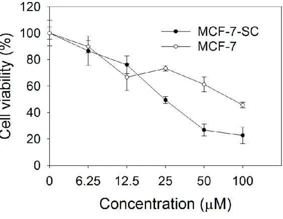
The cell line MCF-7-SC sorted from normal breast cancer MCF-7 with stemness marker CD44high/CD24low was tested for mammosphere formation, drug resistance, and invasiveness. The mammosphere formation, one of the most distinct morphology of cancer stem cells is detected in MCF-7-SC in non-adherent culture (Fig 1A). To compare the drug resistance between MCF-7 and MCF-7-SC, we chose two commercially available drugs, doxorubicin and docetaxel utilized for breast cancer treatment [51]. MCF-7-SC is more resistant than MCF-7 when treated with doxorubicin at highest concentration 40 µM and with docetaxel at highest concentration 5 µM as shown in Fig 1B and 1C, respectively. Recent evidences demonstrating the capacity of cancer stem cells to detach from their primary epithelial sites to a mesenchymal state (epithelial– mesenchymal transition, EMT) to form secondary tumors are accumulated. We now detect the greater migration activity (Fig 1D) and higher expression of EMT markers, Vimentin, Snail in MCF-7-SC, whereas the lack of E-cadherin Epithelial marker in MCF-7-SC (Fig 1E). We next examined the effects of various flavanones with or without methoxyl groups, different number containing and different position of methoxyl groups to identify efficient anti-cancer agents that can induce cell death in breast cancer stem cells MCF-7-SC. Among 13 tested flavanones (Table 1), 2’,3’-dimethoxyflavanone (DMF) showed the highest toxicity with 50% cell death at 50 µM and 80% at 100 µM (Fig 1F). To test whether DMF is specific to cancer stem cell type, we compared the cytotoxicity of DMF toward MCF-7 and SC. MTT assay showed MCF-7-SC is more sensitive to DMF treatment than MCF-7 (Fig 1G) at higher concentration than 25 µM.
10
Figure 1
1A
1B C
11
1D
12
13
1G
Figure 1: 2’,3’-dimethoxyflavanone (DMF) is specific for cancer stem-like cell MCF-7-SC.
(A) MCF-7 and MCF-7-SC cells were cultured in non-adherent plates and observed under microscope. (B) Cell death in response to indicated concentrations of Doxorubicin, and (C) Docetaxel. (D) Wound healing assay for the migration ability of MCF-7 and MCF-7-SC. (E) Western blot analysis with antibodies specific for E-cadherin, Vimentin, Snail, Slug, and the housekeeping protein β-actin, as described in section 2. (F) The cytotoxicity of 13 flavanone compounds. MCF-7-SC were treated with different doses of flavanones (50, 100 µM) for 48 h. (G) The cytotoxicity of DMF in breast cancer. MCF-7 and MCF-7-SC cells were treated with different doses of DMF (0 – 100 µM) for 48 h. The data correspond to the mean ±SD of three independent experiments.
14
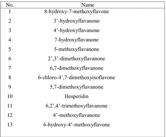
Table 1 List of flavonoids that are examined for their cytotoxicity in MCF-7-SCs
No. Name 1 8-hydroxy-7-methoxyflavone 2 3’-hydroxyflavanone 3 4’-hydroxyflavanone 4 7-hydroxyflavanone 5 5-methoxyflavanone 6 2’,3’-dimethoxyflavanone 7 6,7-dimethoxyflavanone 8 6-chloro-4’,7-dimethoxyisoflavone 9 5,7-dimethoxyflavanone 10 Hesperidin 11 6,2’,4’-trimethoxyflavanone 12 4’-methoxyflavanone 13 6-hydroxy-4’-methoxyflavone
15
3.2. 2’,3’-dimethoxyflavanone induced cell death through apoptotic pathway
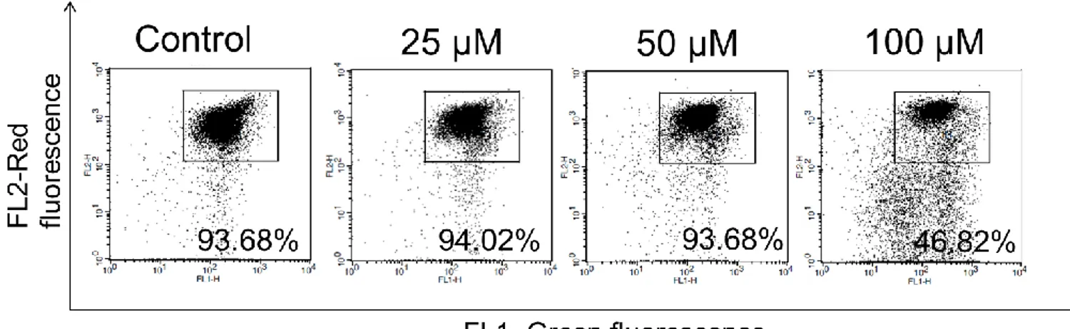
To confirm the cell death which detected by MTT assay in DMF treatment, we tested the DNA damage by cell cycle arrest using Facs analysis. After 48 h incubated with DMF, sub G1 is significantly induced compare with control (Fig 2A). We used Hoechst- DNA dye to observe the cell morphology. DMF induced DNA condensation following apoptotic bodies (white arrows) in dose dependent manner (Fig 2B). Since Hoechst staining showed the sign of apoptosis, we checked the percentage of apoptotic and necrotic cells using Annexin V/ PI staining. When treated cells loss of plasma membrane, the early events in apoptotic pathway is the translocation of phosphatidylserin (PS) from the inner to the outer membrane [52]. As shown in fig 2C, DMF dramatically induced the appearance of Annexin V positive cells at 100 µM, suggesting the occurrence of early and late apoptosis. Consistent with the observation in staining results, western blot showed the increase of cleave-caspase-9, caspase-8, caspase-7, and caspase-3 in dose-dependent (Fig 2D) and time dependent. We also detected the active form of p53 and JNK, the well-known tumor suppressors involved in apoptosis. P53 can indirectly promote the phosphorylation of JNK through increasing the expression level of sertrin-2 [53]. JNK, one of the MAP kinase, also play many roles in regulating cell death. JNK can phosphorylate members of the Bcl-2 families such as phosphorylating Bcl-2 for reducing its affinity with Bax (Bcl-2 associated protein X), hence leading mitochondrial permeabilization [54, 55]. As shown in figure 2D, the increasing of phosphorylation Bcl-2 implied the releasing of Bax from Bcl-2 for the oligomerization subsequent leading to intrinsic apoptotic pathway. In addition, PUMA (P53-upregulated modulator of apoptosis) is up-regulated to induced MOMP and release cytochrome c [56]. The loss of mitochondrial membrane potential (MMP) is also detected confirming the
16
disruption of mitochondria to release apoptotic proteins (Fig 2E).
Figure 2
2A
17
2C
18
2E
Figure 2: DMF induced apoptosis in MCF-7-SC cells. Cells were seeded, incubated for 24 h
and then treated with the indicated concentrations of DMF for an additional 48 h. (A) Treated cells were harvested, fixed with 70% cold ethanol, stained with Propidium Iodine (PI), washed, and analyzed by flow cytometric analysis. (B) Treated cells were fixed and stained with Hoechst 33,342 and observed under a fluorescence microscope. (C) Treated cells were harvested and stained with Annexin V/PI, washed and analyzed by flow cytometry. (D) Western blot analysis antibodies specific for caspase-9, -8, -3, c-PARP, p-p53, p-JNK, Bcl-2, p-Bcl-2, Bax, Puma, β-actin. (E) Treated cells were harvested and stained with JC-1 according to the intructions of the company supplied the kit.
19
3.3 2’,3’-dimethoxyflavanone induced caspase-8 activation through ubiquitination system
Since the MTT assay showed MCF-7-SC is more sensitive than MCF-7 in DMF treatment, we hypothesized that MCF-7-SC may has different level of some pro-apoptotic or pro-survival proteins which play main core in cell death mechanism. Western blot results showed the significant high level of pro-caspase-8 in MCF-7-SC compare with MCF-7 (Fig 3A), implying that caspase-8 may play a critical role in DMF-induced cell death. Recently, caspase-8 is demonstrated that can be activated due to death receptor and the inhibition of intracellular protein degradation machineries. Inhibition of protein degradation can lead to the ubiquitination, accumulation and activation of caspase-8 [57-59]. Indeed, observing in SDS-PAGE, the ubiquitinated-caspase-8 is increased after 24 and 48 h (left), and dose-dependent (right) treated with DMF (Fig 3B). We chose well-known proteasome inhibitor MG-132, which was demonstrated that activated caspase-8 through ubiquitination as a positive control [58]. Immunofluoresence showed the large-sized punctate of caspase-8 in both DMF and MG-132 treatment, implied the accumulation of caspase-8 (Fig 3C). Since the protein ubiquitination and aggregation were observed in DMF treatment, we assumed that DMF is a novel proteasome inhibitor which can inhibit the degradation system to activate caspase-8 oligomerization. As similar as DMF, western blot showed the cleaved form of caspase-8 is also detected in indicated concentrations of MG132 (Fig 3D). Another evident which has been shown to inhibit proteasome activity is the increasing of Reactive oxygen species (ROS) [57, 59, 60]. Indeed, DMF and MG-132 treatment induced ROS production after 48 h (Fig 3E). Together, these data indicated that DMF could be a new proteasome inhibitor, therefore inducing caspase-8 ubiquitination, and self-activation.
20
21
3B
22
3C
23
24
3E
Figure 3: DMF induced caspase-8 activation through ubiquitination system. (A) MCF-7
and MCF-7-SC were performed western blot analysis with antibodies specific for caspase-9, -8, PARP, AKT, mTOR, β-actin. (B) MCF-7-SC were treated with DMF at indicated times (left) or doses (right). Cell lysates were collected and subjected to immunoprecipitation (IP) with antibody specific for caspase-8, and the ubiquitinated proteins were examined by western blot using antibodies specific for caspase-8, ubiquitin, β-actin. MG-132 (40 µM) is utilized as a positive control. (C) MCF-7-SC were treated with DMF (100 µM), or with MG-132 (40 µM) for 24 h in chamber slides. After treating, immunofluorescence assay were performed using mouse IgG Fab 2 Alexafluor 555 antibody specific for caspase-8 (red), and observed under a fluorescence microscope. (D) Immunoblotting analyses using antibodies specific for caspase-8, β-actin after MG-132 treatment at indicated concentrations for 48 h. (E) MCF-7-SC were treated with DMF (left) or MG-132 (right) at indicated concentrations for 48 h. Cells were harvested and incubated with H2DCFH-DA, washed and analyzed by flow cytometry analysis. The data correspond to the mean ±SD of three independent experiments.
25
3.4 Caspase-8 activation is regulated by LC3-ubiquitin-like protein
Since LC3 is reported as an ubiquitin-like protein which directly bind to caspase 8 and lead apoptosis through caspase activation in SKI-I, a pan-sphingosine kinase inhibitor, and proteasome inhibitor bortezomib, MDEG G430F, MG-132 [57, 58, 61] we examined whether DMF triggers the same phenomenon in MCF-7-SC. The conversion of LC3 is required for binding to caspase-8 and mediating protein accumulation, therefore we determined whether LC3 conversion is induced in this cell death. Interestingly, western blot results showed the increasing of LC3 II in dose and time-dependent after treated with DMF (Fig 4A). LC3 II is also well known as a marker for autophagy, so we tested whether this induction due to the autophagic occurrence concomitantly with apoptosis. As a marker for autophagy, acridine orange staining which is used to detect acidic vesicular [62] showed no difference in DMF treatment compared with control in both microscope observation (Fig 4B) and Facs analysis (Fig 4C) in time (upper) and dose-dependent manner (lower). We chose Chloroquine (CQ), a well-known compound can prevent the autophagic flux by inhibiting the fusion of autophagosome and lysosome, hence increasing the LC3 conversion [63]. Co-treatment of CQ and DMF showed no difference of LC3 II compare with DMF treated one (Fig 4D). Altogether, it implied that LC3 conversion is related to apoptotic signaling pathway. To examine the role of LC3 in caspase-8 accumulation process, we performed immunoprecipitation assay to test the interaction of LC3 with caspase-8. In precipitated LC3 samples, caspase-8 is detected in DMF treatment whereas no detection in control (Fig 4E). Co-immunofluorescence assay showed the LC3 puncta (green fluorescence) accumulated to caspase-8 (red fluorescence), implied the co-localization of LC3 and caspase-8 upon DMF treatment (Fig 4F). To demonstrate the important role of LC3 in caspase-8 activation, we co-treated MCF-7-SC with DMF and LC3 conversion inhibitor. 3MA co-treatment increased
26
the ratio of LC3 I/LC3 II, implied the inhibition of 3MA on LC3 lipidation [58, 64]. In the inhibition of LC3 II, the cleaved caspase-8 is reduced concomitantly with down-regulated cleaved-PARP, a substrate of caspase-8 [35] (Fig 4G). For anchoring our hypothesis that DFM is specific for breast cancer-stem cells, we tested the response of MCF-7 which is low caspase-8 level in cells as a negative control. Result showed that DMF did not induced caspase-8 cleavage as well as no altered LC3 conversion compare with no treatment (Fig 4H). Together, these results demonstrated that LC3 play a role in activation of caspase-8 to facilitate cell death in DMF treatment.
27
4A
4B
28
4C
29
4D
30
4F
31
4G
32
Figure 4: Caspase-8 activation is regulated by LC3 activation. (A) MCF-7-SC were treated
with DMF at indicated concentrations for 48 h or indicated times at 100 µM, and performed western blot using antibodies specific for LC3 and β-actin. (B) Acidic vesicular organelles (AVOs) were examined by incubating the cells with 10 µM acridine orange (AO) for 10 min, and observing and imaging them using fluorescence microscope. (C) Quantification of AVOs-positive cells by flow cytometry in dose-dependent (upper), and time-dependent (lower). (D) MCF-7-SC were treated with DMF (100 µM) in the absence or presence of CQ (50 µM). Cells were harvested and performed western blot using indicated antibodies. (E) MCF-7-SC cells were treated with DMF (100 µM) for 24, and 48 h. Cell lysates were collected and subjected to IP with antibody specific for LC3 followed by western blot using indicated antibodies. Input, total cell lysates. (F) Cells were treated with DMF (100 µM) for 24 h in chamber slides, immune-stained with anti-LC3 (green) and anti-caspase-8 (red) antibodies, observed under fluorescence microscope. Note the punctate distribution of LC3 and caspase-8, and their co-localization was detected by collapse of green and red dot (yellow) upon DMF treatment. (G) MCF-7-SC cells were treated with DMF (100 µM) in the absence or presence of 3MA (2mM) for 48 h. Cell lysates were performed western blot using indicated antibodies. (H) MCF-7 cells were treated with DMF dose dependent for 48 h. Cell lysates were performed western blot using indicated antibodies.
33
Fig 5
Figure 5: Suggested mechanism of DMF-induced cell death in breast cancer stem cell
MCF-7-SC. DMF activated p53 through phosphorylating p53. Activated p53 induced Puma, and the phosphorylation of Bcl-2 to disrupt Bcl-2/Bax complex. Bax and Puma facilitate mitochondrial dysfunction, result in caspase-9, -3 activation. These events might lead intrinsic apoptosis. Concomitantly, DMF inhibit the proteasome activity, ubiquitinated caspase-8, result in stabilizing caspase-8, accumulating to the rich ubiquitin region, oligomerization and activation. This accumulation of caspase-8 is facilitated by LC3 conversion.
34
4. DISCUSSION
Currently, around 40% of all breast cancer patients have tumor relapse in conventional therapy. Beside many reasons such as under-treatment of drugs, local sites of tumor growth missed during doing surgery or the prior development of micro-metastases, the stemness property is also the main reason of these recurrences. Hence, there is much interest in identify new chemical or phytochemical which specific target to breast cancer stem cells to reduce the relapse during therapeutic strategies. A better understanding of breast cancer stem cell characteristic is a priority for improvement of patient survival. Like embryonic and adult stem cells, cancer stem cells are not a static population, and that under permissive conditions, possibly dictated by the tumor microenvironment cancer cells transition between stem-like and non-stem-like states [69]. Thus we generated the population isolated from normal breast cancer cells to identify phytochemical can affect specifically on cancer stem cell proliferation. We demonstrated that our new cancer stem-like cells MCF-7-SC is a great model for anti-breast cancer studies with high proliferative, migrative and resistant properties.
The key findings of this report are the new flavanone 2’, 3’-dimethoxyflavanone which is specific for breast cancer stem cells MCF-7-SC and the detailed apoptotic cell death related to autophagic marker LC3. Bearing two methoxyl groups at 2’, 3’ positions, DMF performs a great activity among tested flavanones. Our data support that flavanone which has simple methoxyl groups are more cytotoxic than poly-methoxyl or non-methoxylated flavanone. DMF induced cell death through both intrinsic and extrinsic apoptotic pathways by the detection of caspase-9,-8,-3, and the disruption of Bcl-2 and Bax complex by increasing phosphorylated Bcl-2. Interestingly, we found that MCF-7-SC is more sensitive in DMF treatment than MCF-7 possibly due to the high expression level of caspase-8 in MCF-7-SC. Caspase-8 is well-characterized as a
35
precursor initiate apoptosis. However it also plays an essential role in adhesion and migration. Some reports have revealed that procaspase-8 interact with phosphatidylinositol-3-OH kinase (PI3K) regulatory subunit p85α and c-src to modulate signaling by Rac and extracellular signal-regulated kinase, and promote calpain activation which is important in most or all aspects of cell migration. Surprisingly, the catalytic activity of caspae-8 is not required for caspase-8 dependent promotion of cell motility and adhesion [48, 57, 58]. This opposite function of caspase-8 could explain although caspase-8 expression is compromised in various tumors and highly in breast cancer stem-like cells, it is maintained a potential pro-tumorigenic role. These findings supported our data that MTT result (Fig 1G) showed the sensitive of MCF-7-SC in DMF treatment compare with MCF-7, and the high level of caspase-8 in this cancer stem-like cells without any treatment, confirmed that caspase-8 behave as a unique switch turn on cell death in specific stimuli or enhance tumorigenic property in normal condition. As we mentioned before, the dimerization of caspase-8 is too unstable when release from DISC complex. Therefore, in treatment with doxorubicin or docetaxel, caspase-8 might be degraded by proteasomal degradation system. Since immunoprecipitation results showed the ubiquitination (Fig 3B) and aggregation of caspase-8 (Fig 3C), raising an assumption that DMF may behave as proteasome inhibitor which are demonstrated as one of the mechanism promotes the protein aggregates containing caspase-8 and subsequent caspase-8 activation. MG-132, a well-known proteasome inhibitor showed the same phenomena of caspase-8 ubiquitination, aggregation as DMF treatment (Fig 3B, C, D). This notion supported by our observation on the ROS production which is induced in the presence of proteasome inhibitor. By treating with DMF, it can turn off degradation system to stabilize ubiquitinated-caspase-8 and lead to the caspase-8 oligomerization in rich ubiquitin region where caspase-8 can be fully activated. In DMF treatment, we also
36
observed the activation of tumor suppressor p53 (Fig 2D), which is reported that rapidly accumulated in cells treated with proteasome inhibitors [70, 71]. The stabilization and accumulation of p53 were shown to be activated, hence inducing the downstream signaling. Although other molecules may also be involved in cell death, it is possibly that DMF induced p53 through the inhibition of proteasome. In another case, the apoptotic function of caspase-8 is demonstrated that regulated by FLIP a caspase-8 inhibitor [72]. The hetero-dimerization of caspase-8 with FLIP can enhance the proliferation and migration, as well as drug resistance. We do not exclude one of the possibilities that DMF might target to FLIP to prevent FLIP function or down-regulate its expression level, hence facilitating the homo-dimerization of caspase-8. However, the further experiments need to be investigated.
One of the interesting findings is the increasing of LC3 conversion- a well-known marker for autophagy, whereas no detection of autophagic flux. It raised a question that LC3 may be involved in apoptotic cell death induced by DMF. The LC3 conversion and p62/sequestosome-1 are recently elucidated as main regulators activate caspase-8 aggregation, augmenting full activation of caspase-8. Our data showed the interaction of caspase-8 and LC3, and the co-treatment DMF with LC3 conversion inhibitor 3MA can reduced caspase-8 activation, which are consistent with previous reports that LC3 directly bind to caspase-8 and activate caspase-8 through ubiquitination system [61, 62, 65]. In addition, LC3 is reported not only involved in autophagy and apoptosis, but also play a key role in mediating cytoplasmic vacuolation death [73, 74] and required for the efficient clearance of dead cells [75].The above studies highlight the multi-function of LC3 in many cell death pathways, which is promising for therapeutic target for leading cancer cell death.
37
In summary, we identified DMF- a novel phytochemical which has high toxicity in cancer stem cells. DMF behave as a proteasome inhibitor, which can enrich LC3 lipidation on intracellular membranes and turn on ubiquitination system on caspase-8. Possibly that proteasome inhibitory activity of DMF can stabilize and activate p53, therefore inducing intrinsic pathway related to inactivate Bcl-2 to activate the loss of mitochondrial membrane potential, hence inducing caspase-9, -3. These two events leading to the interaction of caspase-8 and LC3 to facilitate the aggregation and activation of caspase-8, hence induce apoptotic cell death. Furthermore, these results lead a note that LC3 conversion should be carefully interpreted in cell death mechanism studies.
38
REFERENCES
1. Atun, S., et al., Isolation and antimutagenic activity of some flavanone compounds from
Kaempferia rotunda International Journal of Chemical and Analytical Science, 2013. 4(1): p. 3-8.
2. Batra, P. and A.K. Sharma, Anticancer potential of flavonoids. Biotech, 2013. 3: p. 439-459.
3. Levaj, B., et al., Determination of Flavonoid in Pulp and Peel of Mandarin Fruits. Agriculture Conspectus Scientificus, 2009. 74(3): p. 221-225.
4. Tomás-Navarro, M., F. Vallejo, and F.A. Tomás-Barberán, Bioavailability and
Metabolism of Citrus Fruit Beverage Flavanones in Humans. Polyphenols in Human
Health and Disease, 2013. 1: p. 537-551.
5. Choi, E.J., J.I. Lee, and G.H. Kim, Effects of 4',7-dimethoxyflavanone on cell cycle arrest
and apoptosis in human breast cancer MCF-7 cells. Arch Pharm Res, 2011. 34(12): p.
2125-2130.
6. Kaur, K., et al., 3′,4′-Dimethoxyflavone and valproic acid promotes the proliferation of
human hematopoietic stem cells. Stem Cell Research & Therapy, 2013. 4(60): p. 1-8.
7. Shin, S.Y., et al., 5-Methoxyflavanone induces cell cycle arrest at the G2/M phase,
apoptosis and autophagy in HCT116 human colon cancer cells. Toxicol Appl Pharmacol,
2011. 254(3).
8. Wen, X., U.K. Walle, and T. Walle, 5,7-Dimethoxyflavone down-regulates CYP1A1
expression and benzo[a]pyrene-induced DNA binding in Hep G2 cells. Carcinogenesis,
2005. 26: p. 803-809.
9. Walle, T., Methoxylated flavones, a superior cancer chemopreventive flavonoid
subclass? . Seminars in Cancer Biology, 2007. 17: p. 354-362.
10. Walle, T., et al., Cancer chemopreventive properties of orally bioavailable
flavonoids-methylated versus unflavonoids-methylated flavones. Biochem Pharmacol, 2007. 73: p. 1288-1296.
11. Wen, X. and T. Walle, Methylation protects dietary flavonoids from repid hepatic
metabolism. Xenobiotica, 2006. 36: p. 387-397.
12. Wen, X. and T. Walle, Methylated flavonoids have greatly improved intestinal
absorption and metabolic stability. Drug Metab Dispos, 2006. 34: p. 1786-1792.
13. Smalley, M., L. Piggott, and R. Clarkson, Breast cancer stem cells: Obstacles to therapy. Cancer letter, 2013. 338(1): p. 57-62.
14. Cioce, M., et al., Mammosphere forming cells from breast cancer cell line as a tool for
the identification of CSC-like and early progenitor-targeting drugs. Cell Cycle, 2010. 9(14): p. 2878-2887.
15. Stolpe, A.v.d., On the origin and destination of cancer stem cells: a conceptual
39
16. Yuji Toiyama, et al., Increased expression of Slug and Vimentin as novel predictive
biomarkers for lymph node metastasis and poor prognosis in colorectal cancer.
Carcinogenesis, 2013. 0: p. 1-10.
17. Mani, A.S., et al., The epithelial-mesenchymal transition generates cells with properties
of stem cells. Cell, 2008. 133(4): p. 704–715.
18. Al-Hajj, M., et al., Prospective identification of tumorigenic breast cancer cells. Proc Natl Acad Sci USA, 2003. 100: p. 3983–3988.
19. Ginestier, C., et al., ALDH1 is a marker of normal and malignant human mammary stem
cells and a predictor of poor clinical outcome. Cell Stem Cell, 2007. 1(5): p. 555–567.
20. Pham, P.V., et al., Differentiation of breast cancer stem cells by knockdown of CD44:
promising differentiation therapy. Journal of Translational Medicine, 2011. 7: p. 9-209.
21. Martin, S., et al., Soluble CD44 splice variants in metastasizing human breast cancer. International Journal of Cancer, 1997. 74(4): p. 443-445.
22. Barbour, A.P., et al., Expression of the CD44v2-10 isoform confers a metastatic
phenotype: Importance of the heparan sulfate attachment site CD44v3. Cancer Research,
2003. 63: p. 887-892.
23. Tokino, T. and Y. Nakamura, The role of p53-target genes in human cancer. Critical reviews in oncology /hematology, 2000. 33(1): p. 1-6.
24. Moll, U.M. and A. Zaika, Nuclear and mitochondrial apoptotic pathways of p53. FEBS Letters, 2001. 493(2-3): p. 65-69.
25. Maiuri, M.C., et al., Self-eating and self-killing: crosstalk between autophagy and
apoptosis. Nature Reviews Molecular Cell Biology, 2007. 8(9): p. 741-745.
26. Yousefi, S., et al., Calpain-mediated cleavage of Atg5 switches autophagy to apoptosis. Nat Cell Biol, 2006. 8(10): p. 1124-1132.
27. Wirawan, E., et al., Caspase-mediated cleavage of Beclin-1 inactivates Beclin-1-induced
autophagy and enhances apoptosis by promoting the release of proapoptotic factors from mitochondria. Cell Death Dis, 2010. 1: p. 1038-1048.
28. Chen, K.F., et al., Bortezomib overcomes tumor necrosis factor-related
apoptosis-inducing ligand resistance in hepatocellular carcinoma cells in part through the inhibition of the phosphatidylinositol 3-kinase/Akt pathway. J. Biol. Chem, 2009. 284:
p. 11121–11133.
29. Kar, R., et al., A novel role for MAP1 LC3 in nonautophagic cytoplasmic vacuolation
death of cancer cells. Oncogene, 2009. 28: p. 2556–2568.
30. Namgoong, G.M., et al., The prolyl isomerase Pin1 induces LC-3 expression and
mediates tamoxifen resistance in breast cancer. J. Biol. Chemico-Biological
Interactions, 2010. 285(1): p. 23829–2384.
31. Yoshioka, A., et al., LC3, an autophagosome marker, is highly expressed in
gastrointestinal cancers. Int. J. Oncol, 2008. 33: p. 461–468.
32. Tanida, I., T. Ueno, and E. Kominami, LC3 and Autophagy. Methods Mol Biol, 2008.
40
33. Kabeya, Y., et al., LC3, a mammalian homologue of yeast Apg8p, is localized in
autophagosome membranes after processing. EMBO J, 2000. 19: p. 5720–5728.
34. Shvets, E., et al., The N terminus and Phe52 residue of LC3 recruit p62/SQSTM1 into
autophagosomes. J. Cell Sci, 2008. 121: p. 2685–2695.
35. Buchberger, A., B. B, and T. Sommer, Protein quality control in the cytosol and the
endoplasmic reticulum: brothers in arms. Mol. Cell, 2010. 40: p. 238–252.
36. Ding, W.X. and X.M. Yin, Sorting, recognition and activation of the misfolded protein
degradation pathways through macroautophagy and the proteasome. Autophagy, 2008. 4:
p. 141–150.
37. Kirkin, V., et al., A role for ubiquitin in selective autophagy. Mol. Cell, 2009. 34: p. 259– 269.
38. Korolchuk, V.I., F.M. Menzies, and R.D. C, Mechanisms of cross-talk between the
ubiquitin-proteasome and autophagy-lysosome systems. FEBS Lett, 2010. 584: p. 1393–
1398.
39. Hightower, L.E., Heat shock, stress proteins, chaperones, and proteotoxicity. Cell, 1991.
66: p. 191–197.
40. Adams, J., The proteasome: a suitable antineoplastic target. Nat. Rev. Cancer Cell, 2004.
4: p. 349–360.
41. Amaravadi, R.K. and C.B. Thompson, The roles of therapy-induced autophagy and
necrosis in cancer treatmen. Clin. Cancer Res, 2007. 13: p. 7271–7279.
42. Carew, J.S., et al., Targeting autophagy: a novel anticancer strategy with therapeutic
implications for imatinib resistance. Biologics, 2008. 2: p. 201–204.
43. Fehrenbacher, N. and M. Jaattela, Lysosomes as targets for cancer therapy. CAncer Research, 2005. 65: p. 2993–2995.
44. Mathew, R., V. Karantza-Wadsworth, and E. White, Role of autophagy in cancer. Nat. Rev. Cancer, 2007. 7: p. 961–967.
45. Voorhees, P.M. and R.Z. Orlowski, The proteasome and proteasome inhibitors in cancer
therapy. Annu. Rev. Pharmacol. Toxicol, 2006. 46: p. 189–213.
46. Wu, W.K., et al., Proteasome inhibition: a new therapeutic strategy to cancer treatment. Cancer letter, 2010. 293: p. 15–22.
47. Pop, C., et al., Role of Proteolysis in Caspase-8 Activation and Stabilization. Biochemistry 2007. 46: p. 4398-4407.
48. Y, Z., S. X, and R. H, From procaspase-8 to caspase-8: Revisiting structural functions of
caspase-8 Journal of Cellular Physiology, 2010. 225(2): p. 316-320.
49. B., P.A., F.C. D., and K. Sally, Cellular Mechanisms Controlling Caspase Activation and
Function. Cold Spring Harb Perspect Biol, 2013. 5(6).
50. Pham, P.V., et al., Isolation of Breast Cancer Stem Cells by Single-Cell Sorting. Intech, 2012. Chapter 4: p. 59-72.
51. Diéras, V., Review of docetaxel/doxorubicin combination in metastatic breast cancer. Oncology, 1997. 11: p. 31-33.
41
52. Vermes, I., et al., A novel assay for apoptosis. Flow cytometric detection of
phosphatidylserine expression on early apoptotic cells using fluorescein labelled Annexin V. J Immunol Methods, 1995. 184(1): p. 39-51.
53. Kroemer, G., G. Marino, and B. Levine, Autophagy and the Integrated Stress Response. Cell, 2010. 40(2): p. 280-293.
54. Maundrell, K., et al., Bcl-2 undergoes phosphorylation by c-Jun N-terminal
kinase/stress-activated protein kinases in the presence of the constitutively active GTP-binding protein Rac1. J Biol Chem, 1997. 272(40): p. 25238-25242.
55. Matsuyoshi, S., et al., FADD phosphorylation is critical for cell cycle regulation in
breast cancer cells. British Journal of Cancer, 2006. 94: p. 532-539.
56. Jeffers, J.R., et al., Puma is an essential mediator of p53-dependent and -independent
apoptotic pathways. Cancer Cell, 2003. 4(4): p. 321-328.
57. Pan, J.A., et al., Hyperactivation of the mammalian degenerin MDEG promotes
caspase-8 activation and apoptosis. J Biol Chem, 2012. 2caspase-8caspase-8(5): p. 2952-2963.
58. Pan, J.A., et al., Inhibition of Protein Degradation Induces Apoptosis through a
Microtubule-Associated Protein 1 Light Chain 3-Mediated Activation of caspase-8 at Intracellular Membranes. Mol Cell Biol, 2011. 31(3158-3170).
59. Miller, C.P., et al., NPI-0052, a novel proteasome inhibitor, induces caspase-8 and
ROS-dependent apoptosis alone and in combination with HDAC inhibitors in leukemia cells.
Blood, 2007. 110(1): p. 267-277.
60. Conconi, M., et al., Age-related decline of rat liver multicatalytic proteinase activity and
protection from oxidative inactivation by heat-shock protein 90. Arch Biochem Biophys,
1996. 331: p. 232-240.
61. Huang, S., et al., p62/sequestosome-1 Upregulation Promotes ABT-263-induced
Caspase-8 Aggregation/Activation on the Autophagosome. Journal of Biological
Chemistry, 2013. 288: p. 33654-33666.
62. Paglin, S., et al., A novel response of cancer cells to radiation involves autophagy and
formation of acidic vesicles. CAncer Research, 2001. 61: p. 439–444.
63. Degtyarev, M., et al., Akt inhibition promotes autophagy and sensitizes PTEN-null
tumors to lysosomotropic agents. J Cell Biol, 2008. 183: p. 101–116.
64. Young, M.M., et al., Autophagosomal membrane serves as platform for intracellular
death-inducing signaling complex (iDISC)-mediated caspase-8 activation and apoptosis.
J Biol Chem, 2012. 287: p. 12455-12468.
65. He, K., T. Xu, and A. Goldkorn, Cancer cells cyclically lose and regain a drug-resistant
highly-tumorigenic phenotype in culture and in tumor xenografts. Mol Cancer Ther, 2011. 10(6): p. 938-948.
66. Wells, A., A. Huttenlocher, and D.A. Lauffenburger, Calpain proteases in all adhesion
and motility. Int Rev Cytol, 2005. 245: p. 1-16.
42
68. Singha, P.K., et al., Manumycin A inhibits triple-negative breast cancer growth through
LC3-mediated cytoplasmic vacuolation death. Cell Death Dis, 2013. 4.
69. Kar, R., et al., A novel role for MAP1 LC3 in nonautophagic cytoplasmic vacuolation
death of cancer cells. Oncogene, 2009. 28: p. 2556-2568.
70. Martinez, J., et al., Microtubule-associated protein 1 light chain 3 alpha
(LC3)-associated phagocytosis is required for the efficient clearance of dead cells. PNAS, 2011. 108(42): p. 17396-17401.
43
ACKNOWLEDGEMENT
First of all I would present my humble gratitude to my parents, my sisters, brother in law, and the rest of my big family members, who always love and believe me. Their prayers have always been a source of courage at every stage of my life.
I would like to express my deepest sincere thanks to my supervisor, Prof. Kim So-mi. My work would not have been possible without her supports. I always remember the very hard time when I had just arrived here, she spent time to talk with me, encourage me with her sympathy, and patience. That was a big encouragement for me to solve the problems and also a motivation that helped me to improve myself. Under her guidance, I overcame many difficulties in experiments, gained knowledge, and grew an independent thinking. That are a few in lots of things I can mention in here, and for that I am extremely grateful.
I am thankful to Prof. Moonjae Cho for the academic support. This work is corrected and improved followed his kindly suggestions. It is my pleasure to work as a student with him.
I thank to Prof. Dong-Sun Lee, Prof. Pill Sil Song, Prof Dong-kee Jeong, Prof. Tatsuya Unno, Prof. Key-Zung Riu for their teaching, endless help throughout my study. I also thank all Professors, officers, members of Faculty of Biotechnology, College of Applied Life Sciences; Subtropical Horticulture Research Institute; Department of Biochemistry, College of Medicine, Jeju National University for their supports.
I am thankful to my labmates for their always helping presence around me. Especially, I would like to express my gratitude to Jeong Yong oppa, Yeonwoo, Hyeonji uni, Deok Hyeon oppa, HoBong for their suggestions in designing experiments, fixing thesis, and many things beside lab work. I would like to say my thank you to Juhee, Hyerim, Kyong-a, Mo-a for making
44 my stay more nice and joyful here.
I am especially thankful to my Vietnamese friends in Jeju: Thinh brother, Tin, Thuy, Trang sister, Anh Dung, Luong. I would like to mention my friends Nhat brother, Quyen brother, Hung and Dung brother, Nunu, Thanh, Hien sister, Suresh, Kyonghee, JiaMei, Zheng, Vane are a few names whose support were always there for me.
And I want to send my heart felt thank to Kudo and my best friends in Vietnam who always stand by me and trust in me.