Hep3B 간암세포에서 Caspase-3 활성화를 통한 동충하초 열수추출물의 Apoptosis 유도에 관한 연구
김경미1․박 철1,2․서상호2․홍상훈2․이원호1․최영현2,3†
1부산대학교 자연과학대학 생물학과
2동의대학교 한의과대학 한의학과 및
3대학원 바이오물질제어학과
Induction of Apoptotic Cell Death by Aqueous Extract of Cordyceps militaris Through Activation of Caspase-3 in Human Hepatocarcinoma Hep3B Cells
Kyung Mi Kim1, Cheol Park1,2, Sang Ho Seo2, Sang Hoon Hong2, Won Ho Lee1, and Yung Hyun Choi2,3†
1Dept. of Biology, Pusan National University, Busan 609-735, Korea
2Dept. of Oriental Medicine, Dongeui University College of Oriental Medicine and
3Dept. of Biomaterial Control, Dongeui University Graduate School, Busan 614-052, Korea
Abstract
Cordyceps militaris is a medicinal fungus which has been used for patient suffering from cancer in Oriental medicine. It was previously reported that C. militaris extracts are capable of inhibiting tumor growth and inducing apoptosis; however, the anti-poliferative effects of human cancer cells have been poorly understood.
In this study, to elucidate the anti-cancer mechanisms of human cancer cells by treatment with aqueous extract of C. militaris (AECM), we investigated the anti-proliferative effects of AECM in human hepatocarcinoma Hep3B cells. AECM treatment inhibited the growth of Hep3B cells and induced the apoptotic cell death in a concentration-dependent manner such as formation of apoptotic bodies and increased populations of apop- totic-sub G1 phase. The induction of apoptosis by AECM was connected with a proteolytic activation of cas- pase-3 and caspase-8. and concomitant degradation of poly (ADP-ribose) polymerase (PARP) and β-catenin proteins. Furthermore, caspase-3 inhibitor, z-DEVD-fmk, significantly inhibited AECM-induced apoptosis demonstrating the important role of caspase-3 in the observed cytotoxic effect. Taken together, these findings suggest that AECM-induced inhibition of human hepatocarcinoma cell proliferation is associated with the in- duction of apoptotic cell death via activation of caspase-3 and C. militaris may have therapeutic potential in human cancer.
Key words: Cordyceps militaris, Hep3B, apoptosis, caspase-3
†Corresponding author. E-mail: [email protected]
†Phone: 82-51-850-7413, Fax: 82-51-853-4036
서 론
세포의 죽음은 necrosis와 apoptosis로 구분되며, 이것은 세포의 형태학적 및 생화학적인 특성에 의하여 구분될 수 있다. 그 중 apoptosis는 개체의 발생단계나 DNA 손상, 바이 러스 감염 등에 따른 유전적 조절 하에서 일어나는 개체보존 수준에서 손상된 세포들의 제거를 위한 중요한 방어기전이 라는 점에서 생리적 또는 화학적 외상에 의한 세포의 죽음인 necrosis와 구별된다(1,2). 최근 apoptosis 조절에 관여하는 유전자들의 역할에 관한 지식이 증대되면서 apoptosis 조절 의 중요성이 인체 질병과 연관되어 더욱 많은 관심 분야로 인식되어지고 있다. 따라서 apoptosis 유발 해석에 대한 연
구는 특히 암세포의 과다한 증식과 성장억제에 대한 연구에 필연적인 것으로 사료된다. 그중 caspase라고 이름 붙여진 ICE/CED-like protease family는 apoptosis 유발에 가장 중 요한 중심 조절자로서 알려져 있으며, 이들은 proenzyme 형 태로 존재하다가 apoptosis 유도를 활성화시키는 신호에 의 해 활성화된 cysteine-related protease로 되어 직ㆍ간접적 으로 세포 내에 존재하는 많은 표적 단백질의 분해에 관여하 게 된다(3,4).
자낭균강(Ascomycetes), 맥각균목(Clavicitales), 맥각균 과(Clavicipitaceae)에 속하는 동충하초(冬蟲夏草)는 포자 가 곤충의 유충, 번데기 또는 성충 내로 침입하여 기주 안에 서 내생 균핵을 만든 후 밖으로 자실체를 형성하고, 성숙된
자실체는 다시 포자를 형성함으로써 다른 곤충에 기생하는 특이한 생활주기를 가지는 것으로 알려져 있다(5). 따라서 식물체를 기주로 하여 발생하는 영지버섯, 표고버섯, 느타리 버섯 등과는 전혀 다른 동물성과 식물성 유효성분들이 같이 존재하는 버섯이다. 자실체를 형성하는 동충하초속균으로 는 Cordycepes속이 대표적이며, 고대로부터 중국에서 결핵, 천식, 해독, 자양강장제 등의 한방약재로 사용되어온 대표적 인 동충하초는 C. militaris와 C. sinensis이다(6). 최근까지 본 연구와 관련 있는 결과들을 살펴보면, C. sinensis의 추출 물 또는 유효성분들은 백혈병세포 증식억제효과(7,8), 폐암 세포의 전이 억제효과(9), 항염증효과(10) 등이 있는 것으로 보고된 바 있다. 그리고 C. militaris 역시 세포독성 및 유전 독성 억제효과(11), 항산화성 및 항돌연변이 효과(12), 혈관 신생 및 종양생성 억제효과(13), 고형암 성장 억제 및 면역활 성 작용(14), apoptosis 유발에 의한 암세포증식억제효과 (15) 등이 있는 것으로 알려져 있으나, 아직까지 정확한 항암 기전이 밝혀지지는 않았다.
본 연구에서는 동충하초의 항암기전에 대한 부가적인 해 석을 위하여 그동안 연구가 이루어지지 않았던 간암세포의 증식에 미치는 동충하초 추출물의 영향을 조사하였다.
재료 및 방법
암세포배양 및 동충하초 열수추출물의 처리
실험에 사용한 Hep3B 간암세포는 American Type Culture Collection(Rockville, MD)에서 분주 받아 사용하였 으며, 암세포의 배양을 위해 90%의 RPMI-1640 배지(Gibco BRL, Grand Island, NY), 10%의 우태아혈청(fetal bovine serum, FBS) 및 1%의 penicillin 및 streptomycin(Biofluids, Rockville, MD)가 포함된 배지를 사용하여 37oC, 5% CO2
조건 하에서 배양하였다. 본 연구에 사용된 동충하초(C.
militaris)는 대전대학교 한의과대학 부속 한방병원에서 공 급받았으며 선행연구의 방법(15)에 준하여 동충하초 수용액 추출물[aqueous extract of C. militaris(AECM), 수율; 약 15%]을 준비하였으며, 적정 농도로 배지에 첨가하여 녹인 다음, 0.22 μm의 pore size를 가진 주사기용 필터유닛을 사용 하거나 1회용 펌프 필터 유닛을 사용하여 미생물 및 불순물 을 걸러낸 다음, 배지를 갈아주면서 직접 처리하였다.
AECM 처리에 따른 세포의 형태변화는 AECM 처리 48시간 후, 도립 현미경(inverted microscope, Carl Zeiss, Germany) 을 이용하여 200배의 배율로 관찰하였다.
MTT assay를 이용한 세포 증식률의 측정
세포 배양용 6 well plate에 Hep3B 세포를 1×10⁵개/mL로 well 당 2 mL씩 분주하고 24시간 동안 안정화시킨 다음 AECM을 배지에 적정 농도로 처리한 후 48시간 동안 배양하 였다. 48시간 후 배지를 제거하고 tetrazolium bromide salt (MTT, Amresco, Solon, OI)를 0.5 mg/mL 농도가 되게 배
지에 희석하여 2 mL씩 처리 후, 3시간 동안 CO2 incubator에 서 반응시킨 다음 MTT 시약을 깨끗하게 제거하고 dime- thylsulfoxide(DMSO)를 1 mL씩 처리하여 well에 생성된 formazan을 모두 녹인 후 96 well에 200 μL씩 옮겨서 ELISA reader(Molecular Devices, Sunnyvale, CA)로 540 nm에서 흡광도를 측정하였다(15).
DAPI 염색에 의한 핵의 형태변화 관찰
Apoptosis 유발의 증거로 제시되는 apoptotic body의 관 찰을 위하여 정상 및 AECM이 처리된 배지에서 배양된 세포 를 3.7% formaldehyde 용액으로 상온에서 10분 동안 고정하 였다. 고정된 세포를 PBS로 수세 후, 0.2%의 Triton X-100 (Amresco)을 처리하고, 4',6-diamidino-2-phenylindole(DAPI, Sigma, St. Luis, MO) 용액을 이용하여 염색하였다. 상온에 서 15분 정도 염색시킨 후, PBS로 DAPI 용액을 충분하게 세척하고 증류수로 재빨리 세척한 다음 absolute alcohol을 이용하여 탈수과정을 거친 slide glass 위에 mounting sol- ution을 처리한 후 형광현미경(Carl Zeiss, ×400)을 이용하 여 각 농도에 따른 핵의 형태 변화를 관찰하였다(15).
DNA flow cytometry에 의한 sub-G1기 세포 빈도 분석 AECM 처리에 의하여 apoptosis가 유발된 세포의 빈도를 비교하기 위하여 flow cytometry 분석을 실시하였다. 이를 위하여 정상 및 AECM이 함유된 배지에서 자란 세포들을 PBS로 두세 번 씻어내고, 고정액(70% ethyl alcohol, 0.5%
Tween 20)을 첨가하여 4oC에서 고정시킨 후, propidium io- dide(PI, concentration, 50 μg/mL; Sigma)와 10 kunit의 RNase(Sigma)를 처리하고 4oC에서 1시간 동안 염색하였다.
이를 다시 PBS로 두 번 씻어낸 후, nylon mesh로 세포를 하나씩으로 분리시킨 후 DNA flow cytometry(Becton Dickinson, San Jose, CA, USA)에 적용시켜 형광반응에 따 른 histogram을 ModiFit LT(Becton Dickinson) 프로그램으 로 분석하였다(16).
Reverse transcription-polymerase chain reaction 분석
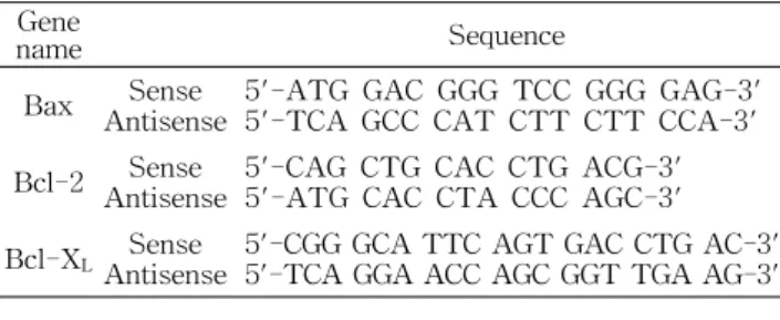
전사수준에서 apoptosis 관련 유전자들의 발현 정도를 비 교하기 위하여 동일한 조건에서 배양된 Hep3B 세포를 대상 으로 RNAzol B를 이용하여 total RNA를 분리하였다. 분리 된 RNA를 정량한 후, oligo dT primer와 AMV reverse transcriptase를 이용하여 2 μg의 RNA에서 cDNA를 합성하 였다. 이 cDNA를 template로 사용하여 관찰 대상 유전자를 polymerase chain reaction(PCR) 방법으로 증폭하였다 (Table 1). 이때 housekeeping 유전자인 glyceraldehyde- 3-phosphate dehydrogenase(GAPDH)를 internal control로 사용하였다. 각 PCR 산물들을 1% agarose gel을 이용하여 전기영동하고 ethidium bromide(EtBr, Sigma)로 염색한 후 UV 하에서 확인하였다(17).
Table 1. Gene-specific primers for RT-PCR Gene
name Sequence
Bax Sense
Antisense
5'-ATG GAC GGG TCC GGG GAG-3' 5'-TCA GCC CAT CTT CTT CCA-3' Bcl-2 Sense
Antisense
5'-CAG CTG CAC CTG ACG-3' 5'-ATG CAC CTA CCC AGC-3' Bcl-XL Sense
Antisense 5'-CGG GCA TTC AGT GAC CTG AC-3' 5'-TCA GGA ACC AGC GGT TGA AG-3'
SDS-polyacrylamide gel 전기영동 및 Western blot analysis
준비된 세포에 적당량의 lysis buffer[250 mM NaCl, 25 mM Tris-HCl(pH 7.5), 5 mM EDTA pH 8.0, 1% NP-40, 0.1 M phenymethylsulfonyl fluoride; PMSF, 1 M 1,4-di- thio-DL-threitol; DTT, protease inhibitor cocktail]를 첨가 하여 4oC에서 30분간 반응시킨 후, 13,500 rpm으로 30분간 원심분리하여 그 상층액을 취하였다. 상층액의 단백질 농도 는 Bio-Rad 단백질 정량 시약(Bio-Rad, Hercules, CA)의 사용법에 따라 동량으로 맞춘 다음 동량의 Laemmli sample buffer(β-melcaptomethanol 5%, Laemmli sample buffer 95%, Bio-Rad)를 섞어서 sample을 만들었다. 이렇게 만든 sample 동량을 sodium dodesyl sulfate(SDS)-polyacryl- amide gel 전기영동으로 분리하였다. 분리된 단백질을 함유 한 acrylamide gel을 nitrocellulose membrane(Schleicher and Schuell, Keene, NH, USA)으로 electroblotting에 의해 전이시킨 후, 10% skim milk를 함유한 PBS-T(0.1%
Tween 20 in PBS)에 담구어 상온에서 2시간 정도 in- cubation하여 비특이적인 단백질들에 대한 blocking을 실시 하고 PBS-T로 15분 정도 세척하였다. 세척 후 1차 항체를 처리하여 상온에서 1시간 이상 또는 4oC에서 over night시 킨 다음 PBS-T로 세척하고 처리된 1차 항체에 적절한 2차 항체를 사용하여 상온에서 1시간 정도 반응시켰다. 다시 PBS-T로 세척하고 enhanced chemiluminoesence(ECL) 용 액(Amersham Life Science Corp., Arlington Heights, IL) 을 적용시킨 다음 암실에서 X-ray film에 감광시켜 특정단 백질의 발현 양상을 비교 분석하였다. 본 실험에 사용된 항 체들은 Santa Cruz Biotechnology Inc.(Santa Cruz, CA)에 서 구입하였으며, 2차 항체로 사용된 peroxidase-labeled donkey anti-rabbit immunoglobulin 및 peroxidase-labeled sheep anti-mouse immunoglobulin은 Amersham Corp.에 서 구입하였다(17).
In vitro caspase 활성의 측정
Caspase의 활성 측정을 위한 colorimetric assay kits는 CLONTECH Lab.(Palo Alto, CA) 및 R&D Systems (Minneapolis, MN)에서 구입하였으며, 제시된 방법에 준하 여 활성의 증감 여부를 조사하였다. 이를 위하여 정상 및 AECM이 처리된 배지에서 48시간 배양된 세포를 모은 뒤
단백질을 추출하고 정량하여 각각 20 μg의 단백질을 fluoro- genic peptide 기질 100 μM이 함유된 extraction buffer[40 mM HEPES(pH 7.4), 20% glycerol(v/v), 1 mM EDTA, 0.2% NP-40 and 10 mM DL-dithiothreitol] 50 μL에 혼합하 였으며, microtiter plate에 다시 extraction buffer에 희석하 여 각 sample 당 총 volume이 100 μL가 되게 하였다. 실험에 사용된 기질은 caspase-3의 경우에는 Asp-Glu-Val-Asp (DEVD)-p-nitroaniline(pNA)이었으며, caspase-8은 Ile- Glu-Thr-Asp (IETD)-pNA이었고, caspase-9는 Leu-Glu- His-Asp(LEHD)-pNA였다. 준비된 plate를 37oC에서 2시 간동안 incubation 시킨 후 ELISA reader를 이용하여 405 nm의 흡광도를 이용하여 반응의 정도를 측정하였다(17).
통계처리
실험결과는 평균과 편차로 표시하였고 분산분석으로 분 석 후 유의성이 나타난 항목에서 유의성 0.05% 수준에서 Student’s t-test를 실시하여 대조군에 대한 유의성을 검정 하였다.
결과 및 고찰
AECM에 의한 Hep3B 세포의 증식억제 및 apoptosis 유발
Hep3B 간암세포에서 동충하초 열수추출물(AECM)에 의 한 항암기전의 해석을 위하여 Hep3B 세포의 증식억제 및 apoptosis 유발의 정도를 조사한 결과는 Fig. 1에 나타낸 바 와 같다. 먼저 Hep3B 세포의 증식에 미치는 AECM의 영향 을 알아보기 위하여 MTT assay를 실시한 결과, 48시간 동 안 정상 배지에서 자란 세포에 비하여 AECM이 함유된 배지 에서 배양된 Hep3B 세포의 경우 AECM의 첨가 농도 의존적 으로 세포의 증식이 감소하였음을 알 수 있었다(Fig. 1A).
즉 0.2 mg/mL의 AECM을 처리하였을 경우에는 약 22% 정 도의 증식억제가 나타났지만 처리 농도의 증가에 따라 증식 억제 정도가 증가하여 2.0 mg/mL의 농도에서는 약 70% 정 도의 증식억제 효과가 나타났다. 이러한 AECM 처리에 따른 Hep3B 세포의 증식억제 현상은 다양한 형태 변화와도 연관 성이 있었는데, 처리된 AECM의 농도가 증가할수록 암세포 의 밀도가 감소되었고, 전체적으로 세포질이 응축되었다. 또 한 세포의 모양이 길어지면서 분지를 형성하였으며, AECM 처리 농도의 증가에 따라 부착력을 상실하고 배지 위로 부유 되는 경향성이 증가되었다(Fig. 1B).
다음은 AECM 처리에 따른 Hep3B 세포의 증식억제 및 형태변형이 apoptosis 유발과 연관성이 있는지의 여부를 확 인하기 위하여 DAPI 염색을 이용한 apoptotic body의 형성 유무를 관찰하였다. Fig. 1C에 나타난 바와 같이 AECM을 처리하지 않았을 경우에는 핵의 모양이 정상적으로 염색된 반면에 처리된 AECM의 농도가 증가할수록 세포 밀도의 감
Relative cell viability (%)
AECM (mg/mL) 0
20 40 60 80 100 120
0 0.4 0.8 1.2 1.6 2
0 0.2 0.4
0.6 0.8 1.0
AECM (mg/mL)
0 0.2 0.4
0.6 0.8 1.0
AECM (mg/mL)
*
AECM (mg/mL)
0.6 0.8 1.0
0 0.2 0.4
AECM (mg/mL)
0.6 0.8 1.0
0 0.2 0.4
AECM (mg/mL)
DNA content
Cell Number
0 0.2 0.4 0.6 0.8 1.0 1.65% 4.53% 5.27% 7.74% 10.84% 41.12%
AECM (mg/mL)
DNA content
Cell Number
0 0.2 0.4 0.6 0.8 1.0 1.65% 4.53% 5.27% 7.74% 10.84% 41.12%
A)
B)
C)
D)
Fig. 1. Growth inhibition and apoptosis induction of human hepatocellular carcinoma Hep3B cells after treatment with aqueous extract of C. militaris (AECM). (A) Hep3B cells were seeded and incubated for 24 hr. The cells were treated with variable concen- trations of AECM for 48 hr. The growth inhibition was measured by the metabolic-dye-based MTT assay. Results represent the mean±SD of triplicate determinations. The significance was determined by a Student’s t-test (*p<0.05 vs. untreated control). (B) After 48 hr incubation with various concentrations of AECM, cell morphology was visualized by a inverted microscopy. Magnification, ×200.
(C) The cells grown under the same conditions as (B) were stained with DAPI for 10 min and photographed with a fluorescent microscope using a blue filter. Magnification, ×400. (D) The cells were fixed and stained with CycleTEST PLUS DNA REAGENT kit, and analyzed by a flow cytometer. The data shown are means of two independent experiments.
소와 더불어 염색질 응축 현상이 증가되어 AECM 처리에 따른 Hep3B 세포의 증식억제는 apoptosis 유도와 밀접한 관련이 있음을 알 수 있었다. 따라서 AECM에 의해 유발된 apoptosis 정도를 정량적으로 분석하기 위하여 apoptosis 유 발 세포군에 해당하는 세포주기 sub-G1기에 속하는 세포들 의 빈도를 flow cytometry를 이용하여 측정한 결과, AECM 을 처리하지 않은 세포의 경우는 sub-G1기에 해당하는 세 포가 약 1.65%에 불과했지만 0.8 mg/mL 및 1.0 mg/mL 처리 군에서는 각각 10.84% 및 41.12%로 현저하게 증가하였음을 확인할 수 있었지만, 세포주기 특이적 arrest 현상은 관찰할 수가 없었다(Fig. 1D).
이상의 결과를 동일 조건에서 수행된 A549 폐암세포의 결과와 비교해 볼 때, A549 암세포의 50% 정도 증식억제 효능을 나타낸 동충하초 열수추출물의 농도가 약 2.0 mg/
mL 정도였으며, 5.0 mg/mL 처리군에서 약 80% 정도의 증 식억제 효과를 나타낸 것에 비하여(16) Hep3B 세포에서는 저농도에서 매우 강력한 증식억제 효과를 보였을 알 수 있 었다. 그리고 혈구암세포에서보다는 감수성이 다소 낮았지 만(17,18), 전반적으로 AECM 처리에 따른 암세포의 증식 억제는 apoptosis 유도와 밀접한 연관성을 지님을 잘 알 수 있었다.
Bcl-2 family 및 caspase의 활성에 미치는 AECM의 영향 Fig. 1의 결과에서 확인된 AECM 처리에 따른 Hep3B 간 암세포의 apoptosis 유발에 관여하는 기전의 해석을 위하여 Bcl-2 family에 속하는 몇 가지 유전자들의 발현 변화를 RT-PRC 및 Western blotting으로 조사하였으며, 아울러 caspases의 발현 및 그들의 활성에 미치는 AECM의 영향을 Western blotting 및 in vitro colorimetric assay로 비교하였다.
Bcl-2 family에 속하는 몇 가지 중요한 인자들은 apopto- sis 유발 조절에 가장 대표적인 유전자로 알려져 있는데, 그 중 Bcl-2나 Bcl-xL은 anti-apoptotic 분자로서 apoptosis의 유발을 억제하는 기능을 가지며, Bax나 Bad 등은 pro- apoptotic 분자로 그들의 발현 증가는 apoptosis의 유발과 관계가 있다(19-21). 이들 두 유전자는 세포 내 소기관 중 mitochondria로부터의 cytochrome c를 유리시켜 cysteine- related protease인 caspase 및 DNA의 단편화와 연관된 en- donuclease 등의 활성을 조절하며(22,23), 서로 dimer의 형 태로 존재하지만 그들의 발현 수준에 변화가 초래되면 apoptosis가 유발되는 것으로 알려져 있다(19-21). 그러나 Fig. 2에 나타낸 결과에서 알 수 있듯이 본 실험에서 조사된 몇 가지 Bcl-2 family에 속하는 유전자들(Bax, Bad, Bcl-2 및 Bcl-xL)의 전사 및 총 단백질 수준에서의 발현은 AECM
AECM (mg/mL) A)
B)
Bax Bcl-2 Bcl-xL GAPDH 0 0.2 0.4 0.6 0.8 1.0
AECM (mg/mL)
Bax Bad Bcl-2
Bcl-xL actin 0 0.2 0.4 0.6 0.8 1.0
A)
B)
Fig. 2. Effects of AECM treatment on the levels of Bcl-2 family members in human Hep3B cells. (A) After 48 hr in- cubation with AECM, total RNAs were isolated and reverse- transcribed. The resulting cDNAs were subjected to PCR with the Bax, Bcl-2 and Bcl-xL primers and the reaction products were subjected to electrophoresis in 1% agarose gel and vi- sualized by EtBr staining. GAPDH was used as an internal control. (B) The cells were lysed and then cellular proteins were separated by SDS-polyacrylamide gels and transferred onto ni- trocellulose membranes. The membranes were probed with the anti-Bax, anti-Bcl-2 and anti-Bcl-xL antibodies. Proteins were visualized using an ECL detection system. Actin was used as an internal control.
처리에 따른 큰 변화를 보여주지 않았다.
한편 대표적인 apoptosis 유발 protease인 caspase family 에 속하는 단백질들은 세포에서 핵과 mitochondria의 외막 에 불활성 상태인 proenzyme 형태로 존재하다가 Bcl-2 및 IAPs family 인자들의 발현 변화와 연관된 apoptosis 유도를 활성화시키는 신호에 의해 활성화된 protease로 전환되어 직접 또는 간접적으로 세포 내에 존재하는 많은 표적 단백질 의 분해에 관여하는 것으로 알려져 있으며(24-26), caspase 의 활성화 자체가 apoptosis의 유발에 대한 또 다른 증거가 될 수 있다(27). 따라서 지금까지 알려진 caspase 중 대표적 인 initiator caspase에 해당되는 caspase-8 및 -9와 effector caspase에 해당되는 caspase-3의 발현 및 활성에 미치는 AECM의 영향을 조사한 결과는 Fig. 3에 나타낸 바와 같다.
결과에서 알 수 있듯이 AECM이 처리된 배지에서 배양된 Hep3B 세포에서는 처리 농도 의존적으로 caspase-3의 활성 이 증가되어 1.0 mg/mL 처리군에서는 대조군에 비하여 약 3배 정도 증가되었다. Caspase-8의 경우도 AECM 처리 농 도의 증가에 따라 활성의 정도가 증가되었지만 최대 농도에
Caspase-3 Caspase-8 Caspase-9 Caspase-3 Caspase-8 Caspase-9
0 0.1 0.2 0.3 0.4 0.5
0 0.2 0.4 0.6 0.8 1.0
AECM (mg/mL)
Caspase activity
B) AECM (mg/mL)
actin 0 0.2 0.4 0.6 0.8 1.0
PARP
b-catenin B)
A)
β-catenin
Fig. 3. Activation of caspases, and degradation of PARP and β-catenin proteins by AECM treatment in human Hep3B cells. (A) Hep3B cells were incubated with AECM for 48 hr and lysed. Aliquots (150 μg proteins) were incubated with DEVD- pNA, IETD-pNA and LEHD-pNA for caspase-3, -8 and -9 ac- tivity, respectively, at 37oC for 3 hr. The released fluorescent products were measured. Data represent the mean of two in- dependent experiments. (B) The cells grown under the same con- ditions as (A) were collected and then lysed. The cellular proteins were separated by SDS-polyacrylamide gels and transferred onto nitrocellulose membranes. The membranes were probed with an- ti-PARP and anti-β-catenin antibodies. Proteins were visualized using ECL detection system. Actin was used as an internal control.
서 1.6배 정도였으며, caspase-9의 경우는 큰 변화를 보여주 지 않았다.
Caspase의 활성화에 동반된 apoptosis가 유발되었을 경 우 특이하게 분해가 일어나는 몇 가지 표적 단백질 중 poly (ADP-ribose) polymerase(PARP) 단백질은 DNA repair나 genomic stability의 유지에 매우 중요한 역할을 하는 것으 로 알려져 있으며, 활성화된 caspase-3에 의하여 단백질의 분해가 일어나면 PARP 기능이 상실되며, 정상적인 세포의 경우 PARP 단백질은 116 kDa의 분자량을 가지지만 apop- tosis가 일어난 경우 85 kDa 크기의 단편이 관찰되거나 주 band의 발현이 감소된다(24,25). 그리고 caspase-3의 또 다 른 표적 단백질 중 하나인 β-catenin은 cell-cell adhesion 및 Wnt signaling에 관여하는 E-cadherin-associated pro- tein으로서 세포 내 골격 유지와 다양한 부착성 세포의 전사 조절에 중요한 역할을 하며 세포 유착과 관계된 apoptosis 조절에 관여한다(28,29). β-catenin은 정상 세포의 경우 92 kDa의 분자량을 가지나 apoptosis가 유발되면 62~72 kDa 의 크기로 단편화가 일어나는 것으로 알려져 있다(30,31).
따라서 AECM의 처리에 의한 Hep3B 세포의 apoptosis에 caspase-3의 활성화가 직접 관여하는지의 여부를 조사하기
Caspase-3 activity 0 0.1 0.2 0.3 0.4 0.5 0.6
% of sub-G1 cells
0 5 10 15 20 25 30 35 40 45 50
- -
+ -
- +
+ + -
- + -
- +
+ +
- -
+ -
- +
+ + -
- + -
- +
+ +
AECM (1.0 mg/mL) z-DEVD-fmk -
-
+ -
- +
+ +
AECM (1.0 mg/mL) z-DEVD-fmk A)
B)
C)
AECM (1.0 mg/mL) z-DEVD-fmk
*
* A)
B)
C)
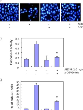
Fig. 4. Inhibition of caspase-3 activity alleviates AECM-in- duced apoptosis in human Hep3B cells. (A) Hep3B cells were pretreated for 1 hr with or without z-DEVD-fmk (50 μM), and then with AECM (1.0 mg/mL) for additional 48 hr. The cells were stained with DAPI for 10 min and photographed with a fluo- rescent microscope using a blue filter. Magnification, ×400 (A).
Caspase-3 activity (B) and DNA contents (C) were determined using MTT assay and a flow cytometer after 48 hr in the presence of the caspase-3 inhibitor z-DEVD-fmk (50 μM) for 1 hr before WECM (1.0 mg/mL) treatment. Data are expressed as mean±SD of three independent experiments. The significance was de- termined by a Student’s t-test (*p<0.05 vs. untreated control).
위하여 PARP 및 β-catenin의 발현에 미치는 AECM의 영향 을 조사한 결과는 Fig. 3A에 나타낸 바와 같다. 제시된 결과에 서 알 수 있듯이 AECM의 처리 농도 증가에 따라 PARP 및 β-catenin 단백질의 단편화 정도가 점차 증가함을 알 수 있었 으며, 이는 AECM 처리에 따른 apoptosis 유도에 caspase-3 이 매우 중요한 역할을 하고 있음을 보여주는 결과이다.
Caspase-3 활성 저해가 AECM에 의한 apoptosis에 미 치는 영향
Fig. 3의 결과에서 AECM에 의한 Hep3B 세포의 apopto- sis 유도에 caspase-3이 매우 중요한 역할을 할 것으로 나타 났기 때문에 AECM 처리에 의한 apoptosis 유도에서 cas- pase-3의 역할을 재확인하기 위하여 caspase-3의 활성을 인위적으로 억제할 경우 AECM에 의해 유발되는 apoptosis 에 어떠한 변화가 나타나는지의 여부를 조사하였다. 먼저 DAPI 염색을 이용하여 핵의 형태를 관찰한 결과는 Fig. 1의 C에서 관찰된 바와 같이, 정상배지에서 자란 Hep3B 세포의 경우는 핵의 형태가 정상으로 염색된 반면에 AECM이 처리
된 배지에서 자란 Hep3B 세포의 경우는 전형적인 apoptotic body가 관찰되었다(Fig. 4A). 그러나 AECM을 처리하기 전 에 caspase-3 선택적 저해제인 z-DEVD-fmk를 1시간 선처 리하였을 경우 정상배지에서 자란 Hep3B 세포와 마찬가지 로 apoptotic body가 관찰되지 않았다. 그리고 AECM에 의 하여 유도되는 caspase-3의 활성에 미치는 caspase-3 저해 제의 영향을 조사한 결과, AECM을 단독으로 처리했을 경우 에는 약 5배 정도의 활성 증가가 관찰되었지만 AECM 처리 전 z-DEVD-fmk를 1시간 선처리하였을 경우에는 거의 대 조군 혹은 z-DEVD-fmk 단독 처리군 수준으로 caspase-3 의 활성이 감소되었다(Fig. 4B). 그리고 AECM을 단독으로 처리했을 경우에는 약 45% 정도의 sub-G1 빈도가 관찰되었 지만 z-DEVD-fmk 선처리에 의하여 sub-G1에 속하는 세 포의 빈도는 현저히 감소하여 약 15% 정도로 나타났다(Fig.
4C). 이상의 결과는 Hep3B 간암세포에서 AECM에 의한 apoptosis의 유도에 caspase-3이 결정적인 역할을 하고 있 음을 의미하는 결과이다.
요 약
본 연구에서는 동충하초(C. militaris)의 항암작용 기전 해석을 위하여 Hep3B 간암세포의 apoptosis 유발에 미치는 동충하초 열수추출물(WECM)의 영향을 조사하였으며, apoptosis 조절에 중요한 몇 가지 유전자들의 발현 및 활성 변화를 조사하였다. AECM 처리에 의한 Hep3B 세포의 증식 억제는 형태적 변형을 동반한 apoptosis 유도와 연관성이 있음을 DAPI 염색을 통한 apoptotic body 출현의 증가 및 flow cytometry 분석에 의한 sub-G1 기에 속하는 세포 빈도 의 증가로 확인하였다. AECM 처리에 의한 apopotosis 유도 에서 Bcl-2 family에 속하는 몇 가지 유전자들의 발현은 큰 변화가 없었으나, caspase-3 및 -8의 활성이 매우 높게 증가 되었으며 이는 PARP 및 β-catenin 단백질의 분해와 연관성 이 있었다. 또한 caspase-3 선택적 저해제인 z-DEVD-fmk 로 caspase-3의 활성을 인위적으로 차단시켰을 경우, AECM 에 의한 apoptois 유도 현상이 유의적으로 감소되어 AECM 에 의한 Hep3B 세포의 apoptosis 유발에 caspase-3이 중요 한 역할을 하고 있음을 알 수 있었다. 본 연구의 결과만으로 동충하초에 의한 간암세포의 증식억제 기전을 명확하게 제 시할 수는 없으나, 이상의 결과들은 동충하초의 생화학적 항암기전 해석을 이해하는데 중요한 기초자료로서 활용될 수 있을 것으로 생각된다.
문 헌
1. Lieberthal W, Koh JS, Levine JS. 1998. Necrosis and apop- tosis in acute renal failure. Semin Nephrol 18: 505-518.
2. Zimmermann KC, Bonzon C, Green DR. 2001. The machi- nery of programmed cell death. Pharmacol Ther 92: 57-70.
3. Evans VG. 1993. Mutiple pathways to apoptosis. Cell Biol Int 17: 461-476.
4. Nagata S. 1997. Apopotosis by death factor. Cell 88: 355- 365.
5. Sung JM, Lee HK, Choi YS, Kim YO, Kim SH, Sung GH.
1997. Distribution and taxonomy of entomopathogenic fun- gal species from Korea. Kor J Mycol 25: 231-252.
6. Buenz EJ, Bauer BA, Osmundson TW, Motley TJ. 2005.
The traditional Chinese medicine Cordyceps sinensis and its effects on apoptotic homeostasis. J Ethnopharmacol 96:
19-29.
7. Chen JL, Greider CW. 2004. Telomerase RNA structure and function: implications for dyskeratosis congenita. Trends Biochem Sci 29: 183-192.
8. Buenz EJ, Weaver JG, Bauer BA, Chalpin SD, Badley AD.
2004. Cordyceps sinensis extracts do not prevent Fas-re- ceptor and hydrogen peroxide-induced T-cell apoptosis. J Ethnopharmacol 90: 57-62.
9. Nakamura K, Yamaguchi Y, Kagota S, Kwon YM, Shinozuka K, Kunitomo M. 1999. Inhibitory effect of Cordyceps sinensis on spontaneous liver metastasis of Lewis lung carcinoma and B16 melanoma cells in syngeneic mice. Jpn J Pharmacol 79: 335-341.
10. Shahed AR, Kim SI, Shoskes DA. 2001. Down-regulation of apoptotic and inflammatory genes by Cordyceps sinensis extract in rat kidney following ischemia/reperfusion.
Transplant Proc 33: 2986-2987.
11. Kim MN, Cui CB, Lee DS, Ham SS. 2001. Cytotoxicity and antigenotoxic effect of Cordycepes militaris extracts. J Korean Soc Food Sci Nutr 30: 921-927.
12. Kim MN, Oh SW, Lee DS, Ham SS. 2001. Antioxidative and antimutagenic effects of the ethanol extract from Cordycepes militaris. Kor J Posthavest Sci Technol 8:
109-117.
13. Yoo HS, Shin JW, Cho JH, Son CG, Lee YW, Park SY, Cho CK. 2004. Effects of Cordyceps militaris extract on angiogenesis and tumor growth. Acta Pharmacol Sin 25:
657-665.
14. Lee H, Lee Y, Park T. 2004. Tumor growth inhibitory and immunomodulatory activities of Cordycepes militaris wa- ter extracts in ICR mice bearing Sarcoma-180 solid tumor.
J Korean Soc Food Sci Nutr 33: 59-65.
15. Hong SH, Kam CW, Park DI. 2004. Induction of apoptotic cell death by an aqueous extract of Cordyceps militaris in A549 human lung carcinoma cells. Kor J Oriental Physiol Pathol 18: 1102-1106.
16. Cho WN, Choi YH, Hwang WD. 2007. Induction of G2/M arrest of the cell cycle by aqueous extract of Cordyceps militaris in A549 human non-small-cell lung cancer cells.
Cancer Prev Res 12: 273-280.
17. Park C, Hong SH, Lee JY, Kim GY, Choi BT, Lee YT, Park
DI, Park YM, Jeong YG, Choi YH. 2005. Growth inhibition of U937 leukemia cells by aqueous extract of Cordyceps militaris through induction of apoptosis. Oncol Rep 13:
1211-1216.
18. Hong SH, Park DI, Seo SH, Choi YH. 2005. Anti-pro- liferative effects by aqueous extract of Cordyceps militaris in human leukemic U937 cells. Kor J Oriental Physiol Pathol 19: 452-458.
19. Antonsson B, Martinou JC. 2000. The Bcl-2 protein family.
Exp Cell Res 256: 50-57.
20. Osford SM, Dallman CL, Johnson PW, Ganesan A, Packham G. 2004. Current strategies to target the an- ti-apoptotic Bcl-2 protein in cancer cells. Curr Med Chem 11: 1031-1039.
21. Donovan M, Cotter TG. 2004. Control of mitochondrial in- tegrity by Bcl-2 family members and caspase-independent cell death. Biochim Biophys Acta 1644: 133-147.
22. Fan TJ, Han LH, Cong RS, Liang J. 2005. Caspase family proteases and apoptosis. Acta Biochim Biophys Sin 37:
719-727.
23. Rupinder SK, Gurpreet AK, Manjeet S. 2007. Cell suicide and caspases. Vascul Pharmacol 46: 383-393.
24. Kaufmann SH, Desnoyers S, Ottaviano Y, Davidson NE, Poirier GG. 1993. Specific proteolytic cleavage of poly (ADP-ribose) polymerase: an early marker of chemo- therapy-induced apoptosis. Cancer Res 53: 3976-3985.
25. Lazebnik YA, Kaufmann SH, Desnoyers S, Poirier GG, Earnshaw WC. 1994. Cleavage of poly(ADP-ribose) poly- merase by a proteinase with properties like ICE. Nature 371: 346-347.
26. Rao L, White E. 1997. Bcl-2 and the ICE family of apoptotic regulators: making a connection. Curr Opin Genet Dev 7:
52-58.
27. Vegran F, Boidot R, Oudin C, Riedinger JM, Lizard-Nacol S. 2005. Implication of alternative splice transcripts of cas- pase-3 and survivin in chemoresistance. Bull Cancer 92:
219-226.
28. Johnson JP. 1999. Cell adhesion molecules in the develop- ment and progression of malignant melanoma. Cancer Metastasis Rev 18: 345-357.
29. Wijnhoven BP, Dinjens WN, Pignatelli M. 2000. E-cadher- in-catenin cell-cell adhesion complex and human cancer.
Br J Surg 87: 992-1005.
30. Fukuda K. 1999. Apoptosis-associated cleavage of β-cat- enin in human colon cancer and rat hepatoma cells. Int J Biochem Cell Biol 31: 519-529.
31. Neufeld KL, Zhang F, Cullen BR, White RL. 2000. APC- mediated downregulation of β-catenin activity involves nuclear sequestration and nuclear export. EMBO Rep 1:
519-523.
(2008년 4월 21일 접수; 2008년 5월 29일 채택)