47(3) : 197∼ 203 (2016)
197
당귀로부터 정제한 Decursin의 인간 급성 단핵구성
백혈병 세포(THP-1 cells)의 세포 독성 및 Apoptosis에 미치는 영향
김남석1·정승일2*·김종석3·오미진4·오찬호1*
1
우석대학교 식품생명공학과,2
(재)전주농생명소재연구원,3
우석대학교 재활학과,4
한국식품연구원Decursin from Angelica gigas Nakai Promotes Cytotoxicity and Induces Apoptosis in THP-1 cells, a Human Acute Monocytic Leukemia
Nam-Seok Kim1, Seung-Il Jeong2*, Jong-Seok Kim3,Mi-Jin Oh4 and Chan-Ho Oh1*
1
Department of Food & Biotechnology, Woosuk University, Wanju-gun, 55338, Korea2
Jeonju Agrobio-Materials Institute (JAMI), Jeonju, 54810, Korea3
Department of Rehabilitation studies, Woosuk university, Wanju, 55338, Korea4
Korea Food Research Institute, Seongnam, 13539, KoreaAbstract − Decursin is a major component of the root of Angelica gigas(Umbelliferae), which has been traditionally used in Korea as a tonic and to treat anemia, hemiplegia, and women’s diseases. The objective of this study is to identify the anti-cancer mechanism induced by decursin on apoptosis of human leukemia and lymphoma cells. Cytotoxicity of decursin on U937, HL- 60, MOLT-4, THP-1 cells showed the significant effects. First of all, IC
50
of decursin on four cell lines was 27.1, 32.4, 17.4, 15.1µM, respectively. So IC50
in THP-1 cells was the smallest among 4 cell lines treated with decursin(15.1µM). In order to understand the apoptosis-mechanism by decursin, we examined the gene expression of bcl-2(anti-apoptotic), bax(pro-apop- totic)and p53(tumor suppressor)after treating the THP-1 cells with decursin(10, 50 and 100 µM). It was found bcl-2 gene was decreased dose dependently, the expression level of bax gene of THP-1 cells treated with 100 µM of decursin was about 3 times higher than those of control, and p53 gene was increased In the same concentration(100µM), p53 gene was increased dose dependent manner. In protein express, bcl-2 and p53 protein showed a tendency to decrease. bax was increased about 4 fold.Therefore decursin is a useful chemotherapeutic agent against leukemia.
Key words − Decursin, Apoptosis, Bcl family, p53, THP-1 cells
백혈병은 미성숙 백혈구의 비정상적인 증식으로 인한 암 으로 4가지 형태로 구분이 된다. 이는 급성 림프아구성 백 혈병(Acute lymphoblastic leukemia; ALL), 만성 림프구성 백혈병(Chronic lymphocytic leukemia; CLL), 급성 골수성 백혈병(Acute myelogenous leukemia; AML) 및 만성 골수 성 백혈병(Chronic myelogenous leukemia; CML)로서 급성 백혈병은 림프아구 나 골수아구 같은 미성숙 혈액세포의 비 정상 적인 증식이 이루어져있으며, 즉시 치료가 필요하다.
반면, 만성백혈병 환자는 즉시 치료를 요하지는 않는다. 2012 년 국내에서는 어린이(0~14세)의 암 중에서도 백혈병 발병 률이 가장 높다(10만명 당 4.1명).
1)
당귀(Angelica gigas Nakai)는 한국, 중국, 일본에서 서식 하는 다년생 식물로, 중국에서는 전통적으로 당귀의 뿌리를 약재로 사용한다. 꽃이 피기 전 당귀의 뿌리를 건조시켜 사 용한 것이 조혈작용에 효과가 있는 것으로 보고 되었다.
2)
몇 가지 암 세포주(HepG2, HeLa, MCF7, SW626)에서 당 귀 유기용매의 추출물은 용매에 따라 다른 효과를 보이지 만 일반적으로 강한 효능을 보였다.3)
2007년에 당귀 뿌리 에서 여러 가지 성분(pyranocoumarin 화합물인 “decursin”,“decursinol angelate”, furanocoumarin 화합물인 “nodakenin”,
“isoimperatorin”, “marmesin”, 및 “xanthoarnol”)가 분리되었 다.
4-6)
Decursin은 당귀뿌리의 주성분으로 국내에서 고대시대부 터 내려오는 전통적인 약재로 여성질환 과 빈혈 치료제로 사용되었다. 당귀에서 분리한 decursin은 해마에서 기억력과
*교신저자(E-mail):[email protected], [email protected] (Tel): +82-63-290-1431, +82-63-711-1050
관련된 아세틸콜린(Acetylcholine)을 분해시키는 acetylcho- linesterase (AChE)의 억제로 인한 기억력 개선효과가 있으 며,
7,8)
염증의 억제, 항전이효과, 인간 전립선 암세포에서 세 포자연사 유도, 인간 신장 표피세포의 세포자연사 억제, 산 화 스트레스의 예방효능이 있다.9-12)
여러 가지 세포주에 대한 항산화, 항균, 혈소판 응집억제, 세포 독성, 항전이효과, 기억력 개선에 효능이 있다.
10,13-19)
최근 세포독성 분야에서는 decursin의 B16F10 melanoma 세 포20)
와 PC12 부신 갈색세포종 세포21)
에 대한 연구가 보고 되어있으며, 백혈병 세포주에 대한 연구는 Kim HH등 (2005)22,23)
은 K562, U937 세포에 세포증식과 분화에 관여 하는 PKC에 대한 연구가 진행되었고, 인간 KBM-5 백혈병 세포에 처리하여 caspase family의 활성화, PARP 분할유도, COX-2가와 survivin의 억제를 시켜 apoptosis를 유도하는 작 용을 하였다.24)
이와같이 여러 효능을 보이는 당귀 decursin의 연구에서 백혈병세포에 대한 연구가 진행되었지만 세포의 미토콘드 리아에 의한 내재성 apoptosis pathway에 작용하는 단백질 로 세포자연사를 일으키기도 하고 억제 시키는 역할을 하 는 단백질 종류를 모두 갖고 있는 Bcl family에 대한 연구 거의 진행되지 않아서 본 연구에서는 decursin의 백혈병과 임파종에 미치는 세포독성 중 Bcl family에 대한 효능에 초 점을 맞추어 연구를 진행하였다.
재료 및 방법
실험재료 − 본 실험에 사용된 당귀(Angelica gigas Nakai) 은 2015년 3월에 광명당(울산)에서 규격품을 구입하였다. 우 석대학교 한의과대학 본초방제학교실 김홍준 교수님으로부 터 감정을 받았으며, 표본은 우석대학교 식품생명공학과 연 구실에 보관 중이다.
세포주 및 재료 − 본 실험에 사용한 세포주로서는 한국세 포주은행(KCLB, Korea)에서 분양받은U937(인간 조직구성 림프종), HL60(인간 급성 전골수성 백혈병), MOLT4(인간 급성 림프아구성 백혈병) 및 THP-1(인간 급성 단구성 백혈 병) 세포를 사용하였으며, RPMI 1640, fetal bovine serum (FBS), phosphate buffered saline(PBS), antibiotics는 Gibco BRL(Gaithersburg, MD). 3-(4,5- dimethylthiazol-2-yl) 2,5- diphenyltetrazolium bromide(MTT)와 Nonidet P-40(NP-40)
는 Amresco 사, DMSO, agarose와 기타 시약은 Sigma- Aldrich 사, 웨스턴 블로팅에 사용한 단클론항체(Bcl-2, Bax, p53와 β-actin 항체)는 Cell Signaling(MA, USA)와 Sigma- Aldrich에서 구입하였다. 그리고 RT-PCR에 사용한 Trizol reagent와 polymerase chain reaction(PCR) kit는 각 Invitrogen (USA)과 Takara(Japan) 사에서 구입하여 사용하였다.
추출 및 분획 − 시료 뿌리(600 g)를 잘게 분쇄하고 2 L의 MeOH로 60
o
C에서 3회 4시간씩 추출한 후 감압농축하여 87.4 g의 추출물을 얻었다. 이 추출물을 증류수와 MeOH 혼 합액(9:1)에 현탁시킨 후 동량의 CHCl3
, EtOAc 및 n-BuOH 를 이용, 순차적으로 분획하여 각각 CHCl3
(9.7 g), EtOAc (2.7 g) 및 n-BuOH(7.8 g)분획물을 얻었다. CHCl3
분획물 일 부(5.80 g)를 용출용매 n-hexane : EtOAc = 97:3 → 60:40 로 silica-gel(230-400 mesh, 100 g) column(Ø=5.0×100 cm) chromatography를 실시하여 총12종의 분획(F01-12)을 얻었 다. 그 중 활성을 측정한 뒤 효능이 우수한 분획 Fr. 4(237 mg)를 prep-HPLC(용매, MeOH, 254 nm) 화합물 1을 얻었 다. 화합물 1은 백색 분말로서1
H-NMR 및13
C-NMR spectrum 등 각종 분광학적 data를 문헌20)
과 비교하여 본 결 과 decursin로 확인되었다.Cell viability 측정 − 계대배양중인 U937, HL60, MOLT4, 및 THP-1 세포를 96 well plate에 5×10
4
cells/well이 되도록 세포수를 조정한 다음, 1% DMSO에 녹인 decursin 시료(최 종농도-10, 50, 100 μM)를 첨가하여 24시간 동안 37o
C의 5% CO2
배양기 내에서 배양하였다. 배양종료 4시간 전에 5 mg/mL 농도로 PBS(pH 7.4)에 희석된 MTT용액 20 μL 를 각 well에 처리하고, 0.1 N HCl에 녹인 10% SDS 100 μL 로 용해시켜 18시간동안 은박지로 빛을 차단하였다. 발색된 각 well의 흡광도를 ELISA reader를 이용해서 570 nm에서 측정하고 대조군의 흡광도와 비교하여 세포생존율을 백분 율로 환산하였다. 각 세포의 IC50
값은 sigmaplot software를 사용하여 분석하였다.25)
RNA 분리 및 유전자 증폭(RT-PCR) − THP-1 세포 (3×10
6
cells/well)에 1% DMSO에 녹인 10, 50, and 100 μM 의 Decursin 시료를 첨가한 후, 24시간 동안 배양하고 RNA 를 분리하였다. Total RNA 분리는 TRIzol reagent를 이용하 였으며 제조회사의 방법에 준하였다.26)
cDNA는 Prime Script II 1st strand cDNA Synthesis kit와 oligo dT primer 를 이용하여 합성하였고, EmeraldAmpGT PCR Master Mix(Takara bio.)를 이용하여 PCR 기기(Dice, Takara bio.) 에서 30 cycles동안 증폭하였다. 각각의 cycle은 94o
C에서 30초간 denaturation시킨 후, 58o
C에서 30초간 annealing시 키고, 72o
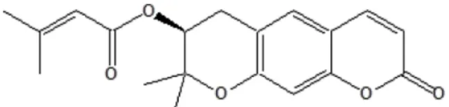
C에서 30초간 extension시켰다. PCR product는 1% agarose gel에서 전기영동하고 LoadingSTAR(DYNE Bio Co.)로 염색하여 UV transilluminator(OPTIMA, JAPAN) 로 관찰하였다. PCR에 사용된 primer는 Table I과 같다.Fig. 1. Chemical structure of decursin.
단백질 분리 및 웨스턴 블로팅 − 계대배양중인 THP-1 세 포(5×10
6
cells/well)에 10, 50, 및 100 μM의 Decursin 시료 를 첨가한 후, 24시간 동안 배양하고 1% NP-40 lysis buffer (+PMSF, pepstatin A, 및 aprotinin protease inhibitor)를 사 용하여 단백질을 추출하였다. 추출한 단백질을 Bradford 법 을 이용해 정량한 후 well 당 10 μg씩 loading한 후 7.5%SDS-polyacrylamide gel을 통해 SDS-PAGE를 시행하였다.
전기영동 후 PVDF membrane(0.2 μm, Milipore)에 transfer (350 mA, 1 hour 30 min)한 다음 1시간 동안 5% skim milk 로 blocking하였다. 그 후 각 1차 항체를 overnight 반응 (1:1000, 4
o
C)하고 TBS-T(0.05% Tween 20)으로 4회 세척 하고 goat anti-rabbit IgG-HRP conjugated antibody(1:5000) 을 1시간 동안 반응시켰다. 다시 TBS-T로 4회 세척 후 PVDF membrane을 ECL법을 이용해 암실에서 X-ray film Table I. Sequence of primer in PCR reactionTemplate Primers PCR product(bp)
GAPDH Forward 5'-GGAGCCAAAAGGGTCATCAT-3' 203
Reverse 5'-GTGATGGCATGGACTGTGGT-3'
Bax Forward 5'-GAGTGTCTCAAGCGCATCGG-3' 297
Reverse 5'-CACCCAACCACCCTGGTCTT-3'
Bcl-2 Forward 5'-ACGAGTGGGATGCGGGAGAT-3' 447
Reverse 5'-TCCACAGGGCGATGTTGTCC-3'
p53 Forward 5'-CGCTGCTCAGATAGCGATGG-3' 410
Reverse 5'-TGGGGAGAGGAGCTGGTGTT-3'
Fig. 2. Inhibitory effect of decursin on the cell proliferation in human lymphoma cells and leukemia cells(U937, HL60, MOLT4, THP-1) in vitro. Decursin were added into several cells. After incubated for 24 hrs, the viability assayed by MTT assay. *; Sig- nificant difference between values for decursin in compared with each control group(*p<0.05, **p<0.01, ***p<0.001).
에 감광시켰다.
통계처리 − 통계처리는 student’s t-test로 하였으며, p<0.05 이하를 유의성이 있는 것으로 판정하였다.
27)
결과 및 고찰
세포생존율(MTT Assay) − 계대배양 한 U937, HL60, MOLT4, THP-1 세포주에 decursin을 첨가해서 세포 생존율 을 측정한 결과, 4가지 세포 모두 농도의존적으로 세포독성 의 효과가 보였으며, 모든 세포에서 IC
50
값이 40 μM 이하 로 나타났다. 그 중에서도, THP-1 세포주에서 세포 독성이 가장 유의적인 효능(IC50
=15.1μM)을 보였다(Fig. 2). 따라 서, decursin를 처리한 경우 1가지 림프종과 3가지 백혈병세 포에서 모두 유의적으로 생존율을 억제하는 효과가 있는 것 으로 나타났다. Park 등(2007)은 당귀에서 decursin을 정제 하여 대장암, 유방암, 간암, 신장암세포에 처리하였을 때 다 른 추출물이나 분획물에 비해 성장 억제 효능을 보였다.14)
Kim 등(2015)은 50 μM의 decursin과 유도체를 U937, TUR 세포에 48시간 동안 처리한 후 trypan blue 염색을 통한 세 포독성을 확인하였으나, 본 결과에서 24시간 동안 처리한 결과 U937세포를 포함한 4가지 백혈병 세포주에서 동일 농 도에서(50 μM) 효능이 더 우수하게 나타났다.23)
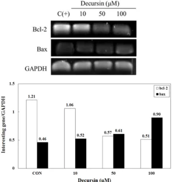
THP-1세포의 세포자연사 관련 유전자(p53, bax, bcl-2 유전자) 발현양상 − THP-1 세포주에 decursin(10, 50, 100 μM)에 대한 apoptosis 관련인자(bcl-2, bax), 종양 억제유전
자(p53)의 발현양상을 RT-PCR을 통해 확인한 결과, decursin 을 첨가한 모든 군에서 처리하지 않은 대조군에 비해 bax 와 p53 mRNA 유전자가 발현이 증가하였고, 특히 decursin 100μM 처리군에서 bax 유전자가 대조군에 비해 유의적으 로 증가하였다. 그리고 bcl-2 유전자(anti-apoptotic gene)는 농도 의존적으로 감소하는 경향을 보였다(Fig. 3). 종양억제 유전자인 p53
28)
은 decursin의 농도의존적으로 발현이 증가 하는 양상을 보였으며 특히 100 μM 처리군에서 대조군보 다 2배 이상 증가하였다(Fig. 4). 본 실험에 사용한 4가지 세 포주 중 세포생존율이 가장 유의적으로 억제되는 THP-1 세 포주에서 세포자연사와 종양억제에 관련 있는 3가지 유전 자(bcl-2, bax, p53)가 모두 대조군에 비해 bcl-2, p53 유전 자는 증가, bax 유전자는 감소되어 세포자연사가 진행되고 있다고 추측된다. Decursin은 2000년대 초반부터 많은 연구 가 진행되어 왔으며 종양을 억제시키는 PKC activator, 종 양세포의 apoptosis G1 arrest와 anti-androgen 효과와 androgen receptor의 길항작용이 있다고 알려져 있고, 이런 anti-androgen효과와 다른 항암 효과기전을 이용하여 전립선 암의 예방과 치료의 목적으로 개발이 활발하다.29,30)
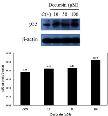
THP-1세포의 세포자연사 관련 단백질(bax, bcl-2, p53 단백질) 발현양상 − THP-1 세포주에 decursin(10, 50, 100 μM)에 대한 apoptosis 관련인자(bcl-2, bax), p53 단백질의 발현양상을 western blot을 통해 확인한 결과, THP-1세포에 서 decursin의 Bcl family에 대한 영향이 발견되었다. 특히 100 μM decursin 처리군에서 bax 단백질이 대조군에 비해
Fig. 3. RT-PCR analysis of bcl-2 and bax genes in THP-1 cells hrs, total RNA was isolated and bcl-2, bax mRNA expression
Fig. 4. RT-PCR analysis of p53 gene in THP-1 cells. THP-1 cells were treated with decursin(10, 50 and 100µM) for 24 hrs, Total RNA was isolated and p53 mRNA expression was determined by RT-PCR.
3배 이상 증가하였고(Fig. 5), 동일한 농도(100 μM)의 decursin 처리군에서는 p53 단백질 발현이 대조군보다 유의 적으로 증가하였다(Fig. 6). Fig. 4에서 확인한 세포자연사와 종양억제 유전자의 양상과 유사하게 단백질에서도 세포자 연사를 촉진, 종양억제를 유도하는 경향을 보여 세포자연사 가 진행되었음을 확인할 수 있었다. Kim 등(2015)은 decursin 은 K562, U937 인간 백혈병세포에서 세포의 성장, 증식에 영향을 주는 PKCα를 감소시켜 세포독성을 증가시키는 것 으로 확인하였다.
22-24)
Yim 등(2005)은 인체 전립선 암세포 에 decursin을 처리하였을 때 높은 성장 억제 효능을 보였 고,11)
Park 등(2007)은 인체 유방암 세포에 처리하였을 때 Hoechst염색으로 핵을 형태학적으로 관찰하고 DNA fragmentation을 정량적으로 분석하였을 때 배양용기에서 세 포가 떨어져 부유되고 세포 밀도가 감소하는 현상을 보였 으며 DNA단편화가 증가하여 apoptosis가 일어난 것을 확인 할 수 있었다.14)
본 연구는 이러한 종양억제인자와 apoptosis 관련 단백질을 분석한 결과라 할 수 있다. 참당귀는 이밖에 도 빈혈이나 어혈 그리고 혈액 순환개선에 많이 사용되는 약재이며,31)
2003년에는 in vivo상에서 sarcoma-180에 의해 종양유도된 mouse에서 양성대조군인 항암제(5-fluorouracil) 에 비해 생존일자가 적었지만 대조군에 비해 수명연장 효 과가 있었고, 종양의 크기도 감소하였다.32)
결 론
당귀로부터 분리 정제한 decursin의 세포에 미치는 영향 을 알아본 결과, 3종류의 백혈병세포와 1종류의 림프종 세 포 모두 높은 독성을 보였으나 그 중 THP-1세포가 가장 독 성이 높았으며(IC
50
: 15.1μM), apoptosis 관련 mRNA 수준 을 확인한 결과 bcl-2는 100 μM 처리군에서 50%이상 감소, bax는 약 2배로 증가하였다. 그리고 종양억제유전자인 p53 은 100 μM에서 2배 이상 증가하였으며 이런 양상은 단백 질 발현 현상에서도 비슷한 경향을 보여 bcl-2는 감소, bax 는 10 μM와 50 μM 처리군은 약 2.5배-3배, 100 μM 처리군 에서는 4배 이상 증가, p53은 100 μM 처리군에서 1.4배 증 가하는 유의적인 경향을 보였다. 결론적으로, 당귀에서 분 리한 decursin은 THP-1세포에서 apoptosis 관련인자인 bax, bcl-2, p53이 관여하여 세포자연사를 일으키고 유의적인 세 포독성을 보였다.인용문헌
1.국립암센터 (2014) Annual report of cancer statistics in Korea in 2012. National Cancer center 1-179.
2. Cho, Y. J. and Hou, W. N. (2005) Effects of dietary bong-ip (Morus alba L.), gam-chei (Glycyrrhiza glabra), sol-ip (Pinus Fig. 5. Expression of decursin on bcl-2 and bax proteins in
THP-1 cells. THP-1 cells were treated with decursin(10, 50 and 100µM)for 24 hrs, bcl-2 and bax proteins were assayed by western blot analysis using specific antibodies.
Fig. 6. Expression of decursin on p53 protein in THP-1 cells.
THP-1 cells were treated with decursin(10, 50 and 100 µM)for 24 hrs, p53 protein were assayed by western blot analysis using specific antibody.
densiflora) and dang-gi (Angelica gigas) on serum compo- sition in rats. Korean J. Food culture 20: 123-129.
3. Han, E. J., Roh, S. B. and Bae, S. J. (2000) Effects of quinone reductase induction and cytoxicity of the Angelica radix extracts. J. Korean Soc. Food Sci. Nutr. 29: 147-152.
4. Kim, J. H., Kim, D. H., You, J. H., Kim, C. H., Kwon, M. C., Seong N. S., Lee, S. E. and Lee H. Y.(2005) Immuno-reg- ulatory activ?ties of various fractions from Ehpedrae sinica Stapf, Rubus Coreanus Miq. and Angelica gigas nakai extracts with untrasonification. Korean J. Medicinal Crop.
Sci. 13: 161-170.
5. Wessler, S., Kleiger, R. E., Cornfield, J. and Teitelbaum, S. L.
(1974) Coumarin theraphy in acute myocardial infarction. A hobson’s choice. Arch. Intem. Med. 134: 774-779.
6. Ristola, P. and Pyorala, K. (1972) Determinants of the response to coumarin anticoagulants in patients with acute myocardial infarction. Ata. Med. Scand. 192: 183-188.
7. Kang, S. Y., Lee, K. Y., Sung, S. H., Park, M. J. and Kim, Y.
C. (2001) Coumarins isolated from Angelica gigas inhibit acetylcholine esterase: structure-activity relationships. J. Nat.
Prod. 64: 683-685.
8. Kang, S. Y., Lee, K. Y., Park, M. J., Kim, Y. C., Markelonis, G. J., Oh, T. H. and Kim, Y. C. (2003) Decursin from Angel- ica gigas mitigates amnesia induced by scopolamine in mice.
Neurobiol. Learn. Mem. 79: 11-18.
9. Kim, J. H., Jeong, J. H., Jeon, S. T., Kim, H., Ock, J., Suk, K., Kim, S. I., Song, K. S. and Lee, W. H. (2006) Decursin inhibits induction of inflammatory mediators by blocking nuclear factor-kappaB activation in macrophages. Mol. Phar- macol. 69: 1783-1790.
10. Son, S. H., Park, K. K., Park, S. K., Kim, Y. C., Kim, Y. S., Lee, S. K. and Chung, W. Y. (2011) Decursin and decursinol from Angelica gigas inhibit the lung metastasis of murine colon carcinoma. Phytother Res. 25: 959-964.
11. Yim, D., Singh, R. P., Agarwal, C., Lee, S. and Chi, H. and Agarwal, R. (2005) A novel anticancer agent, decursin, induces G1 arrest and apoptosis in human prostate carcinoma cells. Cancer Res. 1; 65: 1035-1044.
12. Kim, J. H., Jeong, S. J., Kwon, H. Y., Park, S. Y., Lee, H. J., Lee, H. J., Lieske, J. C. and Kim, S. H. (2010) Decursin pre- vents cisplatin-induced apoptosis via the enhancement of antioxidant enzymes in human renal epithelial cells. Biol.
Pharm. Bull. 33: 1279-1284.
13. Li, L., Li, W., Jung, S. W., Lee, Y. W. and Kim, Y. H. (2011) Protective effects of decursin and decursinol angelate against amyloid β-protein-induced oxidative stress in the PC12 cell line: the role of Nrf2 and antioxidant enzymes. Biosci. Bio- technol. Biochem. 75: 434-442.
14. Park, K. W., Choi, S. R., Shon, M. Y., Jeong, I. Y., Kang, K.
S., Lee, S. T., Shim, K. H. and Seo, K. I. (2007) Cytotoxic effects of decursin from Angelica gigas Nakai in human can- cer cells. J. Korea Soc. Food Sci. Nutr. 36: 1385-1390.
15. Lee, K. Y., Sung, S. H. and Kim, Y. C. (2008) Cognitive- enhancing activity of decursin derivatives. Kor. J. pharma- cogn. 39: 86-90.
16. Lee, S., Shin, D. S., Kim, J. S., Oh, K. B. and Kang, S. S.
(2003) Antibacterial coumarins from Angelica gigas roots.
Arch. Pharm. Res. 26: 449-452.
17. Bae, E. A., Han, M. J., Kim, N. J. and Kim, D. H. (1998) Anti-Helicobacter pylory activity of herbal medicines. Biol.
Pharm. Bull. 21: 990-992.
18. Lee, Y. Y., Lee, S., Jin, J. L. and Yun-Choi, H. S. (2003) Platelet anti-aggregatory effects of coumarins from the roots of Angelica genuflexa and A. gigas. Arch. Pharm. Res. 26:
723-726.
19. Lee, S. H., Lee, Y. S., Jung, S. H., Shin, K. H., Kim, B. K.
and Kang, S. S. (2003) Antioxidant activities of decursinol angelate and decursin from angelica gigas roots. Nat. Prod.
Sci. 9: 170-173.
20. Park, K. W., Choi, S. R., Shon, M. Y., Jeong, I. Y., Kang, K.
S., Lee, S. T., Shim, K. H. and Seo, K. I. (2007) Cytotoxic effects of decursin from Angelica gigas Nakai in human can- cer cells. J. Korean Soc. Food Sci. Nutr. 36: 1385-1390.
20. Kim, B. S., Seo, H., Kim, H. J., Bae, S. M., Son, H. N., Lee, Y. J., Ryu, S., Park, R. W. and Nam, J. O. (2015) Decursin from Angelica gigas Nakai inhibits B16F10 melanoma growth through induction of apoptosis. J. Med. Food 18b 1121-1127.
21. Li, L., Du, J., Zou, L., Xia, H., Wu, T., Kim, Y. and Lee, Y.
(2015) The neuroprotective effects of decursin isolated from Angelica gigas Nakai against amyloid β-protein-induced apoptosis in PC12 cells via a mitochondria-related caspase pathway. Neurochem. Res. 40: 1555-1562.
22. Kim, H. H., Ahn, K. S., Han, H., Choung, S. Y., Choi, S. Y.
and Kim, I. H. (2005) Decursin and PDBu: Two PKC acti- vators distinctively acting in the megakaryocytic differen- tiation of K562 human erythroleukemia cells. Leuk. Res. 29:
1407-1413.
23. Kim, H. H., Bang, S. S., Choi, J. S., Han, H. and Kim, I. H.
(2005) Involvement of PKC and ROS in the cytotoxic mech- anism of anti-leukemic decursin and its derivatives and their structure-activity relationship in human K562 erythroleuke- mia and U937 myeloleukemia cells. Cancer Lett. 223: 191- 201.
24. Ahn, Q., Jeong, S. J., Lee, H. J., Kwon, H. Y., Han, I., Kim, H. S., Lee, H. J., Lee, E. O., Ahn, K. S., Jung, M. H., Zhu, S., Chen, C. Y. and Kim, S. H. (2010) Inhibition of cyclo- oxygenase-2-dependent survivin mediates decursin-induced apoptosis in human KBM-5 myeloid leukemia cells. Cancer Lett. 298: 212-221.
25. Mosmann, T. (1983) Rapid colorimetric assay for cellular growth and survival application to proliferation and cytotoxic assays. J. Immunol. Methods 65: 55-63.
26. Birch, D. E., Kolmodin, L., Laird, W. J., McKinney, N.,
Wong, J., Young, K. K. Y., Zangenberg, G. A. and Zoccoli, M. A.(1996) Simplified Hot-Start PCR, Nature 381: 445-446.
27. Dowdy, S., Weardon, S. and Chilko, D. (2004) Statistics for research (3
rd
ed). Wiley-interscience, USA 285-287.28. Haupt, S., Berger, M., Goldberg, Z. and Haupt, Y. (2003) Apoptosis- the p53 network. J. Cell Sci. 116: 4077-4085.
29. Dy, G. K. and Adjei, A. A. (2002) Novel targets for lung can- cer therapy: part I. J. Clin. Oncol. 20: 2881-2294.
30. Son, C. Y., Baek, I. H., Song, G. Y., Kang, J. S. and Kwon, K. I. (2009) Pharmacological effect of decursin and decursi- nol angelate from Angelica gigas Nakai. Yakhak Hoeji 53:
303-313.
31. Park, K. W., Choi, S. R., Hong, H. R., Kim, J. Y., Shon, M.
Y. and Seo, K. I. (2007) Biological activities of methanol extract of Angelica gigas Nakai. Korean J. Food Preserv. 14:
655-661.
32. Lee, S., Lee, Y. S., Jung, S. H., Shin, K. H., Kim, B. K. and Kang, S. S. (2000) Anti-tumor activities of decursinol ange- late and decursin from Angelica gigas. Arch. Pharm. Res. 26:
727-730.
(2016. 5. 3 접수; 2016. 7. 27 심사; 2016. 8. 16 게재확정)