90
Open reduction and internal fixation of mandibular fracture in an 11-month-old infant: a case report
Tae-Wan Kim1, Eun-Woo Seo2, Seung-Il Song2
1Department of Dentistry, Bundang Jesaeng Hospital, Daejin Medical Center, Seongnam, 2Department of Dentistry, Oral and Maxillofacial Surgery, Ajou University School of Medicine, Suwon, Korea
Abstract(J Korean Assoc Oral Maxillofac Surg 2013;39:90-93)
Mandibular fractures in infants are rare. This case report describes management of a mandibular fracture in an 11-month-old infant using a microplate and screws with open reduction. The surgical treatment was successful. Because the bone fragments were displaced and only the primary incisors had erupted, conservative treatment, such as an acrylic splint and circummandibular wiring, was not recommended. Nine weeks after surgery, the microplate was removed. The results showed complete clinical and radiological bone healing with normal eruption of deciduous teeth.
Key words: Infant, Mandibular fractures, Fracture fixation, Internal
[paper submitted 2013. 2. 20 / revised 2013. 3. 29 / accepted 2013. 3. 30]
to the Department of Oral and Maxillofacial Surgery, Ajou university hospital after falling down from a baby carriage.
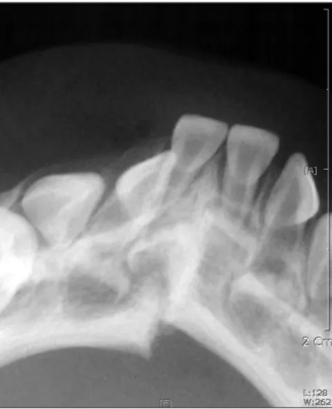
The examination revealed extra-oral swelling and ecchymosis around the area of symphysis. The intraoral examination and standard radiography showed bony displacement between the lower right first and second primary incisor teeth.(Fig.
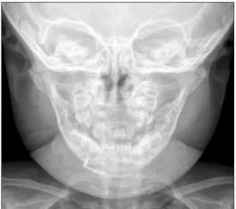
1) Diagnostic computed tomography was performed under sedation, confirming the fracture.(Fig. 2) Specifically, the fracture was visibly displaced, and fragments were freely movable.
A decision was made to treat the fracture with a combina- tion of open reduction and internal fixation with microplate.
Surgery was performed under general anesthesia with sevoflurane, thiopental sodium, fentanyl citrate. After inducement, the patient was intubated with conventional endotracheal tube with 4.0 s cuff via the oral route without difficulty. Lidocaine 2% with 1:100,000 epinephrine was infused in the mandibular anterior vestibule. Horizontal incision was made in the anterior labial vestibule, approxi- mately 3 mm inferior to the mucogingival junction. A mucoperiosteal flap was elevated to expose the fracture line, which ran obliquely from the right second primary incisor to the inferior border of the mandible with step on the buccal cortex and extended to the symphyseal region on the lingual side.(Fig. 3) After gentle reduction of the displaced
I. Introduction
Mandibular fractures in infants (younger than 1 year) are uncommon. In the 0-1 year age group, the frequency has been reported to range from 0.9% to 2.6%1. In terms of treatment, pediatric mandibular fractures are treated with a wide variety of fixation methods. Although conservative treatment such as closed reduction is preferable because displaced fractures have relatively lower incidence in pediatric patients and tooth buds can be damaged, the open reduction-internal fixation method is possible for unfavorably displaced fractures2,3. We report a case of an 11-month-old infant whose displaced fractured mandible was treated with an open surgery method.
II. Case Report
On June 11, 2010, an 11-month-old infant was brought
CASE REPORT http://dx.doi.org/10.5125/jkaoms.2013.39.2.90 pISSN 2234-7550·eISSN 2234-5930
Seung-Il Song
Department of Dentistry, Ajou University School of Medicine, 206 WorldCup- ro, Yeongtong-gu, Suwon 443-749, Korea
TEL: +82-31-219-5328 FAX: +82-31-219-5329 E-mail: [email protected]
This is an open-access article distributed under the terms of the Creative Commons Attribution Non-Commercial License (http://creativecommons.org/licenses/by-nc/3.0/), which permits unrestricted non-commercial use, distribution, and reproduction in any medium, provided the original work is properly cited.
CC
Copyright Ⓒ 2013 The Korean Association of Oral and Maxillofacial Surgeons. All rights reserved.
Open reduction and internal fixation of mandibular fracture in an 11-month-old infant: a case report
91 5-0 chromic. The day after the operation, breastfeeding commenced and continued with good acceptance. Penicillin was administered twice daily at a dose of 50 mg/kg to prevent infection. The patient was discharged on the fourth postoperative day, with the interdental wiring removed about a week after the operation. In the follow-up period, the healing of the fracture was satisfactory.(Fig. 5) No deformities, functional restrictions, or complications such as mouth opening limitation were recorded. Nine weeks after surgery, the microplate was removed under general anesthesia. The fractured wound had healed, and there was evidence of bone growth over the margins of the plate. The lower right primary canine had erupted normally.(Fig. 6)
Total follow-up period was 3 months from the first surgery;
further follow-up was impossible since the patient’s family had immigrated to the United States.
parasymphyseal fracture by interdental wiring, a four-hole L-shaped microplate (overall length of 13 mm, thickness of 0.6 mm; JEIL Medical Co., Seoul, Korea) was adapted to the buccal side of the mandible close to the inferior border to avoid damaging the unerupted primary teeth and permanent tooth germs. After careful drilling, the plate was secured with four 3 mm, low-profile screws.(Fig. 4) The short, low- profile screw was selected to avoid damaging the developing teeth and dental follicles. The incision was sutured with
Fig. 1. Intraoral standard radiograph showing displaced mandibular fractures between the lower right first and second primary incisors.
Tae-Wan Kim et al: Open reduction and internal fixation of mandibular fracture in an 11-month-old infant: a case report. J Korean Assoc Oral Maxillofac Surg 2013
Fig. 2. Coronal computed tomography showing mandibular fracture in the symphysis site.
Tae-Wan Kim et al: Open reduction and internal fixation of mandibular fracture in an 11-month-old infant: a case report. J Korean Assoc Oral Maxillofac Surg 2013
Fig. 3. Intraoperative view of the fractured mandible.
Tae-Wan Kim et al: Open reduction and internal fixation of mandibular fracture in an 11-month-old infant: a case report. J Korean Assoc Oral Maxillofac Surg 2013
Fig. 4. Interdental wiring and four-hole L-shaped microplate with 4 screws was placed on buccal side of mandible.
Tae-Wan Kim et al: Open reduction and internal fixation of mandibular fracture in an 11-month-old infant: a case report. J Korean Assoc Oral Maxillofac Surg 2013
J Korean Assoc Oral Maxillofac Surg 2013;39:90-93
92
Closed reduction with splints fixed with circummandibular wires is the commonly recommended treatment3. Cir-cum- mandibular wiring is advantageous since it neither damages the tooth germs nor requires intermaxillary fixation (IMF).
Moreover, this method does not interfere with condylar growth, but it has limited indications, i.e., it is useful only for anterior mandibular fractures without significant displacement when sufficient eruption of the teeth for fixation is completed.
There are difficulties in IMF-such as the Arch bar-when this method is applied to infants due to the potential risk of condylar growth and imbalanced nutrition during fixation.
Such conservative treatments alone do little to control a significantly displaced fracture of the symphysis11. On the other hand, severely displaced fractures and mobile fragments are common indications for open reduction and internal fixation12. Nonetheless, this method carries the risk of interfering with the eruption of teeth. The developing permanent incisors are located lingual to the primary teeth.
Care should be taken to prevent damage to the tooth germs when a minimally traumatic technique is used in this region with microplates and low-profile screws and when plates are placed at the lower border13.
The titanium microplate and screws were subsequently removed after 9 weeks. According to previous research, plates and screws should be removed as early as 2-3 months after placement14-16. Others maintained rigid fixation for 3 weeks17 or 10 weeks18. The timing for plate removal is still not well- established. The use of an absorbable plate was considered, but the smallest absorbable plate would have been too large and bulky considering the small size of the mandible.
The fracture site was mobile and displaced, requiring rigid
III. Discussion
Mandibular fractures are rare in infants. This has been attributed to the relatively small size of the mandible, resilient nature of the bones, relatively low tooth to bone ratio, and protected environment. For these reasons, facial fractures in infants occur mostly in the form of incomplete fractures or simple fractures without significant displacement4. Mandibular fractures in infants may result from the trauma of short falls. Fractured mandible in infants is diagnosed clinically based on step deformity, hematoma in the floor of the mouth, and mobility of the fractured segments.
The treatment of infant fractures is divided into conserva- tive treatment and surgical treatment. Minor greenstick fractures of the mandible in infants generally do not require immobilization. For more complex fractures, open and closed reduction methods have been used.
The debate on open treatment versus closed treatment of pediatric mandibular fractures remains. Note, however, that recent literature5-8 shows a change in using open reduction and internal fixation (ORIF) in fracture stabilization. The risks of facial growth disturbance in ORIF have not been investigated9. In contrast, non-treatment of unrecognized mandibular fractures leads to high incidence of orthognathic surgery and craniofacial treatment10. The potential damage to tooth roots and follicles can be minimized with a careful technique, which involves placing the fixation screws in the lower mandibular border to avoid the dental follicle.
Fig. 5. Postoperative radiograph showing satisfactory healing of the fracture site.
Tae-Wan Kim et al: Open reduction and internal fixation of mandibular fracture in an 11-month-old infant: a case report. J Korean Assoc Oral Maxillofac Surg 2013
Fig. 6. Intraoral photograph of the 9-weeks after the surgery. The image shows a normally erupted a right lower primary canine.
Tae-Wan Kim et al: Open reduction and internal fixation of mandibular fracture in an 11-month-old infant: a case report. J Korean Assoc Oral Maxillofac Surg 2013
Open reduction and internal fixation of mandibular fracture in an 11-month-old infant: a case report
93
children. J Craniomaxillofac Surg 1993;21:214-9.
6. Posnick JC, Wells M, Pron GE. Pediatric facial fractures: evolving patterns of treatment. J Oral Maxillofac Surg 1993;51:836-44.
7. Siegel MB, Wetmore RF, Potsic WP, Handler SD, Tom LW.
Mandibular fractures in the pediatric patient. Arch Otolaryngol Head Neck Surg 1991;117:533-6.
8. Anderson PJ. Fractures of the facial skeleton in children. Injury 1995;26:47-50.
9. Nørholt SE, Krishnan V, Sindet-Pedersen S, Jensen I. Pediatric condylar fractures: a long-term follow-up study of 55 patients. J Oral Maxillofac Surg 1993;51:1302-10.
10. Demianczuk AN, Verchere C, Phillips JH. The effect on facial growth of pediatric mandibular fractures. J Craniofac Surg 1999;10:323-8.
11. Priest JH. Treatment of a mandibular fracture in a neonate. J Oral Maxillofac Surg 1989;47:77-81.
12. Senel FC, Tekin US, Imamoglu M. Treatment of a mandibular fracture with biodegradable plate in an infant: report of a case. Oral Surg Oral Med Oral Pathol Oral Radiol Endod 2006;101:448-50.
13. Tuovinen V, van Steenis K, Sindet-Pedersen S. Internal fixation of a mandibular fracture in a 6-month-old girl--a case report. Int J Oral Maxillofac Surg 1995;24:210-1.
14. Berryhill WE, Rimell FL, Ness J, Marentette L, Haines SJ. Fate of rigid fixation in pediatric craniofacial surgery. Otolaryngol Head Neck Surg 1999;121:269-73.
15. Haug RH, Foss J. Maxillofacial injuries in the pediatric patient.
Oral Surg Oral Med Oral Pathol Oral Radiol Endod 2000;90:126- 34.
16. Zimmermann CE, Troulis MJ, Kaban LB. Pediatric facial fractures:
recent advances in prevention, diagnosis and management. Int J Oral Maxillofac Surg 2006;35:2-13.
17. Serel S, Can Z, Ersoy A, Sen Z. Management of mandibular fracture using open reduction and internal fixation in a neonate:
case report. J Oral Maxillofac Surg 2005;63:396-9.
18. Walker T, Modayil P, Cascarini L, Collyer J. Dog bite-fracture of the mandible in a 9 month old infant: a case report. Cases J 2009;2:44.
19. Moss ML, Rankow RM. The role of the functional matrix in mandibular growth. Angle Orthod 1968;38:95-103.
20. Vasconcelos BC, Lago CA, Nogueira RV, Gondim DG, Brito Filho A. Mandibular fracture in a premature infant: a case report and review of the literature. J Oral Maxillofac Surg 2009;67:218- 22.
fixation. On the other hand, IMF was contraindicated due to the absence of erupted dentition to support the fixation.
Bone quality and thickness as well as the degree of mobility between the fracture ends, erupted dentition, and possible hindrances to future growth are important considerations that affect the choice of treatment. In such cases, the surgeon should choose the least traumatic procedure. Nevertheless, the mandibular function has important influence on future facial development19. The restoration of mandibular continuity after a fracture is important not only for the immediate function but also for future mandibular development20.
Displaced symphyseal fractures of the mandible in infants may be treated with open reduction when careful positioning of the plates at the lower border of the mandible is performed.
This case with limited follow-up showed no facial growth or tooth eruption problems. Nonetheless, further follow-up would be necessary to observe other problems such as the normal eruption of permanent canine as well.
References
1. Lustmann J, Milhem I. Mandibular fractures in infants: review of the literature and report of seven cases. J Oral Maxillofac Surg 1994;52:240-5.
2. Graham GG, Peltier JR. The management of mandibular fractures in children. J Oral Surg Anesth Hosp Dent Serv 1960;18:416-23.
3. James D. Maxillofacial injuries in children. In: Rowe ML, ed.
Maxillofacial injuries. Vol. 1. Edinburgh: Churchill Livingstone;
1985.
4. Pyo SW. Circum-mandibular wiring for pediatric mandibular fracture: case report. J Korean Assoc Oral Maxillofac Surg 1995;21:619-26.
5. Hardt N, Gottsauner A. The treatment of mandibular fractures in