• 검색 결과가 없습니다.

Larger Testicular Volume Is Independently Associated with Favorable Indices of Lung Function

N/A
N/A
Protected

Academic year: 2021

Share "Larger Testicular Volume Is Independently Associated with Favorable Indices of Lung Function"

Copied!
7
0
0

로드 중.... (전체 텍스트 보기)

전체 글

(1)

Introduction

Chronic obstructive pulmonary disease (COPD) is now considered a systemic disease that affects organs beyond the lungs and airways. The disease is characterized by irreversible airflow obstruction and progressive weight loss, especially loss of lean body mass, which is associated with skeletal muscle dysfunction

1

. Reduced testosterone levels and hypogonadism have been described in COPD

2

. In adult men, this reduction is modest, but a progressive decline in testosterone produc- tion starts between the fourth and sixth decades of life

3,4

. This decline is associated with a simultaneous increase of sex hormone-binding globulin (SHBG) levels, and thus, bioavail-

Larger Testicular Volume Is Independently Associated with Favorable Indices of Lung Function

Tae Beom Kim, M.D.

1

and I-Nae Park, M.D.

2

1

Department of Urology, Gachon University Gil Medical Center, Incheon,

2

Department of Pulmonology, Inje University Seoul Paik Hospital, Seoul, Korea

Background: Men with chronic obstructive pulmonary disease, have reduced endogenous testosterone levels, but the relationship between pulmonary function and endogenous testosterone levels, is inconsistent. Testicular volume is a known indicator of endogenous testosterone levels, male fertility, and male potency. In the present study, the authors investigated the relationship, between testicular volume and lung function.

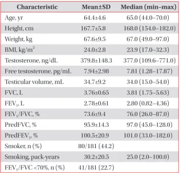
Methods: One hundred and eighty-one South Korean men age 40–70, hospitalized for urological surgery, were retro- spectively enrolled, irrespective of the presence of respiratory disease. Study subjects underwent pulmonary function testing, prior to procedures, and testicular volumes were measured by orchidometry. Testosterone levels of patients in blood samples collected between 7

AM

and 11

AM

, were measured by a direct chemiluminescent immunoassay.

Results: The 181 study subjects were divided into two groups, by testicular volume (≥35 mL vs. <35 mL), the larger testes group, had better lung functions (forced vital capacity [FVC]: 3.87±0.65 L vs. 3.66±0.65 L, p=0.037; forced expiratory volume in 1 second [FEV

1

]: 2.92±0.57 L vs. 2.65±0.61 L, p=0.002; FVC % predicted: 98.2±15.2% vs. 93.8±13.1%, p=0.040;

FEV

1

% predicted: 105.4±19.5% vs. 95.9±21.2%, p=0.002). In addition, the proportion of patients with a FEV
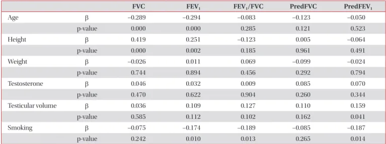
1

/FVC of <70%, was lower in the larger testes group. Univariate analysis conducted using linear regression models, revealed that testicular volume was correlated with FVC (r=0.162, p=0.029), FEV

1

(r=0.218, p=0.003), FEV

1

/FVC (r=0.149, p=0.046), and FEV

1

% predicted (r=0.178, p=0.017), and multivariate analysis using linear regression models, revealed that testicular volume was a significant predictive factor for FEV

1

% predicted (β =0.159, p=0.041).

Conclusion: Larger testicular volume was independently associated, with favorable indices of lung function. These results suggest that androgens, may contribute to better lung function.

Keywords: Respiratory Physiological Phenomena; Pulmonary Disease, Chronic Obstructive; Respiratory Function Tests;

Testis; Testosterone

Address for correspondence: I-Nae Park, M.D.

Department of Pulmonology, Inje University Seoul Paik Hospital, 9 Mareunnae-ro, Jung-gu, Seoul 04551, Korea

Phone: 82-2-2270-0004, Fax: 82-2-2285-2286 E-mail: [email protected]

Received: Dec. 22, 2016 Revised: Mar. 13, 2017 Accepted: May. 8, 2017 Published online: Sep. 1, 2017

cc It is identical to the Creative Commons Attribution Non-Commercial License (http://creativecommons.org/licenses/by-nc/4.0/).

Copyright © 2017

The Korean Academy of Tuberculosis and Respiratory Diseases.

All rights reserved.

(2)

able testosterone may decline more rapidly than total testos- terone

5

.

Several studies have shown that middle aged and elderly COPD patients may develop hypogonadism, and the preva- lence of hypogonadism in men with COPD can range from 22% to 69%. In addition, hypogonadism has been associated with several other systemic manifestations including osteopo- rosis, depression, and muscle weakness

2,6-8

. However, informa- tion regarding the relationship between pulmonary function and endogenous testosterone levels in general populations is inconsistent

5-7,9

. Discrepancies can be mainly attributed to small sample sizes, different patient selection criteria, and circadian variations in testosterone levels. Early morning tes- tosterone levels in young males are, on average, 50% higher than afternoon levels, and reference ranges have usually been derived using morning specimens. Furthermore, testosterone levels can fluctuate substantially on a day-to-day basis. It has been recommended that androgen status should be assessed using more than one measurement

10

.

Testicular volume has been shown to well represent endog- enous testosterone levels, and thus, is considered as an indica- tor of endogenous testosterone level

11-16

, fertility, and potency.

Furthermore, unlike testosterone levels, testicular volumes do not fluctuate markedly, and in fact, are relatively stable.

Testicular volumes have been found to vary widely between individuals in large-scale studies

11,12,17

. Furthermore, like lung function

18

, testicular volume decreases with aging

19,20

. There- fore, in the present study, we have investigated the relation- ship between testicular volume and pulmonary function.

Materials and Methods

1. Study subject selection

One hundred and eighty-one South Korean male patients (aged 40–70 years) hospitalized for urological surgery be- tween April 2014 and March 2016 at a single tertiary aca- demic center were retrospectively reviewed irrespective of the presence of respiratory disease. Testosterone levels of all the patients in blood samples collected between 7

AM

and 11

AM

were measured by a direct chemiluminescent immunoassay (Bayer ADVIA Centaur; assay range, 10–1,500 ng/dL). Regard- ing smoking histories, “never-smokers” were defined as those who had smoked on average <1 cigarette/day for <6 months or had never smoked. For ever-smokers, pack-years were cal- culated to quantify tobacco use; 1-pack-year was equivalent to smoking an average of 20 cigarettes per day for 1 year.

2. Pulmonary function tests

All study subjects underwent spirometry according to American Thoracic Society guidelines

21,22

, before procedures.

Spirometry indices included the forced vital capacity (FVC), forced expiratory volume in 1 second (FEV

1

), and FEV

1

/FVC ratio. Post-bronchodilator responses were not measured.

3. Testicular volume

Patients with conditions known to strongly influence testic- ular volume were excluded from the study. Testicular volumes were measured by an experienced urologist using an orchi- dometry.

4. Statistics

Relationships between the variables were analyzed using Pearson’s linear correlation. Univariate and multivariate analy- ses were performed using linear regression modelling to iden- tify independent predictors of lung factor. The Student’s t test and the chi-square test were used to compare the variables of the two study groups divided by testicular volume (≥35 mL vs.

<35 mL).

The analysis was performed using SPSS version 12.0 (SPSS Inc., Chicago, IL, USA), and p-values of <0.05 were considered statistically significant.

5. Ethics statement

The study was approved by the Institutional Review Board of Gachon University Gil Hospital (GBIRB2016-175) and con- ducted in accordance with the Declaration of Helsinki. The informed consent requirement was waived due to the retro- spective study design.

Results

1. Correlation study

The characteristics of the 181 study subjects are summa- rized in Table 1.

Univariate analysis using linear regression models showed that testicular volume was correlated with FVC (r=0.162, p=0.029), FEV

1

(r=0.218, p=0.003), FEV

1

/FVC (r=0.149, p=0.046), and FEV

1

% predicted (r=0.178, p=0.017) (Table 2).

Multivariate analysis using linear regression models showed that testicular volume significantly predicted FEV

1

% predict- ed (β=0.159, p=0.041) (Table 3).

2. Comparison study between the two study groups (testicular volume ≥35 mL vs. testicular volume <35 mL)

The larger testes group (n=88) had better lung functions

than the smaller testes group (n=93) (FVC: 3.87±0.65 L vs.

(3)

3.66±0.65 L, p=0.037; FEV

1

: 2.92±0.57 L vs. 2.65±0.61 L, p=0.002;

FEV

1

/FVC: 75.5±7.8% vs. 71.9±10.5%, p=0.009; FVC % pre- dicted: 98.2±15.2% vs. 93.8±13.1%, p=0.040; FEV

1

% predicted:

105.4±19.5% vs. 95.9±21.2%, p=0.002) (Table 4). Furthermore, the percentage of patients with a FEV

1

/FVC of <70% was low- er in the larger testes group (15.9% [14/88] vs. 29.0% [27/93], p=0.034) (Table 4).

3. Smoking

Out of the 181 study subjects, 80 (44.2%) were ever-smokers, and the mean smoking exposure of smokers was 30.2±20.5 pack-years (Table 1). Univariate analysis using linear regres- sion models showed that testicular volume was not correlated with smoking (r=–0.094, p=0.216) (Table 2). When patients were divided into two groups according to testicular volume (testicular volume, ≥35 mL vs. <35 mL), proportions of ever- smokers and mean smoking exposure of ever-smokers were not different between the two groups (Table 4).

4. Odds ratio analysis

The odds ratio of ever-smokers in the larger versus the smaller testes group was 1.104, which was not statistically sig- nificant (95% confidence interval [CI], 0.614–1.986). However, the odds ratio of having a FEV

1

/FVC ≥70% in the larger testes group versus the smaller testes group was 2.162, which was Table 1. Characteristics of the study population

Characteristic Mean±SD Median (min–max)

Age, yr 64.4±4.6 65.0 (44.0–70.0)

Height, cm 167.7±5.8 168.0 (154.0–182.0)

Weight, kg 67.6±9.5 67.0 (49.0–97.0)

BMI, kg/m

2

24.0±2.8 23.9 (17.0–32.3)

Testosterone, ng/dL 379.8±148.3 377.0 (109.6–771.0) Free testosterone, pg/mL 7.94±2.98 7.81 (1.28–17.87) Testicular volume, mL 34.7±9.2 34.0 (15.0–54.0)

FVC, L 3.76±0.65 3.81 (1.75–5.63)

FEV

1

, L 2.78±0.61 2.80 (0.82–4.36)

FEV

1

/FVC, % 73.6±9.4 76.0 (26.0–87.0)

PredFVC, % 95.9±14.3 97.0 (45.0–128.0)

PredFEV

1

, % 100.5±20.9 101.0 (33.0–182.0) Smoker, n (%) 80/181 (44.2)

Smoking, pack-years 30.2±20.5 25.0 (2.0–100.0) FEV

1

/FVC <70%, n (%) 41/181 (22.7)

BMI: body mass index; FVC: forced vital capacity; FEV

1

: forced expiratory volume in 1 second; PredFVC: FVC % predicted;

PredFEV

1

: FEV

1

% predicted.

Table 2. Relationships between PFT findings and study variables (univariate analysis using linear regression models) (n=181)

FVC FEV

1

FEV

1

/FVC PredFVC PredFEV

1

TV T

Age r-value –0.405 –0.391 –0.102 –0.107 –0.055 –0.085 0.038

p-value 0.000 0.000 0.170 0.150 0.466 0.253 0.609

Height r-value 0.478 0.353 –0.029 –0.014 –0.040 0.195 0.012

p-value 0.000 0.000 0.696 0.849 0.592 0.008 0.867

Weight r-value 0.286 0.252 0.056 –0.038 –0.003 0.266 –0.029

p-value 0.000 0.001 0.458 0.611 0.968 0.000 0.703

BMI r-value 0.070 0.102 0.086 –0.034 0.023 0.206 –0.053

p-value 0.348 0.172 0.252 0.647 0.757 0.005 0.482

T r-value 0.019 –0.001 –0.016 0.053 0.039 –0.024 -

p-value 0.804 0.985 0.833 0.480 0.606 0.752 -

fT r-value 0.094 0.091 0.030 0.058 0.052 0.027 0.624

p-value 0.212 0.229 0.693 0.445 0.494 0.718 0.000

TV r-value 0.162 0.218 0.149 0.126 0.178 - –0.024

p-value 0.029 0.003 0.046 0.090 0.017 - 0.752

Smoking r-value –0.108 –0.217 –0.213 –0.095 –0.200 –0.094 0.061

p-value 0.153 0.004 0.005 0.208 0.008 0.216 0.421

PFT: pulmonary function test; FVC: forced vital capacity; FEV

1

: forced expiratory volume in 1 second; PredFVC: FVC % predicted; PredFEV

1

:

FEV

1

% predicted; TV: testicular volume; T: testosterone; r-value: Pearson correlation coefficient; BMI: body mass index; fT: free testosterone.

(4)

significant (95% CI, 1.046–4.464) (Table 5).

Discussion

This is the first study undertaken to investigate whether a relation exists between testicular volume and pulmonary

function. It shows testicular volume is positively and indepen- dently associated with better lung functions. Furthermore, the proportion of patients with a FEV

1

/FVC <70% was lower in the larger testes group, and multivariate analysis using linear regression models showed that testicular volume was a sig- nificant predictive factor for FEV

1

% predicted independently of smoking.

Table 3. Multivariate analysis using linear regression models (n=181)

FVC FEV

1

FEV

1

/FVC PredFVC PredFEV

1

Age β –0.289 –0.294 –0.083 –0.123 –0.050

p-value 0.000 0.000 0.285 0.121 0.523

Height β 0.419 0.251 –0.123 0.005 –0.064

p-value 0.000 0.002 0.185 0.961 0.491

Weight β –0.026 0.011 0.069 –0.099 –0.024

p-value 0.744 0.894 0.456 0.292 0.794

Testosterone β 0.046 0.032 0.009 0.085 0.070

p-value 0.470 0.622 0.904 0.260 0.344

Testicular volume β 0.036 0.109 0.127 0.110 0.159

p-value 0.585 0.112 0.102 0.162 0.041

Smoking β –0.075 –0.174 –0.189 –0.085 –0.187

p-value 0.242 0.010 0.013 0.265 0.014

FVC: forced vital capacity; FEV

1

: forced expiratory volume in 1 second; PredFVC: FVC % predicted; PredFEV

1

: FEV

1

% predicted.

Table 4. Characteristics of patients in two study groups

Testicular volume

p-value

≥35 mL (n=88) <35 mL (n=93)

Age, yr 64.0±5.2 64.8±3.8 0.230

Height, cm 168.5±5.7 167.0±5.7 0.079

Weight, kg 69.2±9.8 66.0±9.1 0.024

BMI, kg/m

2

24.3±2.8 23.6±2.7 0.091

Testosterone, ng/dL 382.0±152.6 377.7±145.0 0.844

Free testosterone, pg/mL 8.06±3.25 7.83±2.72 0.618

Testicular volume, mL 42.6±5.3 27.2±4.7 <0.001

FVC, L 3.87±0.65 3.66±0.65 0.037

FEV

1

, L 2.92±0.57 2.65±0.61 0.002

FEV

1

/FVC, % 75.5±7.8 71.9±10.5 0.009

PredFVC, % 98.2±15.2 93.8±13.1 0.040

PredFEV

1

, % 105.4±19.5 95.9±21.2 0.002

Smoking, n (%) 40/88 (45.5) 40/93 (43.0) 0.742

Smoking, pack-years 26.0±17.2 34.2±22.8 0.086

FEV

1

/FVC <70%, n (%) 14/88 (15.9) 27/93 (29.0) 0.034

BMI: body mass index; FVC: forced vital capacity; FEV

1

: forced expiratory volume in 1 second; PredFVC: FVC % predicted; PredFEV

1

: FEV

1

%

predicted.

(5)

A number of chronic illnesses, including COPD, can cause hypogonadism

23

. The mechanism of hypogonadism in COPD is multifactorial, and depends on aging, smoking, obesity, sys- temic inflammation, chronic disease itself, hypoxemia, hyper- capnia, and the taking of glucocorticoids

24

. COPD can affect the hypothalamic-pituitary-testicular axis at multiple levels. In one study, approximately 75% of men with COPD had a low serum testosterone level, a low or below-normal gonadotropin level

7

. Reduced lung function and decline in lung function are associated with morbidity and mortality. However, the effects of sex hormones on lung function are not well understood. In a previous cross-sectional study conducted on 2,197 men, to- tal and free testosterone levels were found to be positively and independently associated with FVC and FEV

1

, and men with severe pulmonary obstruction had lower free testosterone lev- els

9

. However, another study found no relationship between free testosterone level and FEV

1

, residual volume, or total lung capacity

7

. In present study, no relationship was observed between testosterone level and lung function. These discrep- ancies can be attributed to small sample sizes, differences in patient selection, and the inconsistent testosterone levels.

In an autopsy study, the total volume of Leydig cells in the testes of men with a history of chronic bronchitis and emphy- sema of at least 15 years’ duration, and with morphological evidence of the cardiopulmonary effects of hypoxia, was sig- nificantly less than the volume in matched controls

25

. Other chronic conditions, such as, diabetes, stroke, and cancer, have also been associated with late-onset hypogonadism in men, and thus, it appears that a diminished testosterone level is common response to chronic disease

26

. However, our finding of a linear association between testicular volume and pulmo- nary function may suggest a potential link between gonadal and pulmonary functions.

The literature is somewhat inconsistent regarding the re- lationship between smoking and testosterone levels. Most cross-sectional studies indicate that smoking increases total testosterone and that smoking cessation reduces testosterone levels in men. As SHBG levels are elevated in smoking men, it has been suggested that this reported increase in testosterone

levels is a consequence of increased SHBG levels

27,28

. However, Kirbas et al.

28

studied the association between hypogonadism, smoking, and obstructive sleep apnea and found no relation- ship between current smoking and testosterone levels. In the present study, univariate analysis using linear regression mod- els showed that both testicular volume and testosterone levels were not correlated with smoking.

The human testis has been evaluated in term of its en- docrine function, daily sperm output in ejaculates, general appearance of seminiferous tubules, differential cell counts in the testis, and daily sperm production

29

. Although many reports regarding Prader’s orchiometer-derived testicular vol- ume are available, no uniform reference values are available, due to differences in the nature of the populations studied (geographic area, nutritional status, ethnicity, and environ- mental factors)

11,13,30

. The normal range of one testicular vol- ume has been reported to be 14–30 mL

12,13,31,32

, but human testicular volumes are markedly dependent on ethnicity

30,33,34

. For example, testicular volumes have been reported to greater in Caucasian than in Oriental populations

34

. As compared with the results of Auger and Eustache

35

, our study population had smaller total testicular volumes (34.7±9.2 mL vs. 55.7±18.7 mL) (Table 1), and thus, it is not appropriate to compare our Korean data with Caucasian data directly. However, testicular volumes in this study were similar to those found in large- scale studies of healthy young Korean men

11,17

, which means testicular volumes in our study population were within nor- mal limits, as we excluded patients with conditions known to influence testicular volume.

Like lung function

18

, clinically assessed testicular volume varies with age

36

. In a large cohort of males aged 0–28 years, maximum testicular volume was attained at 17–18 and 21–22 years among non-gypsies and gypsies, respectively

37

. Findings of testicular volume declines with aging are not universal, as some have reported a decrease

19,20

and others no change

38

. It has also been reported age, malnutrition and illness exert independent effects on testicular volume

39

, and that age is associated with a reduction in testicular volume only in the eighth decade of life

39,40

. However, recent studies suggest that Table 5. Odds ratio analysis

Testicular volume

OR (95% CI) p-value

≥35 mL <35 mL

FEV

1

/FVC, % ≥70 74 66 2.162 (1.046–4.464) 0.035

<70 14 27

Smoking Ever-smoker 40 40 1.104 (0.614–1.986) 0.741

Never-smoker 48 53

Ever-smoker, pack-years ≥30 16 22 0.545 (0.224–1.325) 0.179

<30 24 18

OR: odds ratio; CI: confidence interval; FEV

1

: forced expiratory volume in 1 second; FVC: forced vital capacity.

(6)

mild testicular volume decline often occurs earlier from the 50 or 60s

41,42

. Reduced testicular volume is associated with hormonal abnormalities, including low testosterone and increased luteinizing hormone (LH) and follicle-stimulating hormone (FSH) levels

12,42

. In aged men, testicular volume, testicular parenchymal weight, and daily sperm production are significantly reduced

29

. In addition, plasma LH, FSH, and estradiol concentrations are elevated whereas plasma testos- terone, free testosterone, and the ability of the testis to secrete testosterone following stimulation are diminished. However, testosterone levels can fluctuate substantially day-to-day and sometimes within days. Therefore, testicular volume, which is relatively stable, represents androgen status better than a single measurement of testosterone level.

Several studies have reported a positive relationship ex- ists between testicular volume and body weight

11,17

. In the present study, height, weight, and body mass index exhibited significant association with testicular volumes by univariate analysis (Table 2). However, body weight was not found to in- dependently predict FEV

1

% predicted by multivariate analy- sis. In fact, only smoking and testicular volume were found to independently predict FEV

1

% predicted (Table 3).

We enrolled the relatively small number of subjects from a specific setting. Furthermore, our results cannot be applied to other populations, given the limited demographics of the study population. Another limitation is that we used pre- bronchodilator spirometric values to evaluate lung function, and thus, we cannot rule out the possibility of transient airflow obstruction in some patients.

Summarizing, although the present study was not conduct- ed using a large-scale, longitudinal design, it shows larger tes- ticular volume was independently associated with favorable indices of lung function, and suggests that androgens may contribute to better lung function.

Conflicts of Interest

No potential conflict of interest relevant to this article was reported.

References

1. Laghi F, Langbein WE, Antonescu-Turcu A, Jubran A, Bam- mert C, Tobin MJ. Respiratory and skeletal muscles in hypo- gonadal men with chronic obstructive pulmonary disease.

Am J Respir Crit Care Med 2005;171:598-605.

2. Kamischke A, Kemper DE, Castel MA, Luthke M, Rolf C, Behre HM, et al. Testosterone levels in men with chronic ob- structive pulmonary disease with or without glucocorticoid therapy. Eur Respir J 1998;11:41-5.

3. Harman SM, Metter EJ, Tobin JD, Pearson J, Blackman MR;

Baltimore Longitudinal Study of Aging. Longitudinal effects of aging on serum total and free testosterone levels in healthy men. Baltimore Longitudinal Study of Aging. J Clin Endocri- nol Metab 2001;86:724-31.

4. Feldman HA, Longcope C, Derby CA, Johannes CB, Araujo AB, Coviello AD, et al. Age trends in the level of serum testos- terone and other hormones in middle-aged men: longitudinal results from the Massachusetts male aging study. J Clin Endo- crinol Metab 2002;87:589-98.

5. Daabis RG, Rehem RN, Hassan MM, Khalil GI. Hypogonad- ism in patients with chronic obstructive pulmonary disease:

relationship with airflow limitation, muscle weakness and systemic inflammation. Alexandria J Med 2016;52:27-33.

6. Van Vliet M, Spruit MA, Verleden G, Kasran A, Van Herck E, Pitta F, et al. Hypogonadism, quadriceps weakness, and ex- ercise intolerance in chronic obstructive pulmonary disease.

Am J Respir Crit Care Med 2005;172:1105-11.

7. Laghi F, Antonescu-Turcu A, Collins E, Segal J, Tobin DE, Jubran A, et al. Hypogonadism in men with chronic obstruc- tive pulmonary disease: prevalence and quality of life. Am J Respir Crit Care Med 2005;171:728-33.

8. Debigare R, Marquis K, Cote CH, Tremblay RR, Michaud A, LeBlanc P, et al. Catabolic/anabolic balance and muscle wast- ing in patients with COPD. Chest 2003;124:83-9.

9. Svartberg J, Schirmer H, Medbo A, Melbye H, Aasebo U. Re- duced pulmonary function is associated with lower levels of endogenous total and free testosterone. The Tromso study.

Eur J Epidemiol 2007;22:107-12.

10. Wheeler MJ, Barnes SC. Measurement of testosterone in the diagnosis of hypogonadism in the ageing male. Clin Endocri- nol (Oxf) 2008;69:515-25.

11. Bahk JY, Jung JH, Jin LM, Min SK. Cut-off value of testes vol- ume in young adults and correlation among testes volume, body mass index, hormonal level, and seminal profiles. Urol- ogy 2010;75:1318-23.

12. Sakamoto H, Ogawa Y, Yoshida H. Relationship between testicular volume and testicular function: comparison of the Prader orchidometric and ultrasonographic measurements in patients with infertility. Asian J Androl 2008;10:319-24.

13. Takihara H, Cosentino MJ, Sakatoku J, Cockett AT. Signifi- cance of testicular size measurement in andrology: II. Cor- relation of testicular size with testicular function. J Urol 1987;137:416-9.

14. Preston BT, Stevenson IR, Lincoln GA, Monfort SL, Pilkington JG, Wilson K. Testes size, testosterone production and repro- ductive behaviour in a natural mammalian mating system. J Anim Ecol 2012;81:296-305.

15. Rey RA, Campo SM, Bedecarras P, Nagle CA, Chemes HE. Is infancy a quiescent period of testicular development? Histo- logical, morphometric, and functional study of the seminif- erous tubules of the cebus monkey from birth to the end of puberty. J Clin Endocrinol Metab 1993;76:1325-31.

16. Ankarberg-Lindgren C, Norjavaara E. Changes of diurnal

(7)

rhythm and levels of total and free testosterone secretion from pre to late puberty in boys: testis size of 3 ml is a transi- tion stage to puberty. Eur J Endocrinol 2004;151:747-57.

17. Ku JH, Kim ME, Jeon YS, Lee NK, Park YH. Factors influenc- ing testicular volume in young men: results of a community- based survey. BJU Int 2002;90:446-50.

18. Fletcher C, Peto R. The natural history of chronic airflow ob- struction. Br Med J 1977;1:1645-8.

19. Stearns EL, MacDonnell JA, Kaufman BJ, Padua R, Lucman TS, Winter JS, et al. Declining testicular function with age:

hormonal and clinical correlates. Am J Med 1974;57:761-6.

20. Baker HW, Burger HG, de Kretser DM, Hudson B, O’Connor S, Wang C, et al. Changes in the pituitary-testicular system with age. Clin Endocrinol (Oxf) 1976;5:349-72.

21. Miller MR, Hankinson J, Brusasco V, Burgos F, Casaburi R, Coates A, et al. Standardisation of spirometry. Eur Respir J 2005;26:319-38.

22. Vestbo J, Hurd SS, Agusti AG, Jones PW, Vogelmeier C, An- zueto A, et al. Global strategy for the diagnosis, management, and prevention of chronic obstructive pulmonary disease:

GOLD executive summary. Am J Respir Crit Care Med 2013;187:347-65.

23. Kalyani RR, Gavini S, Dobs AS. Male hypogonadism in sys- temic disease. Endocrinol Metab Clin North Am 2007;36:333- 48.

24. Balasubramanian V, Naing S. Hypogonadism in chronic ob- structive pulmonary disease: incidence and effects. Curr Opin Pulm Med 2012;18:112-7.

25. Gosney JR. Atrophy of Leydig cells in the testes of men with longstanding chronic bronchitis and emphysema. Thorax 1987;42:615-9.

26. Isidori AM, Lenzi A. Risk factors for androgen decline in older males: lifestyle, chronic diseases and drugs. J Endocrinol In- vest 2005;28(3 Suppl):14-22.

27. English KM, Pugh PJ, Parry H, Scutt NE, Channer KS, Jones TH. Effect of cigarette smoking on levels of bioavailable tes- tosterone in healthy men. Clin Sci (Lond) 2001;100:661-5.

28. Kirbas G, Abakay A, Topcu F, Kaplan A, Unlu M, Peker Y. Ob- structive sleep apnoea, cigarette smoking and serum testos- terone levels in a male sleep clinic cohort. J Int Med Res 2007;

35:38-45.

29. Johnson L. Evaluation of the human testis and its age-related dysfunction. Prog Clin Biol Res 1989;302:35-60.

30. Diamond JM. Ethnic differences: variation in human testis size. Nature 1986;320:488-9.

31. Lenz S, Giwercman A, Elsborg A, Cohr KH, Jelnes JE, Carlsen E, et al. Ultrasonic testicular texture and size in 444 men from the general population: correlation to semen quality. Eur Urol 1993;24:231-8.

32. Arai T, Kitahara S, Horiuchi S, Sumi S, Yoshida K. Relationship of testicular volume to semen profiles and serum hormone concentrations in infertile Japanese males. Int J Fertil Wom- ens Med 1998;43:40-7.

33. Dave DS, Tiwari VD. Normal testicular volume in school children of low socio-economic group. Indian Pediatr 1983;20:901-5.

34. Mittwoch U. Ethnic differences in testis size: a possible link with the cytogenetics of true hermaphroditism. Hum Reprod 1988;3:445-9.

35. Auger J, Eustache F. Second to fourth digit ratios, male genital development and reproductive health: a clinical study among fertile men and testis cancer patients. Int J Androl 2011;34(4 Pt 2):e49-58.

36. Lotti F, Maggi M. Ultrasound of the male genital tract in relation to male reproductive health. Hum Reprod Update 2015;21:56-83.

37. Beres J, Papp G, Pazonyi I, Czeizel E. Testicular volume varia- tions from 0 to 28 years of age. Int Urol Nephrol 1989;21:159- 67.

38. Harman SM, Tsitouras PD. Reproductive hormones in aging men. I. Measurement of sex steroids, basal luteinizing hor- mone, and Leydig cell response to human chorionic gonado- tropin. J Clin Endocrinol Metab 1980;51:35-40.

39. Handelsman DJ, Staraj S. Testicular size: the effects of aging, malnutrition, and illness. J Androl 1985;6:144-51.

40. Sartorius GA, Nieschlag E. Paternal age and reproduction.

Hum Reprod Update 2010;16:65-79.

41. Pilatz A, Rusz A, Wagenlehner F, Weidner W, Altinkilic B. Ref- erence values for testicular volume, epididymal head size and peak systolic velocity of the testicular artery in adult males measured by ultrasonography. Ultraschall Med 2013;34:349- 54.

42. Rastrelli G, Corona G, Lotti F, Boddi V, Mannucci E, Maggi M.

Relationship of testis size and LH levels with incidence of ma-

jor adverse cardiovascular events in older men with sexual

dysfunction. J Sex Med 2013;10:2761-73.

수치

Table 2. Relationships between PFT findings and study variables (univariate analysis using linear regression models) (n=181)
Table 3. Multivariate analysis using linear regression models (n=181)

참조

관련 문서

Ultrasound assessment of diaphragmatic function during acute exacerbation of chronic obstructive pulmonary disease: a pilot study. Santana PV, Cardenas LZ, de Albuquerque

Objective: This study strived to evaluate the relationship between degree of pulmonary emphysema and cardiac ventricular function in chronic obstructive pulmonary disease

Background: The objective of this study was to assess the validity and reliability of the Korean version of chronic obstructive pulmonary disease assessment test (CAT)

Mortality of Dutch coal miners in relation to pneumoconiosis, chronic ob- structive pulmonary disease, and lung function.. Coal workers' pneumoconiosis and exposure to

Conclusion: The total caloric intake of patients with chronic obstructive pulmonary disease showed a positive correlation with the diffusing capacity of the lung,

Background: In chronic obstructive pulmonary disease (COPD) patients, the serum levels of C-reactive protein (CRP) are elevated and an increase of CRP is

Background: The cognitive function is impaired in patients with hypoxemic chronic obstructive pulmonary disease (COPD).. However, there are conflicting

Quantitative CT Assessment in Chronic Obstructive Pulmonary Disease Patients: Comparison of the Patients with and without Consistent Clinical Symptoms and Pulmonary Function