Introduction
Tuberculosis is the leading cause of death from a single in- fectious agent. In 2016, an estimated 10.4 million people fell ill with tuberculosis; and its global mortality was estimated at 1.3 million human immunodeficiency virus (HIV)‒negative people and 0.37 million HIV-positive people
1. Pulmonary tu- berculosis can result in anatomical sequelae, and cause dete- rioration of lung functions
2-4. A study has reported significant pulmonary impairments in more than half of patients treated for tuberculosis
5. The patients with tuberculosis sequelae are thought to be a significant contributor to chronic obstructive pulmonary disease (COPD) population
6.
Unfortunately, there are no treatment guidelines for patients with tuberculosis-destroyed lung. In tuberculosis-endemic
Factors Associated with Indacaterol
Response in Tuberculosis-Destroyed Lung with Airflow Limitation
Tae Hoon Kim, M.D., Ph.D.
1,2, Chin Kook Rhee, M.D., Ph.D.
3and Yeon-Mok Oh, M.D., Ph.D.
11
Department of Pulmonary and Critical Care Medicine, Asan Medical Center, University of Ulsan College of Medicine, Seoul,
2
Department of Internal Medicine, CHA Bundang Medical Center, CHA University, Seongnam,
3Division of Pulmonary, Allergy and Critical Care Medicine, Department of Internal Medicine, Seoul St. Mary's Hospital, College of Medicine, The Catholic University of Korea, Seoul, Korea
Background: Pulmonary tuberculosis can result in anatomical sequelae, and cause airflow limitation. However, there are no treatment guidelines for patients with a tuberculosis-destroyed lung. Recently, indacaterol effectiveness in chronic obstructive pulmonary disease (COPD) patients with Tuberculosis history (INFINITY) study revealed indacaterol provided bronchodilation and symptom improvement in COPD patients with a tuberculosis-destroyed lung.
Methods: We conducted a post-hoc subgroup analysis of the randomized controlled trial, the INFINITY study, to determine factors associated with indacaterol response in a tuberculosis-destroyed lung with airflow limitation. Data from 68 patients treated with inhaled indacaterol, were extracted and analyzed. Factors associated with the response of forced expiratory volume in one second (FEV
1) to indacaterol treatment, were determined using linear regression analysis.
Results: Of 62 patients included, 68% were male, and 52% had history of cigarette smoking. Patients revealed mean FEV
1of 50.5% of predicted value with mean improvement of 81.3 mL in FEV
1after indacaterol treatment for 8 weeks. Linear regression analysis revealed factors associated with response of FEV
1to indacaterol included a short duration of smoking history, and high short-acting bronchodilator response. When patients with history of smoking were excluded, factors associated with response of FEV
1to indacaterol included high short-acting bronchodilator response, and poor health- related quality of life score as measured by St. George’s Respiratory Questionnaire for COPD.
Conclusion: In a tuberculosis-destroyed lung with airflow limitation, short-acting bronchodilator response and smoking history can play a critical role in predicting outcomes of indacaterol treatment.
Keywords: Tuberculosis; Pulmonary Disease, Chronic Obstructive; Indacaterol; Smoking
Address for correspondence: Yeon-Mok Oh, M.D., Ph.D.
Department of Pulmonary and Critical Care Medicine, Asan Medical Center, University of Ulsan College of Medicine, 88 Olympic-ro 43-gil, Songpa-gu, Seoul 05505, Korea
Phone: 82-2-3010-3136, Fax: 82-2-3010-4650 E-mail: [email protected]
Received: Jun. 12, 2018 Revised: Aug. 13, 2018 Accepted: Sep. 11, 2018
cc It is identical to the Creative Commons Attribution Non-Commercial License (http://creativecommons.org/licenses/by-nc/4.0/).
Copyright © 2019
The Korean Academy of Tuberculosis and Respiratory Diseases.
areas, some patients with tuberculosis-destroyed lung may be diagnosed with and treated for COPD because of airflow limi- tations. A recent study suggests that follow-up care after the completion of tuberculosis treatment is important along with an overall improvement in treatment strategies
6. A previous report suggested that the inhaled tiotropium may lead to im- provement in patients with tuberculosis-destroyed lung
7. The indacaterol effectiveness in COPD patients with Tuberculosis history (INFINITY) study showed that, relative to placebo, in- haled indacaterol 150 μg once-daily provided bronchodilation and symptom improvement in COPD patients with tubercu- losis-destroyed lung
8. However, the clinical factors related to the treatment response to indacaterol were not thoroughly explained in the study
8. Thus, the study described here aims to determine the factors associated with the response to inda- caterol treatment in tuberculosis-destroyed lung with airflow limitation.
Materials and Methods
1. Study design and patients
This post-hoc analysis used data collected in the previously reported INFINITY study
8. The objective of this study was to determine the factors associated with response to indacaterol 150 μg once-daily in patients with tuberculosis-destroyed lung and moderate-to-severe airflow limitation. The treatment response was evaluated by the change from baseline in trough forced expiratory volume in 1 second (FEV
1) at week 8 of in- dacaterol treatment.
Briefly, the INFINITY study was a multicenter, randomized, double-blind, placebo-controlled, 8-week trial conducted in South Korea. Eligible patients were aged ≥19 years, had mod- erate-to-severe airflow limitation (post-bronchodilator FEV
1/ forced vital capacity [FVC] <0.7, and post-bronchodilator FEV
1≥30% and <80% of predicted values), and a history of tubercu- losis with no change in the chest radiologic test over the past 1 year, regardless of smoking history. All patients had at least one finding of destroyed parenchyma, including lung volume loss, bronchovascular distortion, fibrosis, and bronchiectasis.
Exclusion criteria included a history of asthma, respiratory infection or COPD worsening within the previous 6 weeks.
After screening, eligible patients were randomized to either a placebo or treatment groups. The treatment group (n=68) re- ceived once-daily indacaterol 150 µg through the Breezhaler device (Novartis Pharma AG, Stein, Switzerland) for 8 weeks.
Because this study is a post-hoc analysis using the data from the previous clinical trial, written informed consent was waived. The study protocol was approved by the Institutional Review Board of Asan Medical Center (2018-0509).
2. Statistical analysis
We compared characteristics of study subgroups using the unpaired t-test for continuous variables and Fisher exact test for categorical variables. We tested the correlation between the response of FEV
1to indacaterol treatment and the sub- types of leukocytes or parameters of lung functions, using the Pearson’s correlation coefficient.
To determine the factors associated with the response of FEV
1to indacaterol treatment, we performed linear regres- sion analysis. Variables identified as significant in univariate analysis were additionally evaluated for the risk-adjusted rela- tionship with the response of FEV
1. The multivariate analysis was processed for seven factors, including age, sex, body mass index (BMI), smoking history (pack-years), spirometry finding (FEV
1, % of predicted value), short-acting bronchodilator re- sponse, and St. George’s Respiratory Questionnaire for COPD (SQRQ-C), using the enter and stepwise backward elimination method.
Statistical analyses were performed using the SPSS version 24.0 (IBM Corp., Armonk, NY, USA). Data are expressed as number (%) or mean±standard deviation, and a p-value of
<0.05 was considered statistically significant.
Results
1. Characteristics of patients
We analyzed 62 patients, who completed the study, among the 68 patients in the indacaterol treatment group. Therefore, baseline characteristics were similar to data of the original report
8. Table 1 summarizes the baseline characteristics of the study population. The mean age of patients was 64.6 years, 67.7% were males, and the overall mean BMI was 21.6 kg/
m
2. Overall, 32 patients (51.6%) had a smoking history with a mean of 34.9 pack-years. There were only four reports of exac- erbations within the previous year (6.5%). Radiologically, lung volume loss (88.7%) was the most common finding, followed by fibrosis (66.1%), bronchovascular distortion (51.6%), and bronchiectasis (50%).
Characteristics of lung function were as follows: mean FVC, 2.58 L (72.3% predicted) and FEV
1, 1.32 L (50.5% predicted).
The reversibility of FEV
1by short-acting bronchodilator was 103.2 mL (mean). After inhaled indacaterol treatment for 8 weeks, the improvement of FEV
1was calculated to be 81.3 mL (mean). The overall mean baseline dyspnea index focal score was calculated to be 7.3; COPD assessment test score, 15.4;
and SGRQ-C score, 37.9.
2. Factors related with the response to indacaterol
Table 2 shows the clinical factors associated with the re-
sponse of FEV
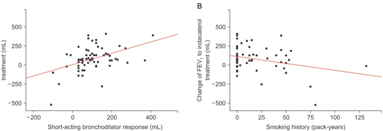
1to indacaterol treatment. In a univariate analy- sis, the response amount of FEV
1to indacaterol treatment was associated with smoking history (p=0.021) and short-acting bronchodilator response (p<0.001). The response of FEV
1by indacaterol treatment demonstrated statistically significant positive correlation with the amount of short-acting broncho- dilator response in FEV
1(ρ=0.385, p=0.002; r=0.443, p<0.001) (Figure 1) and smoking history in pack-years (r=–0.292, p=0.021) (Figure 1). However, age, sex, BMI, FEV
1, and SGRQ- C score did not affect the response of FEV
1to treatment with indacaterol in linear regression analysis. Risk-adjusted analy- sis revealed that indacaterol response was mostly associated with a short smoking history (p=0.018), and high short-acting bronchodilator response (p=0.004). Based on the multivariate analyses using the stepwise backward elimination method, this study also demonstrated that indacaterol response was independently associated with two factors: smoking history (p=0.016), and baseline short-acting bronchodilator response (p<0.001).
3. Factors related with the response to indacaterol:
subgroup analysis according to cigarette smoking Table 3 presents a comparison of smokers and non- smokers, in terms of clinical factors associated with response to indacaterol treatment. In smokers, short smoking history (p=0.019) and high short-acting bronchodilator response (p=0.045) were associated with indacaterol response. How- ever, in nonsmokers, a worse score in health-related quality of life by SGRQ-C (p<0.001), and higher short-acting bron- chodilator response (p=0.001) were related to the response to indacaterol. While 96.9% of smokers were males, only 36.7%
of nonsmokers were males. Thus, we analyzed 42 males in total. Both groups had similar clinical and radiologic features.
However, the smokers had a mean smoking history of 35.4 pack-years, and lower SGRQ-C score (38.8±16.1 vs. 23.5±14.8,
Table 2. Factors associated with the response of FEV
1to indacaterol treatment
Univariate Multivariate
Beta Standard error p-value Beta Standard error p-value
Age –1.524 1.920 0.431 0.019 1.847 0.992
Male sex 42.381 43.969 0.339 –8.367 48.728 0.864
BMI, kg/m
26.579 6.136 0.288 3.389 5.573 0.546
Smoking history, pack-year –1.804 0.764 0.021* –2.169 0.888 0.018*
FEV
1, % pred. –2.017 1.736 0.250 1.487 1.788 0.409
BDR (FEV
1), mL 0.747 0.195 <0.001*** 0.654 0.219 0.004**
SGRQ-C 2.052 1.087 0.064 1.684 1.202 0.167
*p<0.05, **p<0.01, ***p<0.001.
FEV
1: forced expiratory volume in 1 second; BMI: body mass index; BDR: short-acting bronchodilator response; SGRQ-C: St. George’s Respi- ratory Questionnaire for chronic obstructive pulmonary disease.
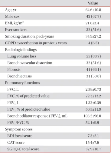
Table 1. Baseline demographic and clinical characteristics Value
Age, yr 64.6±10.8
Male sex 42 (67.7)
BMI, kg/m
221.6±3.4
Ever smokers 32 (51.6)
Smoking duration, pack-years 34.9±27.2 COPD exacerbation in previous years 4 (6.5) Radiologic findings
Lung volume loss 55 (88.7)
Bronchovascular distortion 32 (51.6)
Fibrosis 41 (66.1)
Bronchiectasis 31 (50.0)
Pulmonary functions
FVC, L 2.58±0.73
FVC, % of predicted value 72.3±13.2
FEV
1, L 1.32±0.39
FEV
1, % of predicted value 50.5±11.9 Bronchodilator response (FEV
1), mL 103.2±96.0
FEV
1/FVC, % 52.1±9.9
Symptom scores
BDI focal score 7.3±2.1
CAT score 15.4±7.6
SGRQ-C total score 37.9±18.7
Values are presented mean±standard deviation or number (%).
BMI: body mass index; COPD: chronic obstructive pulmonary disease; FVC: forced vital capacity; FEV
1: forced expiratory volume in 1 second; Bronchodilator response: bronchodilator response by short-acting bronchodilator; BDI: baseline dyspnea index; CAT:
COPD assessment test; SGRQ-C: St. George’s Respiratory Ques-
tionnaire for COPD.
p=0.010). Of the radiologic findings, fibrosis was more com- mon in smokers than nonsmokers (80.6% vs. 36.4%, p=0.019).
4. Relation of radiologic findings with the response to indacaterol
Supplementary Table S1 summarizes the response of FEV
1to indacaterol treatment based on the radiologic findings.
Clinically significant abnormal findings were observed in 52 patients (83.9%). There was no significant difference in the response of FEV
1to indacaterol treatment between patients with abnormal findings (15.0±251.4 mL) and those without abnormal findings (94.0±138.2 mL, p=0.159). In patients with lung volume loss, the response of FEV
1to indacaterol treat- ment was 77.3±153.4 mL; however, there was no difference in patients without lung volume loss (112.9±229.7 mL, p=0.703).
Patients with fibrosis showed a 66.3±122.6 mL change in FEV
1; however, patients without fibrosis had a 110.5±220.0
mL change in FEV
1(p=0.313). There was no significant dif- ference between patients with bronchovascular distortion (100.6±154.9 mL) and those without bronchovascular distor- tion (60.7±168.9 mL, p=0.337). The response of FEV
1to inda- caterol treatment was 66.1±154.8 mL in patients with bron- chiectasis, and this response was similar to patients without bronchiectasis (96.5±170.0 mL, p=0.465). Overall, 15 patients (24.2%) showed all four abnormal radiologic findings with a 42.7±113.6 mL change in FEV
1.
Discussion
This post-hoc analysis of the INFINITY study aimed to identify the characteristics of responders to indacaterol 150 μg once-daily in patients with tuberculosis-destroyed lung with moderate-to-severe airflow limitation. In the current study, the response of FEV
1to indacaterol treatment demonstrated
200 500
250
0
250
ChangeofFEVtoindacaterol treatment(mL) 500
1
Short-acting bronchodilator response (mL)
0 200 400 0
500
250
0
250
ChangeofFEVtoindacaterol treatment(mL) 500
1
Smoking history (pack-years)
25 50 75 100 125