VI The Role of Lung and Diaphragm Ultrasound in Pulmonary Rehabilitation
안태준
가톨릭대학교 의과대학 여의도성모병원 호흡기내과
Patient-tailored treatment is required in pulmonary rehabilitation (PR). For this reason, a comprehensive evaluation of lung and adjacent organ, such as diaphragm, is necessary. The lung ultrasound (LUS) and diaphragm ultrasound (DUS) are emerging diagnostic techniques that the usefulness of those is proved in the various chronic pulmonary diseases. The LUS is a revalued tool which evaluate the status of lung and pleura efficiently without any harm. The DUS is a well-known tool for measuring the factors of diaphragm muscle, such as muscle mass, mobility, contractility, and strength of the diaphragm. Therefore, LUS and DUS are used in PR in various ways. We used LUS to detect comorbid status such as pleural effusion or pneumothorax which are hindrance of exercise. We distinguish the status of exacerbation of underlying diseases by LUS or DUS. These are helpful for determining the proper start timing of PR. Factors which are measured by DUS, such as thickness, thickening fraction, and excursion of diaphragm, are used for assessment and follow-up of outcomes of PR. These are markers of diaphragm dysfunction and atrophy also. We can make plan for exercise such as inspiratory muscle training and we recommend the proper nutritional support to enhance the recovery of muscles. Briefly, LUS and DUS are outstanding tools for performing the PR as patient-tailored way.
Key Words: Pulmonary rehabilitation, Lung ultrasound, Diaphragm ultrasound Corresponding author: Tai Joon An, M.D.
Division of Pulmonary and Critical Care Medicine, Department of Internal Medicine, Yeouido St. Mary’s Hospital, The Catholic University of Korea, 10 63-ro, Yeongdeungpo-gu, Seoul 07345, Korea
Tel: +82-2-3779-1663, Fax: +82-2-780-3132, E-mail: [email protected]
1. 서론
호흡재활은 호흡기 질환의 비약물적 치료 중 하나로1, 그 효과는 만성폐쇄성폐질환(chronic obstructive pul- monary disease, COPD), 천식, 그리고 특발성폐섬유화증(idiopathic pulmonary fibrosis, IPF)과 같은 간질성폐 질환(interstitial lung disease, ILD) 등의 질환에서 확인되었다2-4. 호흡기질환자는 호흡재활을 통하여 호흡곤란 등의 증상 호전, 전신 상태의 개선, 그리고 삶의 질 증진을 이룰 수 있고, 장기적인 관점에서 건강한 삶을 영위할 수 있다5.
이러한 호흡재활의 효과를 높이기 위해서는 환자 및 질병 상태에 대한 포괄적인 평가가 필요하다. 호흡은 폐, 흉벽, 그리고 호흡근의 상호 작용을 통해 조절되기 때문에 호흡재활을 시행할 때에는 폐, 흉막 및 늑막, 흉벽, 그리고 호흡근육의 상태 및 기능에 대한 평가가 필수적으로 수반되어야 한다. 의료진은 이러한 평가결과 를 통하여 환자 맞춤형 호흡재활치료(patient-tailored pulmonary rehabilitation)를 제공할 수 있다4,6.
흉부 기관의 영상 및 기능 평가도구는 다양하다. 흉부 단순촬영(X-ray)은 흉부의 전반적인 정보를 제공하지만 효용에 제한이 있으며, 흉부 전산화단층촬영(Computed tomography, CT)은 폐 및 주변 장기의 입체적인 정보를 제공하는 반면, 방사선 노출로 인한 시행의 제약과 영상 판독에 자원이 소모되는 단점이 있다. 근전도의 경우 호흡근 기능 측정의 표준방법이지만 침습적이고 전문가의 해석이 요구된다. 이러한 도구들의 단점을 보완할 수 있으며, 근래에 많은 연구가 진행되고 있는 영상평가도구가 초음파이다7-9. 초음파는 앞선 검사도구와 달리 인체에 무해하고 반복측정이 가능하며, 실시간으로 결과물을 확인할 수 있고 대체로 직관적이다. 흉부 평가도구 로의 초음파에는 폐 초음파(lung ultrasound, LUS)와 횡격막 초음파(diaphragm ultrasound, DUS)가 있다. 본 review에서는 LUS 및 DUS에 대하여 간략히 알아보고 호흡재활에서의 LUS 및 DUS의 역할에 대해 살펴보고자 한다.
2. 본론
1) 폐 초음파(LUS)
전통적으로 공기로 인한 허상(artifact)은 흉부 초음파 평가의 장애물이었으나 최근 진행된 연구들을 통하여 이에 대한 내용 정리가 이루어지면서 LUS는 비약적인 발전을 이루었다10. LUS는 허상과 실상을 같이 해석하여
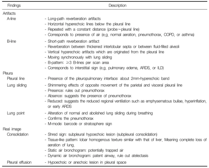
Findings Description
Artifacts
A-line - Long-path reverberation artifacts
- Horizontal hyperechoic lines below the pleural line - Repeated with a constant distance (probe∼pleural line)
- Corresponds to presence of air (e.g. normal aeration, pneumothorax, COPD, or asthma) B-line - Short-path reverberation artifact
- Reverberation between thickened interlobular septa or between fluid-filled alveoli - Vertical hyperechoic artifacts which are originated from the pleural line
- Moving synchronously with lung sliding - B-pattern: ≥3 B-lines per scan area
- Corresponds to interstitial sign (e.g. pulmonary edema, ARDS, or ILD) Pleura
Pleural line - Presence of the pleuropulmonary interface: about 2mm-hyperechoic band Lung sliding - Shimmering effects of opposite movement of the parietal and visceral pleural line
- Presence: rules out pneumothorax
- Absence: suggests the presence of pneumothorax
- Reduced: suggests the reduced regional ventilation such as emphysematous bullae, hyperinflation, or early ARDS
Lung point - Alteration of normal and abolished lung sliding during breathing - Confirms the pneumothorax
- M-mode: barcode or stratosphere sign Real image
Consolidation - Shred sign: subpleural hypoechoic lesion (subpleural consolidation)
- Tissue-like pattern: lobar homogenous texture similar with that of liver. Meaning complete loss of aeration of lung.
- Static air bronchogram: potentially trapped air
- Dynamic air bronchogram: patent airway, rule out atelectasis Pleural effusion - Hypoechoic or anechoic lesion in pleural space
COPD: chronic obstructive pulmonary disease; ARDS: acute respiratory distress syndrome; ILD: interstitial lung disease.
Table 1. Findings of lung ultrasound7,8,10
폐를 평가한다11. LUS의 실상 소견에는 lung consolidation과 pleural effusion 등이 있으며, 허상 소견에는 aera- tion을 의미하는 A-line과 늑막하 소엽간중격(interlobular septa) 사이 또는 허파꽈리사이공간(interalveolar space)의 비후 또는 폐포 내부의 액체 저류에 의한 허상을 지칭하는 B-line이 있다. 흉막선(pleural line)에서 볼 수 있는 소견으로는 호흡에 따른 visceral pleura와 parietal pleura의 상대적 움직임을 확인할 수 있는 lung sliding과 기흉의 존재를 확인할 수 있는 lung point 등이 있다(Table 1).
이러한 소견들을 바탕으로 LUS의 효용성에 대한 연구들이 진행되었다12-16. 한 메타분석에서 LUS는 폐렴을 진단하는 데 있어 높은 통합 민감도(0.88, 95% confidence interval [CI] 0.86∼0.90)와 통합 특이도(0.86, 95%CI 0.83∼0.88)를 보였다17. COPD 또는 천식을 다른 호흡곤란 원인과 감별하는 연구에서도 LUS는 높은 민감도 (89%)와 특이도(97%)를 보였다18. 다른 영상평가도구와 비교한 연구에서는 consolidation을 진단하는 정확도가 LUS는 95%로 확인되었고, 이는 X-ray의 정확도(81%)보다 높았으며 진단의 민감도 또한 LUS가 X-ray에 비하여 유의한 차이를 보였다(70% vs. 48%, p<0.001)19. 또한 LUS는 X-ray나 CT에 비교하여 실시간으로 영상 확인이 가능하였고 폐 및 흉막 등의 실제 움직임을 직관적으로 파악할 수 있는 장점을 보였다20-22. ILD에서의 연구도 다양하게 이루어졌으며23-26, 특히 고해상도 CT의 섬유화 점수와 LUS의 B-line 개수와 늑막의 두께를 비교한 연구에서 ILD의 중증도에 따라 유의한 차이를 보였다. 특히 늑막의 두께가 중증도 구분의 중요한 지표로 확인되 었다(area under ROC curve=0.943, 95%CI 0.797∼0.994; p<0.001)27.
2) 횡격막 초음파(DUS)
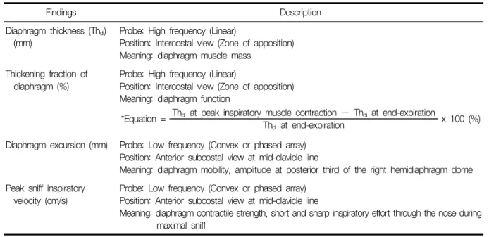
DUS는 주 호흡근인 횡격막의 평가 도구이다. 다른 횡격막 평가 도구인 CT, 자기공명영상, fluoroscopy, 근전 도 등과는 달리, 비침습적이고 실시간 영상을 제공하며 인체에 무해하다. 그러면서도 횡격막의 여러 소견에 대해 신뢰할 만하고 간단하게 평가할 수 있는 도구이다28,29. DUS로 측정하는 횡격막의 소견은 크게 다음과 같다. 횡격막 근육의 두께(diaphragm muscle thickness)를 측정하여 전체적인 횡격막 근육의 질량을 유추할 수 있으며, 흡기와 호기 시의 두께 변화를 통해 근육의 수축력(thickening fraction of diaphragm muscle)을 측정 할 수 있다. 또한 횡격막이 호흡에 따라 움직이는 정도(diaphragm excursion)를 측정할 수 있다(Table 2)30.
COPD와 ILD 등의 호흡기질환에서 횡격막 근육 약화 및 위축 또는 기능 부전의 유병률은 높은 것으로 추정된 다31. COPD 환자를 건강한 성인과 비교한 연구에서는 respiratory distress로 인하여 기저 횡격막 움직임의 증가
Findings Description
Diaphragm thickness (Thdi) (mm)
Probe: High frequency (Linear)
Position: Intercostal view (Zone of apposition) Meaning: diaphragm muscle mass
Thickening fraction of diaphragm (%)
Probe: High frequency (Linear)
Position: Intercostal view (Zone of apposition) Meaning: diaphragm function
*Equation = Thdi at peak inspiratory muscle contraction − Thdi at end-expiration
x 100 (%) Thdi at end-expiration
Diaphragm excursion (mm) Probe: Low frequency (Convex or phased array) Position: Anterior subcostal view at mid-clavicle line
Meaning: diaphragm mobility, amplitude at posterior third of the right hemidiaphragm dome Peak sniff inspiratory
velocity (cm/s)
Probe: Low frequency (Convex or phased array) Position: Anterior subcostal view at mid-clavicle line
Meaning: diaphragm contractile strength, short and sharp inspiratory effort through the nose during maximal sniff
Table 2. Findings of diaphragm ultrasound29,30
와 thickening fraction의 증가가 보였으며32,33, 비축되어 있는 횡격막의 수축력과 air-trapping의 정도 그리고 COPD 중증도가 연관을 보였다30. IPF 환자는 건강한 성인에 비해 diaphragm excursion이 감소하였으며, IPF의 폐기능 지표인 forced vital capacity와 diaphragm excursion의 연관성을 확인하였다34. 그 외에도 호흡곤란증상 점수, 운동능력, 악화상태 구분 등의 영역에서 연구되는 등 현재 호흡기질환자의 심장초음파를 제외한 초음파 분야에서 가장 활발히 연구되고 있다34-37.
3) 호흡재활에서의 폐 초음파와 횡격막 초음파의 역할
앞서 살펴보았듯이 LUS와 DUS는 폐의 구조 및 운동, 흉막 및 늑막, 횡격막 근육의 평가에 사용될 수 있다.
의료진은 이러한 평가를 통하여 환자 맞춤형 호흡재활치료를 제공할 수 있다. 이를 요약하면 Figure 1과 같다.
(1) 호흡재활 시작 전 평가:
첫째, 호흡재활 대상자를 선별할 때 사용한다. LUS는 COPD 또는 ILD 등을 폐부종 등의 다른 질환과 구별할 수 있다. DUS를 통해 진행성 근위약을 보이는 근위축성 측삭경화증 등의 신경근육계 질환이나 횡격막 마비 또는 탈장 등을 감별하여 호흡재활 여부의 결정에 사용하거나 비침습적 환기의 적용 등을 결정할 수 있다.둘째, 환자 및 질병 상태 평가에 사용할 수 있다. 호흡재활을 진행하는 데 있어 동반질환은 호흡재활의 진행에 영향을 준다. LUS를 통해 재활치료 시작 전에 환자에게 동반된 흉수 또는 기흉 여부를 감별하여 치료함으로써 폐의 생리적 상태를 개선할 수 있다. 이를 통하여 치료과정에서 발생할 수 있는 부작용을 줄이고 환자의 치료 순응도를 향상할 수 있다. LUS는 기저질환의 급성악화 여부를 구분할 때 사용하거나, LUS score를 통하여 ILD의 중증도를 구분할 수 있다. 이러한 소견을 통하여 급성악화 호전 시점을 확인할 수 있으며, 이에 따라 치료의 시작이나 강도 조절을 할 수 있다.
셋째, 호흡근의 상태평가에 사용할 수 있다. DUS를 통하여 횡격막 근육의 수축력과 움직임을 평가하여 적합 한 호흡재활 방법을 결정할 수 있다. 예를 들어, DUS를 통해 횡격막 부전을 감별하여 흡기근 트레이닝 적용 여부를 결정할 수 있다. 횡격막 근육의 두께를 횡격막 초음파를 통하여 측정하면 횡격막의 위축이나 근감소증 여부를 추측할 수 있으며, 영양집중치료를 적용하여 근육량 회복을 도모할 수 있다.
(2) 환자 추적 관찰 및 호흡재활 성과 예측:
COPD 환자의 호흡재활 전후로 횡격막의 움직임을 DUS로 측정한 연구들에 따르면 초음파상의 횡격막 움직임의 변화량과 6분보행검사의 변화량이 상관관계를 보였으며(rho=Figure 1. The role of lung and diaphragm ultrasound in pulmonary rehabilitation. LUS: lung ultrasound; DUS: diaphragm ultrasound.
0.49), COPD assessment test 점수의 2점 이상의 호전을 예측하는 도구로 유용성을 보였다(민감도 80%, 특이도 62%). 또한 횡격막 움직임이 소실된 중증의 COPD에서도 호흡재활치료 전후의 움직임 개선 효과를 횡격막 초음파를 통하여 확인할 수 있었다38,39. 이를 통하여 우리는 DUS가 호흡재활 효과의 추적 도구 및 성과 예측도구 로써 의의가 있음을 알 수 있다.
3. 결론
LUS와 DUS는 점차 호흡기질환에서 저변이 확대될 것으로 기대되는 검사도구이다. 특히 폐 및 횡격막 상태에 대한 손쉽고 정확한 평가가 가능하며 실시간으로 결과를 볼 수 있고, 인체에 무해한 특성을 가져 반복적으로 측정할 수 있는 도구이기 때문에 여러 호흡기질환에서 초기 평가 및 추적 관찰에 유용한 도구로 평가된다.
특히, DUS의 경우 호흡기질환에서 자주 확인되는 횡격막 기능 부전이나 횡격막 위축 등을 다른 검사도구와 달리 손쉽게 감별할 수 있는 장점이 있다. 호흡재활에서의 역할도 이와 마찬가지로 점차 증대될 것으로 생각되 며, 정기적인 평가를 통해 patient-tailored treatment의 중요한 평가 도구로의 역할을 기대하는 바이다.
References
1. McCarthy B, Casey D, Devane D, Murphy K, Murphy E, Lacasse Y. Pulmonary rehabilitation for chronic ob- structive pulmonary disease. Cochrane Database Syst Rev 2015;(2):CD003793.
2. Blervaque L, Préfaut C, Forthin H, Maffre F, Bourrelier M, Héraud N, et al. Efficacy of a long-term pulmonary rehabilitation maintenance program for COPD patients in a real-life setting: a 5-year cohort study. Respir Res 2021;22:79.
3. Swigris JJ, Fairclough DL, Morrison M, Make B, Kozora E, Brown KK, et al. Benefits of pulmonary rehabilitation in idiopathic pulmonary fibrosis. Respir Care 2011;56:783-9.
4. Majd S, Apps L, Chantrell S, Hudson N, Eglington E, Hargadon B, et al. A feasibility study of a randomized controlled trial of asthma-tailored pulmonary rehabilitation compared with usual care in adults with severe asthma. J Allergy Clin Immunol Pract 2020;8:3418-27.
5. Troosters T, Casaburi R, Gosselink R, Decramer M. Pulmonary rehabilitation in chronic obstructive pulmonary disease. Am J Respir Crit Care Med 2005;172:19-38.
6. Morisset J, Dubé BP, Garvey C, Bourbeau J, Collard HR, Swigris JJ, et al. The unmet educational needs of patients with interstitial lung disease. Setting the stage for tailored pulmonary rehabilitation. Ann Am Thorac Soc 2016;13:1026-33.
7. Volpicelli G, Elbarbary M, Blaivas M, Lichtenstein DA, Mathis G, Kirkpatrick AW, et al. International evi- dence-based recommendations for point-of-care lung ultrasound. Intensive Care Med 2012;38:577-91.
8. Lichtenstein DA. Lung ultrasound in the critically ill. Ann Intensive Care 2014;4:1.
9. Sferrazza Papa GF, Pellegrino GM, Di Marco F, Imeri G, Brochard L, Goligher E, et al. A review of the ultra- sound assessment of diaphragmatic function in clinical practice. Respiration 2016;91:403-11.
10. Mojoli F, Bouhemad B, Mongodi S, Lichtenstein D. Lung ultrasound for critically Ill patients. Am J Respir Crit Care Med 2019;199:701-14.
11. Lichtenstein DA, Mezière GA, Lagoueyte JF, Biderman P, Goldstein I, Gepner A. A-lines and B-lines: lung ultrasound as a bedside tool for predicting pulmonary artery occlusion pressure in the critically ill. Chest 2009;136:1014-20.
12. Lichtenstein DA, Mezière GA. Relevance of lung ultrasound in the diagnosis of acute respiratory failure: the BLUE protocol. Chest 2008;134:117-25.
13. Sartini S, Frizzi J, Borselli M, Sarcoli E, Granai C, Gialli V, et al. Which method is best for an early accurate diagnosis of acute heart failure? Comparison between lung ultrasound, chest X-ray and NT pro-BNP perform-
ance: a prospective study. Intern Emerg Med 2017;12:861-9.
14. Ticinesi A, Lauretani F, Nouvenne A, Mori G, Chiussi G, Maggio M, et al. Lung ultrasound and chest x-ray for detecting pneumonia in an acute geriatric ward. Medicine (Baltimore) 2016;95:e4153.
15. Touw HR, Parlevliet KL, Beerepoot M, Schober P, Vonk A, Twisk JW, et al. Lung ultrasound compared with chest X-ray in diagnosing postoperative pulmonary complications following cardiothoracic surgery: a pro- spective observational study. Anaesthesia 2018;73:946-54.
16. Vizioli L, Ciccarese F, Forti P, Chiesa AM, Giovagnoli M, Mughetti M, et al. Integrated use of lung ultrasound and chest X-ray in the detection of interstitial lung disease. Respiration 2017;93:15-22.
17. Long L, Zhao HT, Zhang ZY, Wang GY, Zhao HL. Lung ultrasound for the diagnosis of pneumonia in adults:
a meta-analysis. Medicine (Baltimore) 2017;96:e5713.
18. Lichtenstein D. Novel approaches to ultrasonography of the lung and pleural space: where are we now? Breathe (Sheff) 2017;13:100-11.
19. Haggag YI, Mashhour K, Ahmed K, Samir N, Radwan W. Effectiveness of lung ultrasound in comparison with chest X-ray in diagnosis of lung consolidation. Open Access Maced J Med Sci 2019;7:2457-61.
20. Chiumello D, Umbrello M, Sferrazza Papa GF, Angileri A, Gurgitano M, Formenti P, et al. Global and regional diagnostic accuracy of lung ultrasound compared to CT in patients with acute respiratory distress syndrome.
Crit Care Med 2019;47:1599-606.
21. Lichtenstein DA. Lung ultrasound (in the Critically Ill) superior to CT: the example of lung sliding. Korean J Crit Care Med 2017;32:1-8.
22. Sabour S. Lung ultrasound compared to CT in patients with acute respiratory distress syndrome: methodological issue on diagnostic accuracy and agreement. Crit Care Med 2020;48:e262-3.
23. Zanatta M, Benato P, De Battisti S, Pirozzi C, Ippolito R, Cianci V. Pre-hospital lung ultrasound for cardiac heart failure and COPD: is it worthwhile? Crit Ultrasound J 2018;10:22.
24. Volpicelli G, Cardinale L, Garofalo G, Veltri A. Usefulness of lung ultrasound in the bedside distinction between pulmonary edema and exacerbation of COPD. Emerg Radiol 2008;15:145-51.
25. Volpicelli G. Lung ultrasound B-Lines in interstitial lung disease: moving from diagnosis to prognostic stratifi- cation. Chest 2020;158:1323-4.
26. Frongillo E, Gaudioso G, Feragalli B. Ultrasound and interstitial lung disease: use and limitations. Radiol Med 2020;125:66-7.
27. Manolescu D, Oancea C, Timar B, Traila D, Malita D, Birsasteanu F, et al. Ultrasound mapping of lung changes in idiopathic pulmonary fibrosis. Clin Respir J 2020;14:54-63.
28. Spiesshoefer J, Herkenrath S, Henke C, Langenbruch L, Schneppe M, Randerath W, et al. Evaluation of respira- tory muscle strength and diaphragm ultrasound: normative values, theoretical considerations, and practical recommendations. Respiration 2020;99:369-81.
29. Baldwin CE, Paratz JD, Bersten AD. Diaphragm and peripheral muscle thickness on ultrasound: intra-rater reli- ability and variability of a methodology using non-standard recumbent positions. Respirology 2011;16:1136-43.
30. Rittayamai N, Chuaychoo B, Tscheikuna J, Dres M, Goligher EC, Brochard L. Ultrasound evaluation of dia- phragm force reserve in patients with chronic obstructive pulmonary disease. Ann Am Thorac Soc 2020;17:
1222-30.
31. Gea J, Casadevall C, Pascual S, Orozco-Levi M, Barreiro E. Clinical management of chronic obstructive pulmo- nary disease patients with muscle dysfunction. J Thorac Dis 2016;8:3379-400.
32. Baria MR, Shahgholi L, Sorenson EJ, Harper CJ, Lim KG, Strommen JA, et al. B-mode ultrasound assessment of diaphragm structure and function in patients with COPD. Chest 2014;146:680-5.
33. Okura K, Iwakura M, Shibata K, Kawagoshi A, Sugawara K, Takahashi H, et al. Diaphragm thickening assessed by ultrasonography is lower than healthy adults in patients with chronic obstructive pulmonary disease. Clin Respir J 2020;14:521-6.
34. Boccatonda A, Decorato V, Cocco G, Marinari S, Schiavone C. Ultrasound evaluation of diaphragmatic mobility
in patients with idiopathic lung fibrosis: a pilot study. Multidiscip Respir Med 2018;14:1.
35. Alqahtani JS, Oyelade T, Sreedharan J, Aldhahir AM, Alghamdi SM, Alrajeh AM, et al. Diagnostic and clinical values of non-cardiac ultrasound in COPD: a systematic review. BMJ Open Respir Res 2020;7:e000717.
36. Lim SY, Lim G, Lee YJ, Cho YJ, Park JS, Yoon HI, et al. Ultrasound assessment of diaphragmatic function during acute exacerbation of chronic obstructive pulmonary disease: a pilot study. Int J Chron Obstruct Pulmon Dis 2019;14:2479-84.
37. Santana PV, Cardenas LZ, de Albuquerque ALP, de Carvalho CRR, Caruso P. Diaphragmatic ultrasound findings correlate with dyspnea, exercise tolerance, health-related quality of life and lung function in patients with fi- brotic interstitial lung disease. BMC Pulm Med 2019;19:183.
38. Corbellini C, Boussuges A, Villafañe JH, Zocchi L. Diaphragmatic mobility loss in subjects with moderate to very severe COPD may improve after in-patient pulmonary rehabilitation. Respir Care 2018;63:1271-80.
39. Crimi C, Heffler E, Augelletti T, Campisi R, Noto A, Vancheri C, et al. Utility of ultrasound assessment of diaphragmatic function before and after pulmonary rehabilitation in COPD patients. Int J Chron Obstruct Pulmon Dis 2018;13:3131-9.