RBM 표면 임플란트와 산부식 표면 임플란트의 3년 생존율에 대한 비교 연구
조선대학교 치의학전문대학원 구강악안면외과학 교실, 치과보철학교실*
윤대웅․김문섭․장한성․진수영․마득현․정경달․박현춘․김희중*․김학균
이 연구의 목적은 임상적으로 사용되는 RBM과 산부식 임플란트의 3년 생존율을 비교, 분석하기 위한 것이다.
총 152개의 RBM 임플란트를 식립하였고, 이 중 1개의 임플란트에서 실패가 발생하였는데, 원인은 식립체의 파 절이었다. 또한 총 152개의 산부식 표면 임플란트를 식립하였는데, 이 중 7개의 임플란트에서 실패를 하였으며, 원인으로는 과열, 감염, 초기고정 불량 등이었다. RBM 임플란트의 생존율은 99.34%, 산부식 임플란트의 생존율은 95.39%로 두 가지 임플란트 모두 양호한 생존율을 보였으나, RBM 임플란트에서 다소 높은 생존율을 나타내었다.
주요어: RBM, 산부식, 생존율 (구강회복응용과학지 2011:27(4):393~403)
서 론
상실된 치아를 회복시키기 위해 개발된 치과 임플란트는 '연조직의 개재없이 임플란트 표면 과 골조직이 직접 유착한다'는 개념이 여러 학자 들에 의해 증명됨으로써 급속히 발전하게 되었 다. Albrektson은 임플란트의 골유착에 영향을 미 치는 중요 요소로 임플란트 재료의 생체 적합성, 임플란트 매식체의 디자인, 매식체의 표면 특성, 환자의 골질 상태, 외과적 술식, 하중 등 6가지를 제시하였다1). 이 중 매식체의 표면 특성에 대해 살펴보면, Branemark이 개발한 초기 임플란트는 평활한 표면 (machined surface)이었는데, 이는 가 장 오래된 유형의 임플란트로서 우수한 생체 적 합성과 조직 안정성을 가지고 있었으며, 오랜 기
교신저자:김학균
501-825 광주광역시 동구 서석동 421, 조선대학교 치의학전문대학원 구강악안면외과학교실 Tel: +82-62-220-3816, Fax: +82-62-224-9172, E-mail: [email protected] 원고접수일: 2011년 10월 22일, 원고수정일: 2011년 11월 15일, 원고채택일: 2011년 12월 25일
간 동안의 임상 적용을 통하여 성공적인 골 유착 성이 검증되었다1). 그러나 평활한 표면 임플란트 를 골밀도가 낮은 type IV 골에 매식한 경우에는 성공률이 55~85%로 낮아지는 것으로 보고된다
3). 따라서 골유착을 보다 더 향상시키기 위해 표 면 특성의 개선이 요구되었고, 임플란트의 표면 거칠기와 골유착에 대한 관계를 규명하기 위해 많은 연구들이 진행되었다4). 또한 이러한 연구들 을 통해 임플란트이 표면 거칠기가 증가할수록 골과 임플란트 사이의 접촉 면적이 증대되어 골 유착이 더 잘 일어난다는 연구결과가 나옴으로 써, 표면 거칠기를 증가시키기 위한 다양한 표면 처리 기술들이 개발되었다. 이러한 표면 처리 방 법들에는 titanium plasma spray (TPS), resorbable blasting media (RBM), 산부식 처리 (acid-etched),
sandblasted with large grit and acid etched (SLA), 산화처리 (oxidation) 방식 등이 있다.
이 중 RBM 방식은 임플란트 표면에 매질 (media)을 분사(blasting)시켜 표면을 거칠게 만드 는 방법이다. 과거엔 임플란트 표면에 잔존해 있 는 매질이 골유착을 방해하여 잘 사용되지 않았 으나, 현재에는 수산화인회석(hydroxyapatite)처 럼 생체 친화성이 우수한 흡수성 재료를 매질로 사용함으로써 표면 안전성을 확보하였다5). 반면 에 산부식 처리 방식은 산을 이용하여 임플란트 의 표면을 부식시켜 미세한 구멍을 형성함으로 써 거칠게 만드는 방법으로, 현재에는 대부분 두 종류의 산을 이용하는 방식을 채택하고 있다6). 다수의 연구에서 이 두 방식으로 표면 처리한 임 플란트 모두가 특정기간 동안 임상적으로 유의 할만한 높은 생존율을 보일 뿐 아니라, 조직학적 으로도 골-임플란트 접촉율을 증가시킨다고 보 고되고 있다7,8).
본 연구에서는 2회 수술법을 이용하여 식립된 치근형 RBM 표면 임플란트 (이하 RBM 임플란 트)와 산부식 표면 임플란트 (이하 산부식 임플 란트)의 3년간의 생존율을 비교, 분석하고자 하 였다.
연구재료 및 방법 1. 연구대상
2005년 1월부터 2007년 3월 사이에 상실된 치 아의 회복을 위해 내원한 환자 중 RBM 임플란 트 (USII, Osstem, Busan, Korea)와 산부식 임플란 트 (OsseotiteⓇ, Biomet 3iTM, FL, USA)를 식립한 140명의 환자를 대상으로 하였다. 모든 임플란트 는 한 명의 외과의에 의해 식립되었고, 식립한 임플란트는 총 304개였으며, 이중 RBM 임플란 트가 152개, 산부식 임플란트가 152개였다. RBM 임플란트는 55명에서 152개가 식립되었고, 산부 식 임플란트는 85명에서 152개가 식립되었다.
RBM 임플란트가 식립된 환자들의 연령은 만
24-73세로 평균 연령은 47.58세였으며, 산부식 임 플란트가 식립된 환자들은 만 18-67세로 평균 45.97세였다 (Table Ⅰ).
임플란트 식립 전과 후에 미리 정해진 평가항 목에 따라 조사하여 기록하였으며, 환자들은 최 소 24개월 이상 추적 관찰을 시행하였다.
본 연구는 임상시험심사위원회(Institutional Review Board)의 승인을 받았으며(승인번호:
CDMDIRB-0905-33), 피험자들의 서면 동의하에 진행되었다.
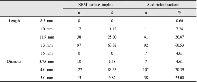
두 종류의 임플란트 모두에서 직경 4mm와 길 이 13mm의 임플란트가 가장 많이 식립되었으며 (Table Ⅱ), 식립 부위로는 상,하악 구치부에서 많 이 식립되었다 (Table Ⅲ).
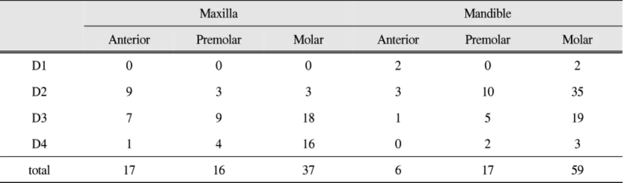
수술 부위의 골질의 상태는 Lekholm과 Zarb의 분류에 따라 수술 시에 평가하여 기록하였다9) (Table Ⅳ~Ⅴ).
임플란트 보철은 RBM 임플란트에서는 fixed bridge가, 산부식 임플란트에서는 single crown이 가장 많았으며, 대합치로는 두 종류의 임플란트 모두에서 자연치가 가장 많았다 (Table Ⅵ~Ⅶ).
2. 연구방법
모든 임플란트는 식립 후 submerged 되었으며, 식립 부위의 상태에 따라 2-6 개월 후에 2차 수 술을 시행하여 노출시켰고, 2차 수술 후 1-2개월 이내에 보철치료가 종료되었다. 임플란트 식립 전과 후에 미리 정해진 평가항목에 따라 조사하 여 기록하였으며, 환자들은 최소 36개월 이상 추 적 관찰을 시행하였다.
임플란트 실패는 Albreksson 등10)이 제시한 임 플란트 성공기준과 또 Buchs 등11)이 제안한 기준 을 참고로 하여 다음 사항이 존재하는 경우를 실 패로 간주하였다.
(1) 임플란트 동요
(2) 임플란트가 제거된 경우 (3) 임플란트 파절
RBM surface implant Acid-etched surface implant
Gender women men women men
No. of implants 72 80 57 95
No. of patients 23 32 32 53
Range of age 24 to 73 18 to 67
Mean age 47.58 45.97
Table Ⅰ. Patients characteristics
RBM surface implant Acid-etched surface
n % n %
Length 8.5 mm 0 0 1 0.66
10 mm 17 11.18 11 7.24
11.5 mm 38 25.00 41 26.87
13 mm 97 63.82 92 60.53
15 mm 0 0 7 4.61
Diameter 3.75 mm 10 6.58 7 4.61
4.0 mm 127 83.55 107 70.39
5.0 mm 15 9.87 38 25.00
Table Ⅱ. Distribution of Implants by length and diameter
RBM surface implant Acid-etched surface
n % n %
Anterior 12 7.89 17 11.18
Maxilla Premolar 21 13.82 16 10.53
Molar 30 19.74 37 24.34
Anterior 7 4.61 6 3.95
Mandible Premolar 18 11.84 17 11.18
Molar 64 42.11 59 38.82
Table Ⅲ. Distribution of implants by site
Maxilla Mandible
Anterior Premolar Molar Anterior Premolar Molar
D1 0 0 1 0 3 6
D2 1 6 9 4 9 31
D3 5 11 13 3 4 21
D4 6 4 7 0 2 6
total 12 21 30 7 18 64
Table Ⅳ. Distribution of bone quality in site of RBM surface implant
Maxilla Mandible
Anterior Premolar Molar Anterior Premolar Molar
D1 0 0 0 2 0 2
D2 9 3 3 3 10 35
D3 7 9 18 1 5 19
D4 1 4 16 0 2 3
total 17 16 37 6 17 59
Table Ⅴ. Distribution of bone quality in site of acid-etched surface implant
Prosthesis RBM surface implant Acid-etched surface implant
n % n %
Single crown 48 29.61 78(6 failure) 51.32
Fixed bridge 98(1 failure) 64.47 66(1 failure) 43.42
Overdenture 6 3.95 8 5.26
Table Ⅵ. Distribution of implants by type of prostheses
(4) 지속적인 동통이나 불편감
모든 환자의 보철 치료 완료 후 1년이 경과한 다음, 진료 기록부 및 방사선 사진을 토대로 위 와 같은 기준을 이용하여 임플란트 생존율을 비 교하였고, 실패의 유형과 원인을 분석하였다.
결 과
RBM 임플란트의 경우 55명의 환자에서 152개 의 임플란트가 식립되었으며, 임상적, 방사선학 적으로 실패한 임플란트는 1개로 99.34%의 생존
Opposing dentition RBM surface implant Acid-etched surface implant
n % n %
Natural tooth 82 53.95 92(6 failure) 60.53
Crown 33 21.71 21 13.82
Implant 25 16.45 31(1 failure) 20.39
Removable denture 12 7.89 8 5.26
Table Ⅶ. Distribution of opposing dentitions
No. of implants No. of failed implant Survival rate(%)
RBM surface implant 152 1 99.34%
Acid-etched surface implant 152 7 95.39%
Table Ⅷ. Implant failure and survival rate
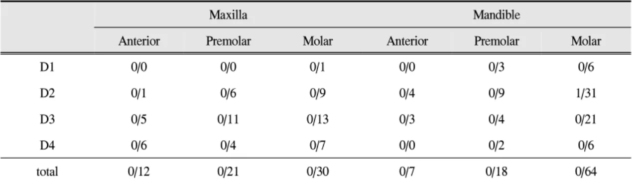
Maxilla Mandible
Anterior Premolar Molar Anterior Premolar Molar
D1 0/0 0/0 0/1 0/0 0/3 0/6
D2 0/1 0/6 0/9 0/4 0/9 1/31
D3 0/5 0/11 0/13 0/3 0/4 0/21
D4 0/6 0/4 0/7 0/0 0/2 0/6
total 0/12 0/21 0/30 0/7 0/18 0/64
Table Ⅸ. Distribution of the failed RBM surface implant by site & bone quality
율을 보였다. 반면에, 산부식 임플란트는 85명의 환자에서 152개가 식립되었고 그 중 7개가 실패 하여 95.39%의 생존율을 보였다 (Table Ⅴ).
실패한 임플란트가 식립된 부위별로 살펴보 면, RBM 임플란트의 경우엔 하악 대구치 부위에 서 1개가 실패하였으며 골질은 2형이었다 (Table
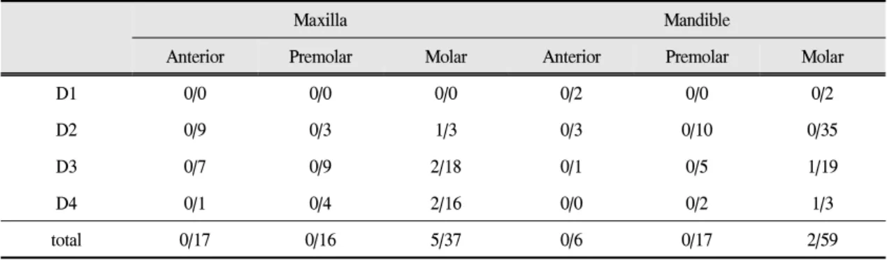
Ⅸ). 산부식 임플란트는 상악 구치부에서 5개가 실패하였고, 하악 구치부에서 2개가 실패하였으
며, 골질은 주로 3형과 4형이었다 (Table Ⅹ).
실패한 임플란트의 보철 유형을 살펴보면, 산 부식 임플란트에서는 7개의 실패 중 6개가 단일 치아 수복의 경우에 발생하였다 (Table Ⅺ).
대부분의 실패한 임플란트에서 대합치는 자연 치열인 경우가 가장 많았다 (Table Ⅻ).
RBM 임플란트에서는 직경 4mm 임플란트 가 저작 중 매식제의 파절로 실패하였고, acid-
Maxilla Mandible
Anterior Premolar Molar Anterior Premolar Molar
D1 0/0 0/0 0/0 0/2 0/0 0/2
D2 0/9 0/3 1/3 0/3 0/10 0/35
D3 0/7 0/9 2/18 0/1 0/5 1/19
D4 0/1 0/4 2/16 0/0 0/2 1/3
total 0/17 0/16 5/37 0/6 0/17 2/59
Table Ⅹ. Distribution of the failed acid-etched surface implants by site & bone quality
Prosthesis RBM surface implant Acid-etched surface implant
single crown 0/48 6/78
Fixed bridge 1/98 1/66
Overdenture 0/6 0/8
Table Ⅺ. Distribution of the prostheses type in the failed implants
Opposing dentition RBM surface implant Acid-etched surface implant
n % n %
Natural tooth 82 53.95 92(6 failure) 60.53
Crown 33 21.71 21 13.82
Implant 25 16.45 31(1 failure) 20.39
Removable denture 12 7.89 8 5.26
Table Ⅻ. Distribution of opposing dentitions of the failed implants
No. age/sex diameter×length Bone quality Site factor
1 49/M 4×13mm D2 #46 fracture of fixture
Table ⅩⅢ. Analysis of the failed RBM surface implant
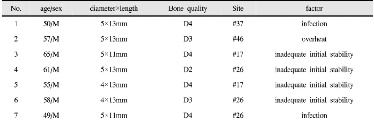
No. age/sex diameter×length Bone quality Site factor
1 50/M 5×13mm D4 #37 infection
2 57/M 5×13mm D3 #46 overheat
3 65/M 5×11mm D4 #17 inadequate initial stability
4 61/M 5×13mm D2 #26 inadequate initial stability
5 55/M 4×13mm D4 #17 inadequate initial stability
6 58/M 4×13mm D3 #26 inadequate initial stability
7 49/M 5×11mm D4 #26 infection
Table ⅩⅣ. Analysis of the failed acid-etched surface implants
etched surface 임플란트에서는 직경 5mm 임플란 트가 5개, 직경 4mm 임플란트가 2개 실패하였으 며, 실패 원인으로는 초기고정 불량이 4례, 감염 이 2례, 과열이 1례였다 (Table ⅩⅢ & ⅩⅣ).
고 찰
골 유착성 치과 임플란트는 기존의 고정성 또 는 가철성 보철물의 단점인 치조골의 흡수나 저 작 효율의 감소 등 환자의 불만족을 해결할 수 있는 획기적인 치료법으로 인정받게 되었다2). 하 지만 치과 임플란트의 실패가 많은 역효과를 유 발하면서, 실패율을 감소시키기 위한 노력이 지 속적으로 이루어지고 있으며, 이를 위해 식립된 임플란트의 성공 및 실패에 대한 분석을 통한 연 구가 활발히 진행되어 왔다. 이러한 치과 임플란 트가 보편화되고 그만큼 성공적인 치료 방법으 로 인정받게 된 것은 임플란트의 디자인, 표면 처리 방법, 임플란트의 생역학 등에 걸쳐 전반적 인 발전이 있었기 때문이다.
우선, 성공적인 임플란트와 골과의 융합을 얻 기 위해서는 초기 안정성이 필수적인 요구조건 이며 이에 술자의 경험과 술기, 임플란트의 적절 한 디자인과 표면처리가 중요하다. 이중 임플란 트의 표면처리 방식에는 많은 발전이 있어왔는
데, 이는 표면처리를 하지 않은 매끈한 1세대 임 플란트의 표면적을 넓히기 위한 것들이었다. 임 플란트 표면에 티타늄 powder를 고온으로 용융 하여 부착시킨 TPS (Titanium Plasma Sprayed) 표 면, 또는 titanium sphere를 소결해서 부착시킨 표 면의 임플란트들이 2세대에 해당된다. 하지만 이 들은 임플란트 표면의 부착물들이 골유착 후 표 면과 분리되어 임플란트의 실패를 유발하였다.
이와는 반대로 임플란트 표면에 매질(media)을 분사시켜 표면이 패이게 함으로써 표면적을 넓 히는 방식의 3세대 임플란트가 개발되었으나, 매 질이 표면에 잔존하여 임플란트의 실패를 유발 하기도 하였다2). 이에 이러한 문제점을 해결하기 위해 흡수성 매질 (Resorbable Blasting Media;
RBM)을 사용하게 되었다.
RBM 임플란트는 3세대에 해당되며, 흡수성 매질을 이용하여 임플란트의 거칠기와 표면적을 증가시켰다. Wennerberg12) 등은 적절한 거친 표 면을 가질 수 있게 RBM 표면 처리된 임플란트 가 removal torque, value, 골 접촉률 등에 있어서 평활면 임플란트 보다 우수한 결과를 보였다고 보고하였다. blasting을 이용하여 표면처리를 하 였을 경우에 분사되는 입자들의 영향으로 표면 적이 증가되며 요철 효과로 인하여 골과의 결합 력이 증가되며 거친 표면으로 세포의 반응이 활
성화되는 잇점을 갖는다. RBM 임플란트 표면 처 리 방식에 이용되는 매질로는 생체 친화성이 우 수한 알루미나(Al2O3), 산화타이타늄(TiO2), 산화 칼슘(Ca3PO4), 수산화인회석(HA) 등이 있다.
많은 연구들에서 조직형태계측학적인 분석을 통하여 임플란트의 표면 거칠기를 증가시키는 것은 골과 임플란트의 접촉을 증가시키는데 있 어 효과가 있는 것으로 보고하고 있다13-14). 임플 란트 표면의 거칠기를 증가시키기 위한 RBM 방 식을 이용한 임플란트에 관한 연구에서 Piattelli 등15)은 토끼의 대퇴골에 machined 표면 임플란트 와 RBM 방식으로 처리한 임플란트를 식립하고 8주 후에 조직형태계측학적으로 비교한 결과 RBM 표면 처리 임플란트가 machined 표면 임플 란트에 비하여 골아세포와 성숙한 골이 더 많이 임플란트에 직접 접촉되어 있는 것을 관찰하였 음을 보고하였다. Maurizo 등14)은 가토의 슬관절 에 RBM 표면 처리 임플란트와 machined 표면 임 플란트를 식립하였는데 RBM 표면 처리 임플란 트가 machined 표면 임플란트에 비하여 골 접촉 률이 높았으며 골 형성도 빨랐다고 보고하였다.
Sanz 등16)은 조직형태계측학적으로 평활면 임플 란트와 RBM 임플란트를 비교하였는데 평활면 임플란트는 51%의 골-임플란트 접촉률을 보였으 며 RBM 표면 처리 임플란트는 62.3%의 골-임플 란트 접촉률을 보여 RBM 임플란트가 골-임플란 트 접촉률이 높았음을 보고하였다. 이때, RBM 임플란트 주위에서 칼슘을 함유한 부위의 분포 와 농도가 더 높은 것으로 보와 RBM 임플란트 가 초기 골 형성이 많이 되며 골 형성의 속도도 더 빠른 것으로 생각된다고 하였다. 본 연구에서 RBM 임플란트의 성공률은 99.34%로 높은 성공 률을 보였다.
본 연구에서 사용된 OsseotiteⓇ 임플란트는 타 이타늄 표면을 황산과 염산으로 열이중산부식 (thermal dual acid etching) 처리 한 것이다17,18). 이 는 표면 처리를 하지 않은 평활한 표면과는 달리 표면구조가 산과 골들로 조밀하게 구성되어 있
으며, 산과 산 간격이 1-3μm, 산과 골의 간격이 5-10μm로 되어 있어, 계면접촉의 면적이 두배 이 상 넓다. 또한 1μm 이하의 지름을 가지고 있는 섬유 혈병이 견고하게 결합될 수 있으며, 결합된 섬유 혈병을 따라서 조골세포가 모여서 골 생성 을 한다고 한다19). 따라서 불량한 골질에서도 골 결합이 우수하며, 골치유 시기가 촉진되어 수술 후 6주만에 골 결합을 이룰 수 있다고 보고된 바 있다20). Testori 등21)은 OsseotiteⓇ를 임플란트 치 료환자에게 적용하여 4년간 추적 조사한 결과 그 성공률이 98.7% 라고 보고하였다. Sullivan 등
17)은 OsseotiteⓇ의 6년간 누적 성공률이 96.6%라 하였으며, 흡연자, 골질이 불량한 경우, 길이가 짧은 임플란트, 또는 시술 후 조기부하를 가한 증례 등에 있어서 성공률의 차이가 보이지 않았 다고 보고하였다. 본 연구에 사용된 OsseotiteⓇ 임플란트는 95.39%의 성공률을 보여 이전의 보 고들 보다 다소 저조한 성공률을 보였다.
결 론
1. 총 152개의 RBM 임플란트를 식립하였고, 이 중 실패는 1개의 임플란트에서 발생하였다.
실패한 임플란트는 하악 제1대구치 부위에 식 립된 증례였으며 식립체의 파절로 인하여 실 패하였다. 그 밖의 임플란트는 관찰된 기간 중 양호한 양상을 보였으며, 99.34%의 생존율을 나타냈다.
2. 총 152개의 산부식 임플란트를 식립하였고, 이중 7개의 임플란트가 실패를 하였으며, 95.39%의 생존율을 나타냈다. 실패의 원인은 과열, 감염, 초기고정 불량 등으로 추정되었다.
본 연구는 서로 다른 표면처리를 시행한 두 가 지 임플란트 시스템의 임상적 결과에 대한 후향 적 연구로서, 두 가지 임플란트 모두 양호한 생 존율을 보였으나, RBM 임플란트에서 다소 높은 생존율을 나타내었다.
연구비 지원 및 사의
이 논문은 2011년 조선대학교 학술연구비의 지원을 받아 연구되었음.
참 고 문 헌
1. Quirynen M, Bollen CM, PaPaioannou W et al. The influence of titanium abutment surface roughness on plaque accumulation and gingivitis: Short-term observations. Int J oral Maxillofac Implants 1996;11:
169-178.
2. Alvreksson T, Qarb G, Worthington P et al : The long-term efficacy of currently used dental implants:
A review and proposed criteria of success. Int J oral Maxillofac Implants 1986;1:11-25.
3. Hutton JE, Heath MR, Chai JY et al : Factors related to success and failure rates at 3-year follow-up in a multicenter study of overdentures supported by Branemark implants. Int J Oral Maxillofac Implants 1995;10:33-42.
4. Martinez H, Davarpanah M, Missika P et al. Optimal implant stabilization in low density bone. Clin Oral Implants Res 2001;12:423-432.
5. Piattelli M, Scarano A, Paolantonio M et al. Bone response to machines and resorbable blast material titanium implants: An experimental study in rabbits.
J Oral Implantol 2002;28:2-8.
6. Trisi P, Lazzara R, Rao W et al. Bone-implant co ntact and bone quality: Evaluation of expected and actual bone contact on machined and osseotite implant surface. Int J Periodontics Restorative Dent 2002;22:533-545.
7. Choi JY, Koh SW, Ryu HW. Clinical study on survival rate of osseointegrated implants. J Korean Assoc Maxillofac Plast Reconstr Surg 2009;31:
306-313.
8. Jeon HR, Kim MR, Lee DH et al. Four-year survival rate of RBM surface internal connection non- submerged implants and the change of the peri-implant crestal bone. J Korean Assoc Maxillofac Plast Reconstr Surg 2009;31:237-242.
9. Lekholm U, Zarb GA. Patient selection and preparation. In: Branemark, PI, Zarb, GA, Albrektsson, T(eds). Tissue-integrated prostheses. Chicago: Quintessance publishing Co; 1985:199-209.
10. Albrektsson T, Zarb G, Worthington P et al. The long-term efficacy of currently used dental implants:
a review and proposed criteria of success. Int J Oral Maxillofac Implants 1986;1:11-25.
11. Buchs AU, Hahn J, Vassos DM. Interim clinical study report :a threaded, hydroxyapatite-coated implant- five-year post-restoration safety and efficacy.
J Oral implantol 1995;21:266-274.
12. Wennerberg A, Albrektsson, Lausmaa J. Torque and histomorphometric and removal torque study of screw- shaped titanium implants and 75um sized particles of Al2O3. J Biomed Mater Res 1996;30:
251-260.
13. Buser D, Scjenk RK, Steinemann S et al. Influence of surface characteristics in bone integration titanium implants. A stomor phometic study in miniature pigs.
J Biomed Mater Res 1991;25:889-902.
14. Maurizio P, Antonio S, Michele P et al. Bone reponse to machined and resorbable blast material titanium implants: An experimental study in rabbits. Oral implantol 2002;28:2-8.
15. Piattelli M, Scarano A, Paolantonio M et al. Bone response to machines and resorbable blast material titanium implants: An experimental stydy in rabbits.
J Oral Implantol 2002;28:2-8.
16. Sanz A, Oyarzum A, Farias D et al. Experimental study of bone response to a new surface treatment of endosseous titanium implants. Implant Dent 2001;10:
126-131.
17. Sullivan DY, Sherwood RL, Mai TN. Preliminary results of a multicenter study evaluating a chemically enhanced surface for machined commercially pure titanium implants. J Prosthet Dent 1997;78:379-386.
18. Klokkevold PR, Nishimura RD, Adachi M et al.
Osseointegration enhanced by chemical etching of the titanium surface. A torque removal study in the rabbit. Clin Oral Implants Res 1997;8:442-447.
19. Wong M, Eulenberger J, Schenk R et al. Effect of surface topology on the osseointegration of implant
materials in trabecular bone. J Biomed Mater Res 1995;29:1567-1575.
20. Nishimura K, Itoh T, Takaki K et al. Periodontal parameters of osseointegrated dental implants. A 4-year controlled follow-up study. Clin Oral Implants Res 1997;8:272-278.
21. Testori T, Wiseman L, Woolfe S et al. A prospective multicenter clinical study of the Osseotite implant:
four-year interim report. Int J Oral Maxillofac Implants 2001;16:193-200.
3-Year Survival Analysis of RBM and Acid-Etched Surface Implants
Dae-Woong Yoon, Moon-Seob Kim, Soo-Young Jin, Han-Seung Jang, Deuk-Hyun Mah, Gyeong-Dal Jeong, Hyun-Chun Park, Hee-Jung Kim*, Hak-Kyun Kim
Department of Oral and Maxillofacial Surgery, Department of Prosthodontics*, School of Dentistry, Chosun University
The purpose of this study was to analyze and compare survival rates of resorbable blast media(RBM) surface and acid-etched surface implants being usually used in clinics. RBM surface implants (USII, Osstem, Busan, Korea) or acid-etched surface implants (Osseotite®, Biomet 3i[TM], FL, USA) were placed in edentulous area of 140 patients between January of 2005 and March of 2007. The number of implants was 304, and 152 out of them were RBM surface implants while another 152 were acid-etched surface implants. According to the evaluation items, the survey was performed before and after the implants installations. The 3-year survival rates of both kind of implants were calculated. 1. Total of 152 RBM surface implants were placed. Among them, one implant was failed, which was implanted in the posterior mandible with D2 bone quality. The failure was resulted from fracture of the fixture. Others showed good results and survival rate of RBM surface implant was 99.34%. 2. Total of 152 acid-etched surface implants were placed. Seven implants of them were failed, thus, survival rate was 95.39%. The causes of the failures were considered as infection, overheat and the lack of initial stability. In this research, both implants showed good 3-year survival rate, although RMB surface implant represented a better result.
Key words: resorbable blasting media, acid-etched, survival rates
Correspondence to : Dr. Hak-Kyun Kim
Department of Oral & Maxillofacial Surgery, School of Dentistry, Chosun University.
421 Seosuk-Dong, Dong-Gu, Gwangju, 501-825, Korea
Tel: 82-62-220-3816, Fax: 82-62-224-9172, Email: [email protected]
Received: October 22, 2011, Last Revision: November 15, 2011, Accepted: December 25, 2011