www.jpis.org
pISSN 2093-2278 eISSN 2093-2286 Copyright © 2010 Korean Academy of PeriodontologyThis is an Open Access article distributed under the terms of the Creative Commons Attribution Non-Commercial License (http://creativecommons.org/licenses/by-nc/3.0/).
Bone-added osteotome sinus floor elevation with simultaneous placement of non-submerged sand blasted with large grit and acid etched implants:
a 5-year radiographic evaluation
Jee-Hee Jung1, Seong-Ho Choi2, Kyoo-Sung Cho2, Chang-Sung Kim2*
1Department of Periodontology, Yonsei University College of Dentistry, Seoul, Korea
2Department of Periodontology, Research Institute for Periodontal Regeneration, Oral Science Research Center, Yonsei University College of Dentistry, Seoul, Korea
Purpose: Implant survival rates using a bone-added osteotome sinus floor elevation (BAOSFE) procedure with simultaneous placement of a non-submerged sand blasted with large grit and acid etched (SLA) implant are well documented at sites where native bone height is less than 5 mm. This study evaluated the clinical results of non-submerged SLA Straumann implants placed at the time of the BAOSFE procedure at sites where native bone height was less than 4 mm. Changes in graft height af- ter the BAOSFE procedure were also assessed using radiographs for 5 years after the implant procedure.
Methods: The BAOSFE procedure was performed on 4 patients with atrophic posterior maxillas with simultaneous placement of 7 non-submerged SLA implants. At least 7 standardized radiographs were obtained from each patient as follows: before sur- gery, immediately after implant placement, 6 months after surgery, every year for the next 3 years, and after more than 5 years had passed. Clinical and radiographic examinations were performed at every visit. Radiographic changes in graft height were calculated with respect to the implant’s known length and the original sinus height.
Results: All implants were stable functionally, as well as clinically and radiographically, during the follow-up. Most of the ra- diographic reduction in the grafted bone height occurred in the first 2 years; reduction after 2 years was slight.
Conclusions: The simultaneous placement of non-submerged SLA implants using the BAOSFE procedure is a feasible treat- ment option for patients with severe atrophic posterior maxillas. However, the grafted bone height is reduced during the healing period, and patients must be selected with care.
Keywords: Dental implantation, Maxillary sinus, Radiography.
INTRODUCTION
The placement of implants in the posterior maxilla is limit- ed occasionally by insufficient bone volume as a result of al- veolar atrophy or pneumatization of the maxillary sinus. This
clinical problem can be resolved by sinus augmentation us- ing surgical procedures such as onlay augmentation of the alveolar crest [1,2], Le Fort I osteotomy with an interpositional bone graft [3,4], lateral-approach sinus augmentation [5-7], or osteotome sinus augmentation [8-11].
Received: Feb. 10, 2010; Accepted: Mar. 24, 2010
*Correspondence: Chang-Sung Kim
Department of Periodontology, Research Institute for Periodontal Regeneration, Yonsei University College of Dentistry, 134 Shinchon-dong, Seodaemun-gu, Seoul 120-752, Korea
E-mail: [email protected], Tel: +82-2-2228-3186, Fax: +82-2-392-0398
vation procedure employing simultaneous grafting and the immediate placement of implants [8]. Using the Summers osteotome kit [8,9], which was specifically designed for this procedure, the pre-existing crestal bone is displaced toward the sinus floor as the osteotomes are inserted. Various graft materials and implants can be used in this surgical proce- dure. However, a minimum native bone height is required to get initial stability of the implant, and at least 5 mm of alveo- lar ridge height under the sinus is recommended for an im- plant that is 10 mm or longer [9]. Clinical case reports and studies on the bone-added osteotome sinus floor elevation (BAOSFE) procedure with simultaneous placement of im- plants show a relatively high survival rate in non-submerged sand blasted with large grit and acid etched (SLA) implants (94-98%) [10-15], but implant survival rates drop significantly when native bone height is 4 mm or less. Therefore, there are only a few clinical case reports involving sites with less than 4 mm of native bone height.
This report evaluates the clinical results of non-submerged SLA implants placed at the time of the BAOSFE procedure at sites where native bone height was less than 4 mm. Changes in graft height after the BAOSFE procedure were assessed ra- diographically for 5 years after the implant procedure.
MATERIALS AND METHODS
Patients
Four consecutive patients (2 women and 2 men, mean age of 61) with severe atrophy of the alveolar process in the pos- terior maxilla were treated at the Department of Periodon- tology, Yonsei University College of Dentistry. The patients showed no signs or symptoms of sinus or intraoral disease.
All four patients underwent the BAOSFE procedure with si- multaneous placement of a total of 7 Straumann SLA im-
The patients provided informed consent to participate in this clinical evaluation. The evaluation was approved by the Insti- tutional Review Board at Yonsei University Dental Hospital (IRB No. 2-2009-0024).
Surgical techniques
All patients’ medical histories were reviewed at an initial examination in order to rule out any local or systemic diseas- es that might contraindicate the surgical procedures. The pa- tients received oral hygiene instructions and whole-mouth scaling prior to the surgery.
The BAOSFE procedure was performed using a Summers Osteotome kit, (3i Implant Innovations, Palm Beach Gardens, USA), as described by Summers [8,9]. Briefly, an incision was made under local anesthesia of lidocaine 2% with 1:80,000 epinephrine (Kwangmyung Pharmaceutical, Seoul, Korea) at the edentulous area to be treated. After the crestal incision was made, full-thickness buccal and palatal flaps were re- flected. Site preparation was begun using the Summers #1 and #2 osteotomes. When the bone was too dense for hand instrumentation, 2-mm twist drilling was used to reach the cancellous bone, stopping 1 mm below the floor of the sinus.
The preparation site was widened using #2 and #3 Summers osteotomes. Prepared bone graft material with beta-tricalci- um phosphate, Cerasorb (Curasan AG, Kleinostheim, Germa- ny), and demineralized freeze-dried bone, Dembone (Pacific Coast Tissue Bank, Los Angeles, USA), which acts as a shock absorber, was added to the preparation site with a carrier. El- evation of the maxillary sinus membrane was achieved using the #3 osteotome that was used previously to force the graft ahead of its tip to achieve the sinus floor up-fracture. At this stage, the integrity of the sinus membrane was confirmed by the Valsalva maneuver. Finally, the non-submerged Strau- mann SLA implants were place into the osteotomy site. Pri- Table 1. Radiographic measurements for each patient.
Patient
no. Site
tooth no. Bone quality
Implant NBH
(mm) GBH0
(mm) GBH6
(mm) GBH12
(mm) GBH24
(mm) GBH36
(mm) GBH60
(mm) Reduction24
(mm) Reduction60
D (mm) L (mm) (mm)
1 27 D2 4.1 10 3.6 8.6 8.4 6.4 6.4 6.4 6.4 2.2 2.2
2 18 D3 4.1 10 4.0 8.0 7.6 7.6 6.8 6.6 6.1 1.1 1.8
3 15 D2 4.1 10 3.1 9.9 10.5 9.7 7.4 7.3 7.2 2.5 2.7
16 D2 4.8 10 4.0 9.0 8.8 8.7 7.0 6.9 6.2 2.0 2.8
4 16 D3 4.1 10 3.3 9.0 8.2 6.7 6.7 6.7 6.7 2.3 2.3
17 D3 4.1 10 2.1 9.2 8.7 8.1 7.9 7.9 7.9 1.3 1.3
18 D3 4.1 10 3.7 6.9 6.0 6.9 6.7 6.3 6.3 0.1 0.6
Mean 3.4 8.6 8.3 7.7 7.0 6.9 6.7 1.6 1.9
The mean total reduction in grafted bone height was 1.9 mm. Reduction was greatest during the first 2 years (1.6 mm).
Subscript numbers indicate the number of months elapsed since the surgery.
D: diameter, L: length, NBH: native bone height, GBH: grafted bone height.
was achieved using monofilament suture material, Ethilon (Johnson & Johnson Int., Edinburgh, UK). All surgical proce- dures were performed by C. S. Kim.
Postoperatively, patients were instructed to rinse their mouth twice a day with a 0.12% chlorhexidine solution, Hex- amedin (Bukwang Pharmaceutical Co., Seoul, Korea) for 2 weeks after surgery. Antibiotics were prescribed for 7 days, and sutures were removed after 10 days. After a mean heal- ing period of 7 months, all patients were rehabilitated with fixed crowns or bridges.
Follow-up
After inserting the implants, the patients received follow-up care at 1 and 2 weeks, at 3, 6, and 9 months, and every 6 months thereafter. Clinical and radiological evaluations were performed using standardized radiographs according to the following schedule: prior to surgery, immediately after sur- gery, 6 months after surgery, and then every year after sur- gery up to 5 years.
Radiographic analysis of the grafted bone height
Using a scanner, HP scanjet 7400c (Hewlett Packard, Palo
age analysis program Image-Pro Plus (Media Cybernetics, Silver Spring, USA) was used for linear analysis of the radio- graphs. The magnification of each radiograph was corrected using the known actual length of the inserted implants so that an accurate graft height could be obtained. The radio- graphs from the same patient were blinded in terms of which time point they represented. Native bone height, grafted bone height, and implant height were measured on each ra- diograph as described in Fig. 1.
RESULTS
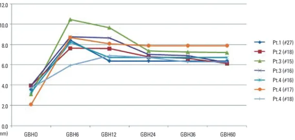
Radiographic examination showed that the sinus floor was elevated immediately after surgery in all patients. Table 1 shows the radiographic measurements for each patient. The mean native bone height was 3.4 mm. The average gain in the grafted bone height of the implants was 8.6 mm (range, 6.9-9.9 mm). The grafted bone area was easily distinguished from the sinus floor on the radiographs. Clinical and radio- graphic examination during the initial healing period showed normal healing in all patients. At 6 months, radiographic eval- uation showed the maturation of the grafted bone, including increased density and sinus floor remodeling. Although the change in grafted bone height varied from patient to patient, there were marked differences in bone height immediately after the surgery versus 2 years after surgery. The mean re- duction in grafted bone height, which was gradual, was 1.6 mm (85% of the mean total reduction) during the first 2 years.
(Fig. 2) In contrast, subsequent grafted bone height reduc- tion was minimal: After 2 years, the mean bone height was further reduced by 0.3 mm (15% of the mean total reduction).
In case of patient no.4, significant radiographic remodeling of grafted bone occurred also during the first 2 years. And the mean reduction was minimal between 2 and 6 years (Fig. 3).
Thus, the total mean reduction in the grafted bone height was 1.9 mm 5 years after surgery. All implants were function- ally stable, and crestal bone remodeling was minimal.
DISCUSSION
This report evaluated the clinical results of non-submerged SLA implants placed simultaneously in sites with less than 4 mm of native bone height using the BAOSFE procedure. Us- ing radiographs, this report also assessed changes in the grafted bone height during the long-term (5-year) healing period.
All implants were maintained successfully for over 5 years.
The results suggested that simultaneous placement of non- submerged Straumann SLA implants using the BAOSFE pro- Figure 1. Schematic drawing of the measured parameters. (A) Na-
tive bone height: the distance from the alveolar crest to the floor of the maxillary sinus at the implant site, which was calculated as the mean of the mesial and distal native bone heights. Grafted bone height: the distance from the floor of the maxillary sinus to the bor- der of the grafted bone at the implant site, which was calculated as the mean of the mesial (B) and distal (B�) grafted bone heights. (C) The implant height: the distance from the apex to the head of the fixture.
cedure is a feasible treatment option for patients with atro- phic posterior maxillas. However, radiographic reduction of the grafted bone height was observed, especially during the first 2 years of the healing period, although there was some variation among the patients. Therefore, patients must be chosen carefully and the clinicians should consider that some reduction will occur. There was some variation in results among patients, depending on the follow-up time, inclusion
criteria, surgical and prosthetic techniques, and other factors;
however, the BAOSFE procedure with simultaneous place- ment of an implant shows a predictable survival rate ranging from 95-100% [6,10,11]. The 1-step approach using the BAOS- FE procedure has the advantage of being less invasive, and this technique can enhance the bone quality of the implant site from type III or IV to type II. Reducing the surgical and healing times can be achieved because coordinated consoli- Figure 3. Radiographic evaluation of changes in grafted bone height using a series of radiographs from patient no. 4. The radiographs show remodeling of the grafted bone during the first post-surgery examination. (A) Prior to surgery, (B) immediately after surgery, (C) six months after surgery, (D) one year after surgery, (E) two years after surgery, (F) three years after surgery, (G) five years after surgery, (H) six years after surgery.
E F G H
A B C D
Figure 2. Periodical changes of grafted bone height based on the radiographic analysis. Results show that the notable radiographic changes occurred during the first 2 years after surgery. Numbers indicate the number of months elapsed since the surgery. Pt: patient, #: tooth num- ber by F.D.I. numbering system, GBH: grafted bone height.
Pt.1 (#27) Pt.2 (#18) Pt.3 (#15) Pt.3 (#16) Pt.4 (#16) Pt.4 (#17) Pt.4 (#18) 12.0
10.0 8.0 6.0 4.0 2.0
(mm)0.0 GBHO GBH6 GBH12 GBH24 GBH36 GBH60
period is expected. Moreover, little difference has been re- ported between the survival rate of implants placed at the time of grafting versus those placed after a delay [16]. Differ- ences in implant design and surface characteristics may in- fluence the survival rate of different types of implants [11].
The superiority of SLA surface implants in conjunction with the osteotome sinus floor elevation technique has been doc- umented in many studies [17,18]. Regarding the extent of bone retention, some studies have reported that the SLA sur- face is superior to a machined-surface implant [19,20]. More- over, the survival rate of SLA-surface implants in the sinus- augmented maxilla is markedly higher than that of the ma- chined-surface implants [21].
The survival rate of implants is also influenced by the qual- ity and quantity of the native bone [11,12,22]. In particular, the survival rate is clearly reduced when the native bone height in an implant site is 4 mm or less [11]: It is difficult to achieve primary stability of the implant, and there is a higher possi- bility that the Schneiderian membrane will tear [23]. Howev- er, this is somewhat controversial. Peleg et al. [24] evaluated the efficacy of augmentation of the maxillary sinus using a lateral approach with simultaneous placement of hydroxyap- atite surface implants in patients with 3-5 mm of residual bone height. In 63 patients, all 160 implants were stable dur- ing the 2- to 4-year follow-up periods. Together with previous studies, these results show that using rough surface implants in the augmented sinus area results in a predictable progno- sis. Therefore, a 1-step procedure involving both grafting of the maxillary sinus and simultaneous placement of rough surface implants might be a feasible treatment option for pa- tients with as little as 5 mm of native bone height.
In this report, the height of the grafted bone was reduced markedly by an overall mean of 1.6 mm during the course of the short-term healing period, i.e. the first 2 years. During the long-term healing period, i.e. over 5 years, the height of the grafted bone was reduced by an overall mean of 1.9 mm.
Dimensional changes in the height of augmented grafts in the sinus have been documented in clinical and radiographic studies [25,26]. At the Sinus consensus conference in 1996, there was a report on 100 patients and 145 sinus-grafting sites that were evaluated using panoramic radiographs over a 3-year period. All graft materials resulted in a radiographic reduction ranging from 0.79-2.09 mm. However, it was not determined whether this reduction in graft height occurred in the initial healing period or was part of an ongoing heal- ing process. Hallman et al. analyzed 30 maxillary sinuses in 20 patients who were grafted with a mixture of autogenous bone and bovine hydroxyapatite, and reported that a small (<10%) but statistically significant dimensional reduction was
[27]. Other studies on the reduction of sinus grafts using X- rays have also been performed; most of these studies show agreement with the results of this report in that that shrink- age of the grafted materials and reduction in grafted bone height were observed during the initial healing period after the BAOSFE procedures were performed [28-30]. Hatano et al. [31] assessed long-term changes in the sinus-graft height after a maxillary sinus floor augmentation with simultaneous placement of implants. Those results showed that the graft height decreased during the first 2-3 years after augmenta- tion, but all subsequent changes were minimal.
While small, this report suggests that simultaneous place- ment of non-submerged SLA implants using the BAOSFE procedure is a feasible treatment option for patients with se- verely atrophic posterior maxillas. However, the grafted bone height is reduced during the healing period, and clinicians should expect some radiographic reduction of the grafted bone height, especially in the first few years after the proce- dure.
CONFLICT OF INTEREST
No potential conflict of interest relevant to this article was reported.
ACKNOWLEDGMENTS
This study was supported by a faculty research grant of Yonsei University College of Dentistry for 2009 (6-2009- 0040).
REFERENCES
Jensen J, Simonsen EK, Sindet-Pedersen S. Reconstruc- 1.
tion of the severely resorbed maxilla with bone grafting and osseointegrated implants: a preliminary report. J Oral Maxillofac Surg 1990;48:27-32.
Adell R, Lekholm U, Grondahl K, Branemark PI, Lindstrom 2.
J, Jacobsson M. Reconstruction of severely resorbed eden- tulous maxillae using osseointegrated fixtures in immedi- ate autogenous bone grafts. Int J Oral Maxillofac Implants 1990;5:233-46.
Isaksson S. Evaluation of three bone grafting techniques 3.
for severely resorbed maxillae in conjunction with imme- diate endosseous implants. Int J Oral Maxillofac Implants 1994;9:679-88.
Kahnberg KE, Nystrom E, Bartholdsson L. Combined use 4.
of bone grafts and Branemark fixtures in the treatment of severely resorbed maxillae. Int J Oral Maxillofac Implants
Fugazzotto PA. Maxillary sinus grafting with and without 5.
simultaneous implant placement: technical consider- ations and case reports. Int J Periodontics Restorative Dent 1994;14:544-51.
Fugazzotto PA, Vlassis J. Long-term success of sinus aug- 6.
mentation using various surgical approaches and grafting materials. Int J Oral Maxillofac Implants 1998;13:52-8.
Blomqvist JE, Alberius P, Isaksson S. Two-stage maxillary 7.
sinus reconstruction with endosseous implants: a pro- spective study. Int J Oral Maxillofac Implants 1998;13:758- 66.
Summers RB. A new concept in maxillary implant surgery:
8.
the osteotome technique. Compendium 1994;15:152-8.
Summers RB. The osteotome technique: Part 3--Less in- 9.
vasive methods of elevating the sinus floor. Compendium 1994;15:698-704.
Zitzmann NU, Scharer P. Sinus elevation procedures in 10.
the resorbed posterior maxilla: comparison of the crestal and lateral approaches. Oral Surg Oral Med Oral Pathol Oral Radiol Endod 1998;85:8-17.
Rosen PS, Summers R, Mellado JR, Salkin LM, Shanaman 11.
RH, Marks MH, et al. The bone-added osteotome sinus floor elevation technique: multicenter retrospective re- port of consecutively treated patients. Int J Oral Maxillofac Implants 1999;14:853-8.
Bruschi GB, Scipioni A, Calesini G, Bruschi E. Localized 12.
management of sinus floor with simultaneous implant placement: a clinical report. Int J Oral Maxillofac Implants 1998;13:219-26.
Winter AA, Pollack AS, Odrich RB. Placement of implants 13.
in the severely atrophic posterior maxilla using localized management of the sinus floor: a preliminary study. Int J Oral Maxillofac Implants 2002;17:687-95.
Horowitz RA. The use of osteotomes for sinus augmenta- 14.
tion at the time of implant placement. Compend Contin Educ Dent 1997;18:441-52.
Komarnyckyj OG, London RM. Osteotome single-stage 15.
dental implant placement with and without sinus eleva- tion: a clinical report. Int J Oral Maxillofac Implants 1998;
13:799-804.
Tong DC, Rioux K, Drangsholt M, Beirne OR. A review of 16.
survival rates for implants placed in grafted maxillary si- nuses using meta-analysis. Int J Oral Maxillofac Implants 1998;13:175-82.
Ferrigno N, Laureti M, Fanali S. Dental implants place- 17.
ment in conjunction with osteotome sinus floor eleva- tion: a 12-year life-table analysis from a prospective study on 588 ITI implants. Clin Oral Implants Res 2006;17:194- 205.
screstal sinus floor elevation: report of a case series. Int J Periodontics Restorative Dent 2006;26:151-9.
Wennerberg A, Albrektsson T, Johansson C, Andersson B.
19.
Experimental study of turned and grit-blasted screw- shaped implants with special emphasis on effects of blast- ing material and surface topography. Biomaterials 1996;
17:15-22.
Ogawa T, Ozawa S, Shih JH, Ryu KH, Sukotjo C, Yang JM, 20.
et al. Biomechanical evaluation of osseous implants having different surface topographies in rats. J Dent Res 2000;
79:1857-63.
Pinholt EM. Branemark and ITI dental implants in the 21.
human bone-grafted maxilla: a comparative evaluation.
Clin Oral Implants Res 2003;14:584-92.
Cavicchia F, Bravi F, Petrelli G. Localized augmentation of 22.
the maxillary sinus floor through a coronal approach for the placement of implants. Int J Periodontics Restorative Dent 2001;21:475-85.
Fugazzotto PA. Augmentation of the posterior maxilla: a 23.
proposed hierarchy of treatment selection. J Periodontol 2003;74:1682-91.
Peleg M, Mazor Z, Garg AK. Augmentation grafting of the 24.
maxillary sinus and simultaneous implant placement in patients with 3 to 5 mm of residual alveolar bone height.
Int J Oral Maxillofac Implants 1999;14:549-56.
Buchmann R, Khoury F, Faust C, Lange DE. Peri-implant 25.
conditions in periodontally compromised patients fol- lowing maxillary sinus augmentation: a long-term post- therapy trial. Clin Oral Implants Res 1999;10:103-10.
Raghoebar GM, Timmenga NM, Reintsema H, Stegenga 26.
B, Vissink A. Maxillary bone grafting for insertion of en- dosseous implants: results after 12-124 months. Clin Oral Implants Res 2001;12:279-86.
Hallman M, Hedin M, Sennerby L, Lundgren S. A pro- 27.
spective 1-year clinical and radiographic study of implants placed after maxillary sinus floor augmentation with bo- vine hydroxyapatite and autogenous bone. J Oral Maxillo- fac Surg 2002;60:277-84.
Geurs NC, Wang IC, Shulman LB, Jeffcoat MK. Retrospec- 28.
tive radiographic analysis of sinus graft and implant place- ment procedures from the Academy of Osseointegration Consensus Conference on Sinus Grafts. Int J Periodontics Restorative Dent 2001;21:517-23.
Bragger U, Gerber C, Joss A, Haenni S, Meier A, Hashorva 29.
E, et al. Patterns of tissue remodeling after placement of ITI dental implants using an osteotome technique: a lon- gitudinal radiographic case cohort study. Clin Oral Im- plants Res 2004;15:158-66.
Diserens V, Mericske E, Mericske-Stern R. Radiographic 30.
observations. Clin Implant Dent Relat Res 2005;7:70-8.
Hatano N, Shimizu Y, Ooya K. A clinical long-term radio- 31.
graphic evaluation of graft height changes after maxillary
nograft mixture and simultaneous placement of dental implants. Clin Oral Implants Res 2004;15:339-45.