서 론
Brånemark(1985)에 의해 임플란트가 소개된 이후로 치아 결손부위 수복에 임플란트가 성공적으로 사용되어 왔다. 임 플란트의 재료로 사용되는 순수 티타늄의 경우 골과 생물학 적 친화도가 높다고 알려져 있으며 순수 티타늄 표면에 형성 되는 산화층이 골과의 결합을 촉진시키는 것으로 보고되었다
수산화인회석 코팅 임플란트와 sandblasted, large-grit and acid-etched implant의 안정성에 대한 임상연구
박현춘1ᆞ김수관1,*ᆞ오지수1ᆞ정민채1ᆞ이성규1ᆞ정미애2ᆞ김정선3ᆞ김승희3
1조선대학교 치의학전문대학원 구강악안면외과학교실, 2강원대학교 치위생학과, 3광주보건대학교 치위생학과
A study of stability of hydroxy-apatite coated implants and sandblasted, large-grit and acid-etched implants
Hyun-Chun Park1, Su-Gwan Kim1,*, Ji-Su Oh1, Min-Chae Jeong1, Sung-Kyu Lee1, Mi-Ae Jeong2, Jeong-Sun Kim3, Seung-Hee Kim3
1
Department of Oral and Maxillofacial Surgery, School of Dentistry, Chosun University, Gwangju,
2Department of Dental Hygiene, Kangwon National University, Chuncheon,
3Department of Dental Hygiene, Gwangju Health College, Gwangju, Korea
ABSTRACT
Purpose: The purpose of this study was to compare the stability of two types of surface treated implants.
Materials and Methods: Participants in the experimental group were divided into two groups; the hydroxyapatite (HA)-coated
implant group and the sandblasted with large grit and acid etched (SLA) implant group. For each group, implants were installed at various sites from anterior teeth to posterior teeth. The stability of each implant was then evaluated and compared. Stability of each implant was evaluated using Osstell-mento and Periotest immediately after installation, and six weeks and 12 weeks after installation. Paired t-test and independent samples t-test were used in comparison and analysis of each measurement.Results: Measurements using Osstell-mento were found to increase with time in both the HA-coated implant and SLA implant
groups. At 12 weeks after installation, only HA-coated implants showed statistical significance. However, using Periotest, no statistical significance was observed. In comparisonof HA-coated implants and SLA implants, no significant difference was observed between the two groups.Conclusion: HA-coated implants and SLA implantsinstalled at various sites with excellent initial stability showed successful
osseointegration. With excellent initial stability, surface treatment of implants does not appear to have a signifi cant effect on the stability of the implant.Key Words: Dental implant, Hydroxyapatite, Coating
Received Jan 23, 2012
Revised version received 1st, Feb 28, 2012; 2nd, Mar 2, 2012 Accepted Mar 8, 2012
Corresponding author: Su-Gwan Kim
Department of Oral and Maxillofacial Surgery, College of Dentistry, Chosun University, 421 Seosuk-dong, Dong-gu, GwangJu 501-825, Korea
Tel: 82-62-220-3815, Fax: 82-62-228-7316
E-mail: [email protected]
(Kasemo & Lausamm, 1988; Adell et al., 1990). 순수한 티타늄 을 이용한 기계절삭형 임플란트가 초기에 성공적인 골유착 을 이룬다고 보고되어 왔으나 골량이 부족하고 골질이 좋지 않은 경우 기계절삭형 임플란트의 한계가 있었다(Esposito et al., 1998). 골질과 골량이 부족한 경우 초기 임플란트 고정을 얻기 어려워 임플란트의 미세한 동요가 생기게 되고 이에 따 라 실패율이 높아질 수 있었다(Lekholm & Zarb, 1985; Jaffi n &
Berman, 1991).
여러 연구자들은 골질이 불량한 경우 다양한 표면처리를 통해 임플란트가 골유착에 영향을 받을 수 있음을 보고하였 다(Gottlander & Albrektsson, 1991; Wennerberg et al., 1993).
산부식(sand blasted with large grit and acid etching, SLA) 방 법의 경우 임플란트 표면에 큰 분화구를 형성시킨 후 산부식 을 통하여 작은 분화구를 형성하여 임플란트 표면을 거칠게 함으로써 골유착을 증진시킬 수 있다고 하였다(Gottlander &
Albrektsson, 1991). 최근 많은 제품들이 SLA 표면처리 방법 을 이용하고 있다. 수산화인회석 피복(hydroxyapatite [HA]
coating)의 경우 HA 입자를 임플란트 표면에 부착시킴으로 써 거칠기를 증가시키게 되며 기계절삭형 표면보다 HA 피복 된 표면이 골모세포(Osteoblast)가 더 잘 부착될 수 있다고 하 였다(Yoshinari et al., 1991; Overgaard et al., 1996). 하지만 HA coated 임플란트의 경우 HA가 박리되는 문제점이 있어 장기 간의 예후가 좋지 않다고 알려져 왔는데, 이러한 문제를 극복 하기 위해 최근에 많은 방법이 개발되어 이러한 박리가 많이 줄었으며 골질이 불량한 부분에서 좋은 성공률을 보인다고 보고되고 있다(Yoshinari et al., 1994).
임플란트의 안정성 및 골유착 정도를 측정하는 방법으로 는 많은 검사들이 알려져 왔으나 적용방법이 다양하기 때문 에 표준화가 어렵다. 그 중 Periotest (Siemens AG, Bensheim,
Germany)의 경우 임플란트의 동요도를 측정하여 안정성을 평가할 수 있다고 알려져 있으나 지대치에 대한 각도와 높이 및 측정기와의 거리에 민감하게 영향을 받아 정확한 측정에 한계가 있으며 주로 임플란트에 하중을 가하는 시기를 결정 하는데 이용되고 있다(Friberg et al., 1999; Glauser et al., 2001).
또한 Osstell-mentor (Integration Diagnostics AB, Göteborg, Sweden)을 이용한 공진주파수 분석을 통해 안정성을 평가할 수 있으며 이는 최근 여러 연구를 통해 안정성의 정량적 측정 에 유용함이 알려져 왔다(Meredith, 1997).
이번 연구에서는 최근 불량한 골질에서 좋은 성공률을 보이 고 있는 HA coated 임플란트와 SLA 임플란트가 식립된 성인 에서 Osstell-mento와 Periotest를 이용하여 임플란트에 부하 를 가하기 전까지 안정성을 비교평가하여, 표면처리에 따른 임플란트의 안정성의 변화를 비교하고자 한다.
재료 및 방법
12명(남자 8명, 여자 4명)의 특이한 전신질환이 없는 성인 을 대상으로 25개의 임플란트를 식립하였다. 각각 5명의 환자 에게 SLA 임플란트(Dentium, Seoul, Korea) 14개, 7명의 환자 에게는 HA coated 임플란트(Dentis, Daegu, Korea) 11개를 식 립하였다. 사용한 임플란트는 직경 3.7, 4.1, 4.3 mm, 길이 8, 10, 12 mm였으며, 모두 국소마취 하 전층 판막 거상 후 일반적 인 1회 법으로 식립하였다. 식립 부위는 견치에서 대구치까지 상ᆞ하악 다양했으며 식립 직후 각각 Osstell-mento를 이용한 ISQ 측정과 Periotest를 시행하였다. 식립 후 약 6, 12주 간격으 로 Osstell-mento와 Periotest를 시행하였다. Osstell-mento와 Periotest는 각각 협측 및 설측을 측정하여 그 평균값을 측정값 으로 정하고 SPSS for Windows (ver. 10.0; SPSS Inc., Chicago,
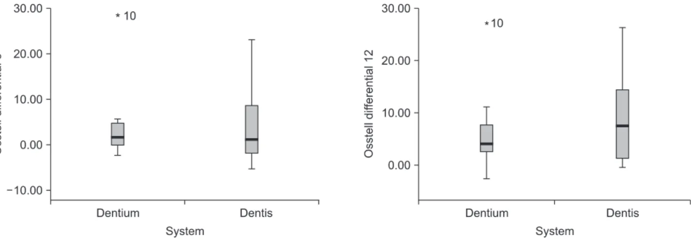
Fig. 1. The means of ISQ value at 6, 12 months.
IL, USA)의 paired t-test와 independent t-test를 이용하여 통계 분석을 시행하였다.
결 과
총 12명의 환자에게 25개의 임플란트를 식립하였으며 평 균 연령은 56세(45-62세)였다. SLA 임플란트 식립 환자 분 석 시 주기적으로 Periotest를 시행하지 않은 3개의 임플란트 는 통계처리에 포함시키지 않았다. 식립 직후, 6주, 12주의 Osstell-mentor 측정 결과, HA coated 임플란트를 식립한 집 단과 SLA 임플란트를 식립한 집단 모두에서 시간이 경과할 수록 측정값이 증가하는 양상이 관찰되었으나 통계적 유의성 은 HA coated 임플란트 식립 후 12주 뿐이었다(Fig. 1, Table 1).
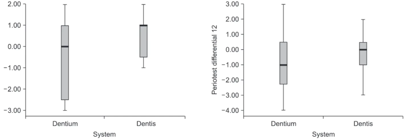
Periotest의 경우 HA coated 임플란트를 식립한 집단과 SLA 임플란트를 식립한 집단 모두에서 측정치의 증가양상이 뚜 렷하지 않았으며 통계적으로 유의성을 나타내지 않았다(Fig.
2, Table 2). HA coated 임플란트를 식립한 집단과 SLA 임플란 트를 식립한 집단을 서로 비교하였을 경우 Osstell-mento와 Periotest 모두 유의적 차이를 나타내지 않았다.
고 찰
임플란트 식립은 임플란트의 부분 무치악 또는 전악 무치 아 수복 시 매우 일상적인 술식이 되었으며 최근 95% 이상의 성공률을 보이고 있다(Brånemark, 1985). 하지만 골량이나 골 질이 불량한 경우 성공률이 매우 저하됨이 보고되었다(Jaffi n
& Berman, 1991). 불량한 골질을 가지는 상악 무치악 구치부 에 임플란트를 식립할 때의 성공률을 높이기 위해 많은 연구 자들은 임플란트 디자인이나 표면처리방법을 개발하였다.
Glauser 등의 연구에 따르면 골질이 불량할 경우 임플란트의 디자인과 표면처리 방법에 따라 임플란트의 골유착이 영향을 많이 받을 수 있다고 하였다(Glauser et al., 2001).
최근 주로 이용되고 있는 임플란트의 표면처리 방법에는 SLA, 산화처리, HA coating 등의 방법이 있으며 이번 연구에 서는 SLA와 HA coating 처리한 임플란트를 비교하였다. SLA 임플란트의 경우 큰 입자를 가지는 Al2O3 등의 물질을 가지고 sand blasting 처리한 후 다시 염산(HCl)과 황산(H2SO4)을 적 절한 비율로 혼합한 용액에 산부식 처리를 하는 방법을 말한 다. 이는 임플란트 표면에 큰 분화구를 형성하고 미세한 분화 구를 추가로 형성함으로써 임플란트의 거칠기를 증가시키며 골유착을 빠르게 촉진시킨다고 알려져 있다(Buser et al., 1991;
Piattelii et al., 1998). HA coated 임플란트의 경우 임플란트 표
Table 1. ISQ Value Depending upon Timing Tested
Implant type At installation 6 months 12 months SLA implant
HA coated implant
75.29±7.56 72.41±8.91
78.79±4.71 77.09±4.87
80.21±3.64 81.55±2.84*
Values are presented as mean±standard deviation. *p-value=0.001.
SLA: sand blasted with large grit and acid etching, HA: hydroxyapa- tite.
Fig. 2. The means of Periotest value at 6, 12 months.
Table 2. Periotest Value Depending upon Timing Tested
Implant type At installation 6 months 12 months SLA implant
HA coated implant
−4.27±1.85
−4.00±1.41
−4.91±1.04
−4.40±0.89
−5.00±0.94
−4.14±1.36
Values are presented as mean±standard deviation. SLA: sand blasted
with large grit and acid etching, HA: hydroxyapatite.
면에 HA 입자를 도포하는 방법으로 임플란트 표면 거칠기 를 증가시켜 초기 고정력을 증가시키고 빠른 골유착을 유도 할 수 있다(Deporter et al., 1986; Buser et al., 1991). 이번 연구 에서 사용된 SLA 임플란트의 경우도 좋은 초기 고정력과 단 기간 안정성이 관찰되었다. HA coating의 경우 과거 plasma spraying을 이용하여 시행하였으나 HA 피막의 박리에 의해 장기간의 안정성이 떨어지는 문제점이 존재하였다. 최근에는 ion-sputering (Yoshinari et al., 1991), ion-plating (Overgaard et al., 1996)등이 개발되어 박리현상을 감소시킴으로써 장기 간의 안정성이 크게 증가하였다. 이번 연구에서 사용한 HA coated 임플란트의 경우 resorbable blasting media (RBM) 처 리된 임플란트 표면에 상온 초박막 피복방법을 이용하여 제 작되었으며 우수한 초기 고정력과 안정성을 보이고 장기간의 부하에도 HA 피막의 박리가 일어나지 않는다고 알려져 있다.
이번 연구에서는 장기간의 안정성은 평가하지 못하였으나 우 수한 초기 고정 및 단기간 안정성은 확인할 수 있었다.
임플란트 안정성의 정량적 측정성을 위하여 여러 방법이 소 개되었으며 이번 연구에서는 Osstell-mento과 Periotest를 이 용한 평가를 시행하였다. Osstell-mento (Meredith, 1997)의 경 우 임플란트 안정성의 정량적 평가에 유용한 것으로 알려져 있으며 Periotest (Glauser et al., 2001)의 경우 지대치에 대한 각 도와 거리 등에 의해 민감하게 반응하므로 주로 임플란트에 하중을 가하기 전에 동요도를 측정할 때 주로 사용된다고 알 려져 있다. 이번 연구에서 Osstell-mento을 이용한 안정성 측 정 결과 SLA 임플란트와 HA coated 임플란트 모두 시간경과 에 따라 안정성이 증가하였다. Periotest의 경우 시간이 경과함 에 따른 증가양상이 관찰되지 않았으나 −4 이상의 좋은 안정 성을 보였다. 이는 초기 측정값이 약 −4 정도로 좋은 초기 고 정을 보였기 때문이라고 생각해 볼 수 있다. 그리고 SLA 임플 란트와 HA coated 임플란트를 서로 비교하였을 경우 안정성 의 증가에 대해 유의적 차이를 나타내지 않았지만, HA coated 임플란트에서만 12주 후 통계적 유의성을 보였다. 이는 실제 적으로 두 집단 모두에서 안정성은 증가했으나 임플란트 숫 자가 너무 작아서 나타난 결과로 생각된다. 따라서 Periotest 를 이용한 초기 고정도 평가 시 초기 고정이 좋았기 때문에 안 정성에 임플란트의 표면처리가 영향을 주지 않았다고 생각할 수 있다. 앞선 연구에서도 골질이 좋은 상태에서는 표면처리 를 하지 않은 기계절삭형 임플란트와 SLA, HA coated 임플란 트 모두 좋은 성공률을 보이고 우수한 안정성을 보였으므로 이번 연구의 결과와 일치한다고 할 수 있다(O’Sullivan et al., 2000).
이번 연구의 경우 연구대상의 임플란트 수가 적으며 연구기 간이 짧아 장기간의 안정성 평가에 한계가 존재하며, 모든 임
플란트가 하중을 가하지 않았고, 식립 부위 또한 초기 안정성 이 높게 측정될 만큼 골질이 불량하지 않았으며 식립 부위도 한정되지 않았다. 또한 안정성의 평가에서, 방사선 사진 및 조 직학적 평가 등을 하지 않았기 때문에 종합적인 안정성 평가 라고 할 수 없다는 한계가 있었다.
향후 정확한 임플란트 표면처리에 따른 안정성의 차이를 평 가하기 위해서는 실제적으로 초기안정도가 불량한 골질을 가 지는 부위로 식립 부위를 한정시키며, Osstell-mento 측정과 함께 다른 측정법의 개발이 필요할 것으로 생각된다.
감사의 글
이 논문은 2010년도 재단법인 조선대학교 치과대학교육문 화재단 특수목적기금의 지원을 받아 연구되었음.
참 고 문 헌