Ⅰ. 서 론
결손된 치아를 타이타늄 임플란트로 수복한 경우에 관한 첫 논문
1)이 30 여년 전 발표된 이후 지금까지 팔천 편
2)이 넘는 논문이 발표되었다. 요즈음 임플란트 유지 고정성 및 가철성 보철물은 높은 예견성을 가지는 치료 방법이며
3)또 한 임플란트 치료를 받은 환자의 90 % 이상이 기능 및 심
미적 관점에서 치료에 만족하고 있다.
2)그러므로 전 세계적 으로 많은 임플란트 시스템이 개발되어 시장에 나와있으며 한국에서만 50 개 이상의 임플란트 시스템과 2천억 시장 규 모로 보고된 바 있다.
4)이러한 현실을 감안해 볼 때 임상의 들이 다양한 조건에서 임플란트를 선택할 때 각기 다른 시 스템에 따른 장단점과 장기간 경과 관찰 결과는 매우 중요 하다.
전혜란
1∙김명래
1,2∙이동현
2∙신정섭
2∙강나라
1,21
이화여자대학교 임상치의학대학원 임플란트치의학과,
2이화여자대학교 의학전문대학원 치과학교실 구강악안면외과
RBM 표면처리 내부연결형 비매립 임플란트의 4년 생존율과 주변골 흡수에 관한 임상 및 방사선학적 연구
FOUR-YEAR SURVIVAL RATE OF RBM SURFACE INTERNAL CONNECTION NON-SUBMERGED IMPLANTS AND THE CHANGE OF THE PERI-IMPLANT CRESTAL BONE
Hyeran Jeon
1, Myungrae Kim
1,2, Donghyun Lee
2, Jungsub Shin
2, Nara Kang
1,21
Ewha Womans University Graduate School of Clinical Dentistry Implant Dentistry
2
Ewha Womans University School of Medicine Department of Oral and Maxillofacial Surgery
Implant-supported fixed and removable prostheses provide a proper treatment modality with reliable suc- cess. The SS Ⅱ
�Implants is a one-stage nonsubmerged threaded titanium implants with Resorbable Blasting Media (RBM) surface developed by Osstem company (Busan, Korea) in October of 2002.
This study is to evaluate the survival rate of the SS Ⅱ
�Implants for 4 years using radiographic parame- ters and to review the retrieved implants by the cytotoxicity tests.
Since September 2003, 439 SS Ⅱ
�implants had been used for 173 patients at Ewha Womans University Medical Center in Korea. Patients consisted of 91 females (52.6 %) and 82 males (47.4 %). The patients’
mean age was 42 ± 16 years, ranging from 21 to 83 years. The follow-up period ranged from 9 to 46 months (mean F/U 24.2 ± 10.2 months).
The results are as follows;
1. Of 439 implants, 17 implants were removed and 4-year cumulative survival rate was 96.1%.
2. 82.3% of 17 failed implants were founded during healing phase, and 94.1% of failed fixtures were removed within 5 months after implantation.
3. Crestal bone around the implants was resorbed to 1 mm in 89.0%, to 1 - 2 mm loss of the marginal bone in 8.3%, and the bone loss over 2 mm was occurred in 2.7%.
4. Microscopic examination of the retrieved implants disclosed Grade 0 cytotoxicity in 4 and Grade 1 cytotoxicity in 2 of 6 groups divided according to LOT numbers. Inhibition rate with optical density was acceptable as low as ISO standard.
Key words: SS Ⅱ
�Implant, Survival rates, Cytotoxicity tests
Abstract
SS Ⅱ
�임플란트(오스템, 부산, 대한민국)는 일회법 수술 을 기본으로 하는 치은 레벨의 internal octagon connec- tion과 straight body를 가진 임플란트로 2002년 10월 출 시되었다. Self tapping이 가능하여 초기 고정력이 우수하 며 수산화인회석(hydroxyapatite (HA;Ca
10(PO
4)
6(OH
2)) 으로 blasting 처리된 Resorbable Blasting Media (RBM) 표면으로 생체 친화성이 우수하다고 소개되었다.
본 연구의 목적은 임상 및 방사선학적, 세포 독성 검사를 이용한 SS Ⅱ
�임플란트의 단기간 경과 관찰 결과를 보고하 고자 한다.
Ⅱ. 연구 재료 및 방법
1. 연구 대상
2003년 9월부터 이화의대 목동 병원에서 임플란트 수술 을 받은 환자 중 SS Ⅱ
�임플란트 시스템을 이용하여 시술 받은 173 명의 환자에게 이식된 439 개의 임플란트를 대상 으로 하였다. 모든 환자들에게 구강 및 방사선 검사, 전신 병력 검사를 시행하였으며 절대적 금기증에 해당하는 환자 들은 다른 방법으로 상실치를 수복하였다.
총 173명의 환자들은 91명(52.6%)의 여성과 82명 (47.4%)의 남성으로 구성되었으며 연령은 21세 이상 83 세로 평균 42 ± 16세 이었다. 추적 관찰의 기간은 9개월 이상 46개월까지 평균 24.2 ± 10.2 개월이었다.
2. 연구 방법
환자의 진료기록으로 임플란트의 종류(직경과 길이), 식 립위치, 보철물의 종류, 경과를 조사하고, 디지털 파노라마 및 구내표준 x-선사진으로 임플란트 주변 변연골의 높이 변 화를 비교 계측하였다.
1) 생존율에 대한 기준
임플란트의 생존율(기능유지율; Survival Rate)은 Buser 등
5,6)과 Cochran 등
7)의 기준에 따라 (1) 각 임플란 트가 임상 검사시 동요도가 없고, (2) 동통이나 주관적인 이상 감각이 없고, (3) 임플란트 주위 염증이 없으면서, (4) 임플란트 주위에 지속적인 방사선 투과상이 없이 구강 내에 서 기능하고 있는 것으로 기준하였다.
2) 방사선학적 평가
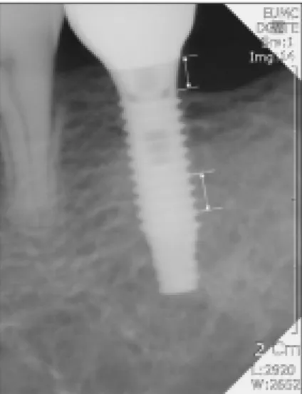
파노라마 및 치근단 사진(PiViewSTAR, Infinitt North America Inc., Phillipsburg, USA)을 통해 방사선학적 평 가를 시행하였으며 식립 후 6개월, 1년 및 2년째의 치조골 흡수를 측정하였다. 측정은 컴퓨터 프로그램을 이용하여
1/100mm 단위까지 임플란트의 장축과 평행하게 각 임플 란트의 근원심 간격(microgap)에서 첫번째 골-임플란트 접 촉부위까지 측정하고, 나사간 거리(Pitch) 간격이 0.8 mm 임을 감안하여 실제 흡수량을 환산하였다. 첫 번째 골-임플 란트 접촉부위가 방사선 사진상에서 불분명할 경우 측정 대 상에서 제외하였다.
3) 세포 독성 검사
식립 후 2-3개월 사이에 제거된 4개를 포함하여 제거된 임플란트의 표면 검사를 위해 세포독성 검사를 의뢰하였다.
비슷한 시기에 생산된 임플란트를 LOT 번호에 따라 6개의 그룹으로 구분하여 세포형태를 관찰하고 흡광도 및 저해율 를 측정하였다.
4) 통계 분석
GraphPad Prism version 5.00 (GraphPad Software, San Diego, California, USA)를 이용한 Kaplan-Meier 및 log rank (Mantel-Cox) 테스트를 시행하여 생존율을 평가하고 상하악 간의 생존율 차이를 비교하였다.
Ⅲ. 연구 결과
1. 사용된 임플란트의 분포
식립된 SS Ⅱ
�임플란트의 경우 직경은 3.3mm, 4.1mm, 4.8mm 3가지 종류 중 4.1 mm가 87.2% 이었 고, 임플란트의 길이는 7mm, 8.5mm, 10mm, 11.5mm, 13mm, 15mm 중 10mm, 11.5mm, 13mm가 89.6%이 었다. (Table 1과 2 참조). 식립 위치에 따른 분포는 Fig. 2 와 같다.
Fig. 1. A computer-assisted calibration was carried out for
each single site by evaluating the given distance between
several threads (pitch: 0.8 mm).
보철물의 종류는 195개(상악 64개, 하악 131개)의 수복 물 중 단일치 수복 78개(40.0%), 다수치아 수복(4개 치아 까지) 101개(51.8%), 완전무치악 고정성 수복 9개 (4.6%), 피개의치(overdenture) 7개(3.6%)이었다.
(Table 3)
2. 임플란트의 생존율과 실패한 임플란트의 제거 시기 총 439개의 임플란트 중 17개(3.9%)를 제거하였으며 4
년 누적 생존율은 96.1 % 였다(Fig. 3, 상악 : 94.7%, 하 악 : 97.0%). 상악과 하악의 임플란트 생존율의 차이는 통 계적 유의성이 관찰되지 않았다(p>0.05). 제거한 임플란트 는 14개가 보철 전에 실패하였고 보철 후에는 3개가 제거되 었다. 실패한 임플란트의 94.1%는 식립 후 5개월 이내에 제거되었고, 9개월 이후에 제거한 임플란트는 없었다.
제거된 임플란트의 94.1 %가 Lekholm과 Zarb의 분류
8)Type 4 상태의 골에 식립되었고, 제거한 임플란트의 특징 은 Table 4에 제시되었다.
Fig. 2. Distribution of implants by locations
Table 1. Distribution of implants by diameter (mm)
Diameter No. implants %
3.3 7 1.6
4.1 383 87.2
4.8 49 11.2
Total 439 100.0
Table 2. Distribution of implants by length (mm)
Length No. implants %
8.5 11 2.5
10 104 23.7
11.5 182 41.5
13 107 24.4
15 35 8.0
Total 439 100.0
Table 3. Details of the prosthetic rehabilitations in the mandible and in the maxilla
Maxilla Mandible Total
Single crowns 26 52 78
Fixed partial dentures
29 72 101
(up to 4 teeth)
Fully edentulous 6 3 9
Fixed partial dentures
Overdenture 3 4 7
Total 64 131 195
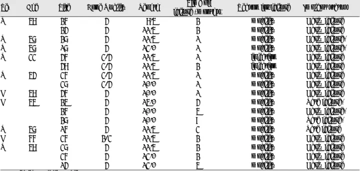
Table 4. Failure analysis
Sex Age Site Bone Quality Length Time of
Reason for failure Implant status failure (months)
M 58 26 4 8.5 2 mobility Early failure
24 4 11.5 2 mobility Early failure
M 57 27 4 11.5 1 mobility Early failure*
M 57 17 4 13.0 1 mobility Early failure*
M 33 46 3-4 11.5 1 infection Early failure
48 3-4 11.5 2 infection Early failure
M 54 36 3-4 11.5 1 mobility Early failure
37 3-4 10.0 1 mobility Early failure
F 58 46 4 10.0 1 mobility Early failure
F 55 25 4 15.0 4 mobility Late failure*
26 4 10.0 5 mobility Early failure*
27 4 10.0 9 mobility Late failure*
M 57 16 4 11.5 3 mobility Late failure
F 66 36 2-3 11.5 2 mobility Early failure
M 58 37 4 11.5 2 mobility Early failure
36 4 13.0 2 mobility Early failure
14 4 13.0 5 mobility Early failure
* suspected surface problem
3. 임플란트 주위 변연골의 방사선학적 평가
362개의 SS Ⅱ
�임플란트 중 89.0 %에서 1 mm 이하의 치조골 흡수가 관찰되었으며, 8.3 %에서 1-2 mm, 2.7 % 에서 2 mm 이상의 치조골 흡수가 관찰되었다.
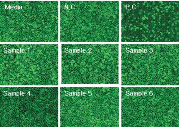
4. 제거한 임플란트 표면의 세포 독성 검사 결과
제거한 임플란트와 비슷한 시기에 생산된 임플란트를 LOT 번호에 따라 6군으로 분류하였다. 임플란트 표면의 세포형태가 Fig. 4에서 관찰된다. 분류 1, 2, 4, 5 군들은 Grade 0으로 분류되었으며, 3군과 4군은 느슨하게 부착되 Fig. 3. The 4-year cumulative survival rate: (p>0.05).
Fig. 4. Microscopic tests; Grade 0 in sample 1, 2, 4 and 5, Grade 1 in sample 3 and 4, N.C: negative control, P.C: positive control
Table 5. Reactivity Grades for Elution Test Grade Reactivity Conditions of all cultures
0 None Discrete intracytoplasmic granules; no cells lysis
1 Slight More than 20% of the cells are round, loosely attached, and without intracytoplasmic granules;
Occasionally lysed cells are present
2 Mild More than 50% of the cells are round, devoid of intracytoplasmic granules; extensive cell lysis and empty areas between cells
3 Moderate Greater than 70% of the cell layers contain rounded cells and/or are lysed
4 Severe Nearly complete destruction of the cell layers
어 있고 때때로 분리된 세포들이 관찰되는 Grade 1에 해당 되었다. 세포독성을 평가하는“ISO 10993-5”규정에 의하 면 Grade 0, 1, 2는 독성이 없다고 판정한다.
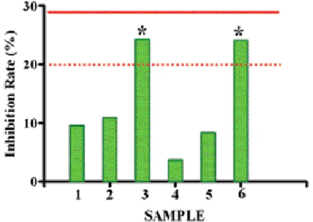
5. 흡광도와 저해율 평가
ISO 10993-5 시험 방법 및 기준에 따르면 저해율이 29 % 이하일 때 세포 독성이 없다고 판정하는데, 제조회사의 규 정은 20 % 이하로 좀 더 엄격한 편이다. 3군과 6군에서 각 각 24.26 %와 24.11 %로 제조회사의 규정에 따르면 부적 합하게 나타났다(Fig. 5).
Ⅳ. 고 찰
임플란트 치아를 이용한 결손치아의 보철수복은 높은 예 견성 및 성공률을 가지는 치료 방법으로 환자와 술자 모두 에게 만족감을 주는 술식이다. 국내의 치과 임플란트 시장 규모는 업계 추정 약 2천억원의 시장이라 추산하고 있으며 국내에서 임플란트 시스템을 연구 개발 판매하거나 수입 판 매하고 있는 업체만도 약 50 여 개에 달한다.
4)앞으로도 임 플란트 판매업체와 제품들이 계속 늘어갈 전망 가운데 임플 란트 치아에 관한 장기적인 성공률, 오랜 임상 데이터, 신뢰 할 수 있는 회사의 제품, 경제성 등은 임플란트의 선택 시 매우 중요한 고려사항이다.
SS Ⅱ
�임플란트는 일회법 시술을 바탕으로 한 비매립형 (non-submerged type) 임플란트로서 internal octagon connection과 straight body의 안정적 연결 구조를 가진 다. Wennerberg
9,10)는 표면 거칠기가 Ra 1.3 ~ 1.5μm일 때 최적의 골유착이 이루어진다고 보고하였다. 이번 연구에 서 사용된 SS Ⅱ
�임플란트의 표면 거칠기는 Ra 1.2 ~ 1.8μm 이며 생체 친화성이 우수한 수산화인회분(Hdroxyl- apatite powder)을 입힌 RBM(Resorbable Blasting
Media) 표면을 갖는다.
많은 연구에서 골-임플란트 접촉계면에서의 전단 응력은 거친 임플란트 표면에서 증가된다고 보고하였다.
9,11-13)평활 면 임플란트에서는 혈병이 표면으로부터 먼쪽으로 골화 (distance osteogenesis)가 진행되는 반면 거친면 임플란 트에서는 표면과 직접 접촉한 상태에서 혈병이 직접 골화된 다(contact osteogenesis).
14)접촉성 골화(Contact osteo- genesi)에서는 골형성 세포의 이주를 촉진시켜 임플란트 표 면에서의 조기 골형성이 가능하다. 따라서 거친면 임플란트 의 골전도성은 임플란트 표면에서의 골형성 속도를 증가시 켜 임플란트의 식립과 기능 부하의 기간을 감소시킬 수 있 다.
15)치과 임플란트는 의료기자재의 하나로 제품의 사용 및 판 매를 위해서 물리적 특성에 관한 시험, 기계적 특성에 관한 시험, 생물학적 안전성에 관한 시험 등을 거쳐야 하는데 생 물학적 안전성 시험에 세포독성 시험이 포함되어 있다. 세 포독성 시험은 세포배양 기술을 이용하여, 재료나 기자재 또는 그 추출물에 의한 세포의 용해(세포괴사), 세포 성장 저해 및 그 외 세포에 미치는 영향(효소의 변화, 세포의 유 전자에 대한 재료의 영향)에 대하여 알아보는 실험이다. 이 번 연구에서는 임플란트 표면의 세포형태 관찰 결과 독성이 없다고 판정되었으며 흡광도 및 저해율 검사 결과 ISO 규 정(저해율 29% 이하)에는 적합하였지만 회사의 내부 규정 (저해율 20% 이하)에는 부적합하였다. 따라서 이 시험 결 과를 바탕으로 이후 임플란트의 제작 과정 중 세척 공정에 사용되는 증류수의 순도를 상향 조정하고 생산 LOT 별로 모든 표본을 선택하여 세포 독성 검사를 실시하기로 하였다 는 회신을 받은 바 있다.
본 연구에서는 총 439개의 임플란트 중 17개가 제거되었 으며 4년 누적 성공율은 96.1 %였다. 실패한 17개의 임플 란트 중 14개(82.4 %)가 초기 치유 기간 동안 제거되었고, 문헌상에서도 일단 골유착이 일어나면 실패가 발생할 가능 성은 급격히 감소한다고 보고한 바 있다.
16)하악(97.0 %) 에 비해 상악(94.7 %)에서 더 낮은 생존율이 관찰되었으며 이는 골질의 불량과 제한된 골 양에 기인하는 것으로 사료 되었다.
15)다른 일회법 시술 임플란트 시스템의 생존률과 비교할 때 Romeo 등
17)이 보고한 ITI
�임플란트의 단일치 수복의 7년 누적 생존률 96.77 %, Bischof M 등
18)의 wide neck ITI
�임플란트의 1, 2, 5년 생존률 각 98.8 %, 97.7 %, 97.89 % 과 큰 차이가 없으며, Buser 등
5)의 8년간 다기관 2,359개 의 ITI
�임플란트를 대상으로 한 96.7 %의 생존률을 고려 하면 이번 SS Ⅱ
�임플란트의 4년간 96.1 %의 생존율은 신뢰할 만한 임플란트로 여겨진다. 그러나 다 기관에서의 계속적인 장기간 경과 관찰 및 혼란변수(confounding fac- tor)를 통제한 전향적 연구가 더욱 필요하다고 사료된다.
Fig. 5. Optical density and Inhibition rate ; improper inhibi-
tion rates were observed at sample 3 and 6 (according to
the company regulation;20% but acceptable to ISO;29%)
Ⅴ. 결 론
2003년 9월부터 이화의대 목동병원에서 173 명의 환자에 게 식립되었으며 9개월 이상 추적 관찰된 439 개의 SS Ⅱ
�임플란트의 4년 누적 생존율과 임플란트의 변연골 흡수 및 제거한 임플란트에 대한 세포학적 관찰을 하고 다음과 같은 결론을 얻었다.
1. SS Ⅱ
�임플란트의 4년 누적 생존율(기능유지율)은 96.1 %이었다.
2. 상악에서는 94.7 %, 하악에서는 97.0 %의 생존율을 보였다. (p>0.05).
3. 17개의 제거된 임플란트 중 82.3%(14개)가 보철 전에 실패하였고 보철 후에는 17.7%(3개)가 제거되었다.
4. 실패한 임플란트의 94.1%는 식립 후 5개월 이내에 제 거되었고, 9개월 이후에 제거한 임플란트는 없었다.
5. 식립 1년 후 평균 골흡수량은 89%에서 1 mm 이하, 8.3 %에서 1-2 mm, 2.7 %에서 2 mm 이상의 치조 골 흡수가 관찰되었다.
6. 실패한 임플란트 표면의 세포독성 검사 결과, 흡광도는 Grade 0와 1, 저해율은 6군 중 4 군에서 10% 이하, 2 군에서 24% 정도를 보임으로써 ISO규정에 적합하 였다.
References