D
ental implant based on the original work by Bra�nemarket al1-4has been documented widely and proven to be a very reliable treatment choice in therestoration of edentulous jaws.
Reducing treatment period in implant den- tistry is a matter of main concerns. There are so many factors affecting the success rate of imme-
BONE RESPONSE OF THREE DIFFERENT SURFACE IMPLANTS : HISTOMORPHOMETRIC AND
RESONANCE FREQUENCY ANALYSIS IN DOGS
Woo-Seok Song, D.D.S., Yung-Soo Kim, D.D.S., M.S.D., Ph.D., M.Sc(O.S.U.), Chang-Whe Kim, D.D.S., M.S.D., Ph.D., Kyung-Soo Jang, D.D.S., M.S.D., Ph.D., Young-Jun Lim, D.D.S., M.S.D., Ph.D.
Department of Prosthodontics, Graduate School, Seoul National University
Statement of problem. Reducing treatment time in implant dentistry is a matter of main con- cern. There are so many factors affecting the success rate of immediate or early loaded implant for the initial bone response. The especially microscopic properties of implant surfaces play a major role in the osseous healing of dental implant.
Purpose. The aims of this study were to perform a histologic and histomorphometric com- parison of the healing characteristics anodically roughened surface, HA coated surface and RBM surface implant, and to compare of ISQ values measured by OsstellTMfor resonance frequency analysis in dogs mandible during 2 weeks.
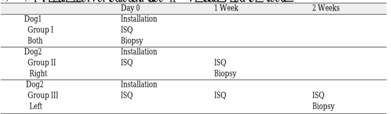
Material and method. Bone blocks from 2 dogs were caught after covered healing for 0 day(2 h); Group I, 1 week; Group II and 2 weeks; Group III . One longitudinal section was obtained for each implant and stained with hematoxylin and eosin. Histomorphometric analysis was done with Kappa Imagebase system to calculate bone-to-implant contact and bone volumes inside the threads. ISQ values were measured in every time of surgery schedule.
Conclusion.
The experiment revealed that :
1. The percentages of bone-to-implant contact on the fixture in each group were not significantly different(P > 0.05).
2. The percentages of bone area inside the threads on the fixture in each group were not sig- nificantly different(P > 0.05).
3. The ISQ level showed clinical stability of each fixture during 2 weeks(all ISQ level ≥71).
Key Words
Surface, Histomorphometry, ISQ, RFA, Anodic oxidation, RBM, HA
J Korean Acad Prosthodont : Volume 42, Number 6, 2004
diate or early loaded implants. And the macro- scopic and microscopic features of implant sur- faces have been described as one of the major fac- tors of osseointegration.4The osseous healing characteristics of various surface structures have been investigated in several experimental and clin- ical studies.5-7
The implant surfaces and types most frequently described in the literature and made may be subdivided into implants with roughened surfaces by coating[e.g. resorbable blast media (RBM), hydroxyappatite coated (HA)], implants with roughened surfaces with electrochemical modi- fication (anodic oxidation) of commercially pure titanium implants and implants with roughened surfaces without coating [e.g. sand-blasted or acid-etched]. These surfaces were known to pro- mote initial healing capacity by rougheness.8-10
The evaluation of the clinical outcome of implant therapy has mainly been made through clinical and radiographic examinations, which have been aimed at locating failed implants and ongoing peri- implant bone resorption. However, with resonance frequency analysis(RFA), it is now possible to mea- sure the degree of implant stability at any time dur- ing the course of implant treatment and loading.11 The technique is now commercially available as OsstellTM (Integration Diagnostics, Sa¨vedalen, Sweden) and a new unit to describe implant sta- bility quotient(ISQ). ISQ is recorded as a number between 1 and 100, 100 is representing the high- est degree of stability. Each transducer is calibrated by the manufacturer, which makes all measure- ments directly comparable.
The aims of this study were to perform a his- tologic and histomorphometric comparison of the healing characteristics of anodically roughened surface (TiUnite, Nobel Biocare, Go¨teborg, Sweden), HA coated surface (Replace select, Nobel Biocare, Go¨teborg, Sweden) and RBM surface (Kimplant, Avana�, Osstem, Busan, Korea) , and to compare of ISQ values measured by OsstellTMfor reso-
nance frequency analysis in dogs mandible dur- ing 2 weeks.
MATERIALS AND METHODS
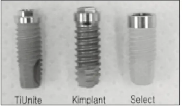
Fixtures (Fig.1)
1) RBM surface (Kimplant* , Avana�, Osstem, Busan, Korea)
Diameter 4.0mm×10mm straight form (* : project name )
2) Anodically roughened surface (TiUnite**, Nobel Biocare, Go¨teborg, Sweden)
Diameter 4.0mm×10mm MK III
3) HA coated surface (Replace Select**, Nobel Biocare, Go¨teborg, Sweden)
Diameter 4.3mm×10mm straight form (** : commercially available )
Experimental animals and Anesthesia Two adult healthy beagle dogs weighing 10Kg were used. One is adult female(dog1) and anoth- er is adult male(dog2).
The dogs were anesthetized with combination of ketamine10mg/kg (Yu-han, Gunpo, South Korea) and Rompun 0.15mL/kg (Bayer Korea, Ansan, South Korea) intramuscularly. The partially extracted mandibles were prepared for the sites of fixtures and washed and decontaminated with Betadine (Sung Kwang, Buchun, South Korea) and prior to surgical draping. One milli-
Fig. 1.Fixtures.
liter of 2% lidocaine (Yu-han; 1:100,000) was administered local to the implant sites.
Surgical procedures and implant placement A controlled surgical technique was used to place 9 implants (Dog1; 6 fixtures/ Dog2; 3 fix- tures). Using sterile surgical techniques, an inci- sion was made in the mucosa to expose alveolar bone, and the mucoperial steal flap was elevated.
The implant site was drilled in usual manner, by using drills of each own system with increas- ing diameters under constant irrigation with sterile saline.
The order of installation was TiUnite-Kimplant- Select (from the anterior site).
Group Ⅰ : Fixtures submerged during 2 hours (0 day)
Group Ⅱ : Fixtures submerged during 1 week Group Ⅲ : Fixtures submerged during 2 weeks
ISQ measurements
ISQ measuring by OsstellTM(Integration Diagnostics, Sa¨vedalen, Sweden) was done on each schedules of experiment.(Table. I) Each transducer is calibrated by the manufacturer, which makes all measurements directly compa- rable. In this study, transducer “Type F13 L5;100132”for Replace select and “ Type F1 L 8.5;
10050”for external hexagon(Kimplant and TiUnite) were used.
Preparation of specimens and histomorpho- metric analysis
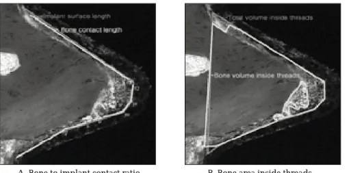
Every fixture was removed from the mandible as a form of block. The specimens and sur- rounding tissues were washed in saline solu- tion and fixed in 4% paraformaldehyde and 0.1 % glutaraldehyde in 0.15mlo/L cacodylate buffer at 4 ℃ and pH 7.4. The specimens were further dehydrated in ascending concentration of alcohol rinses and infiltrated with glycolmethacrylate resin (Technovit 7200 VLC, Kulzer & Co, Wehrheim, Germany). After polymerization, the specimens were sectioned longitudinally at about 100μm and ground to a final thickness of about 25 μm (EXAKT 310, GMBH & Co, Germany) as described by donath.12One section was obtained for each implant and stained with hematoxylin and eosin.. In histomorphometric analysis, Kappa imagebase system was used. To calculate bone- to-implant contact ratio, first we draw the con- nection of straight line along the fixture surface and then draw the line that was thought to be the junction of bone and fixture surface (line tool). Each point of continuous line was numbered in order (1, 2, 3…) and the ratio was calculated by Microsoft Excel. In calculation bone volumes inside threads, polygonal tool of system was used, which con- nected the boundary line of bone, vacant space, and threads. And then the ratio was calculated by Microsoft Excel.
Table I. Schedule for implant installation, ISQ measurement and biopsy
Day 0 1 Week 2 Weeks Dog1 Installation
Group I ISQ
Both Biopsy
Dog2 Installation
Group II ISQ ISQ Right Biopsy Dog2 Installation
Group III ISQ ISQ ISQ Left Biopsy
Statistics
Mean values of bone contact ratio and bone area in each group were calculated and subjected to a repeated measure ANOVA to test for signifi- cant differences. Statistical testing was carried out at the 5% significance level.
RESULTS
All nine implants in bone blocks were ana- lyzed. No implant exhibited clinical mobility.
Further the mucosa exhibited only minor signs of inflammation with no pus discharge or severe
A - C : 0 Day (Group I ) ; A : Kimplant B : TiUnite C : Select D - F : 1 Week (Group II) ; D : Kimplant E : TiUnite F : Select G - I : 2 Weeks (Group III) ; G : Kimplant H : TiUnite I : Select Fig. 2.
A B C
D E F
G H I
inflammation(uneventful). But the post-operative wound healing of the all implants showed dehis- cence by incision for measuring ISQ.
1. Histological finding
Fig. 2 A-I illustrated a ground-section of an implant with surrounding hard tissues from biopsy sampled. The peripheral portions or the
pitches were in close contact with the surround- ing bone tissue, and thus hereby mechanical sta- bility for the implant during the initial phase of wound healing was provided. In specimens, the various cells within this coagulum could be iden- tified (Arrow;A-C; Group I). The gap between thread and bone of 1 week and 2 weeks specimens were more close contact than that of 0 day spec- imens.
A. Bone to implant contact ratio B. Bone area inside threads Fig. 3. Histomorphometric evaluation of bone-to-implant contacts and bone area inside threads : using Kappa Imagebase program.
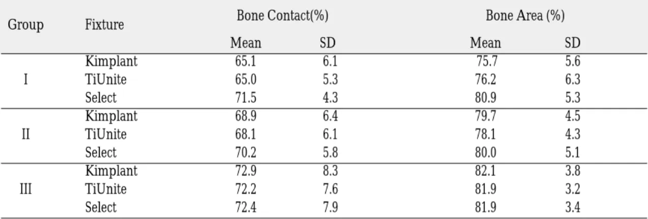
Table II. Results from histomorphometry
A.Total Mean value of every pitch from each fixtures (Kimplant n=12 / TiUnite n=14 / Replace Select n=13)
Group Fixture Bone Contact(%) Bone Area (%)
Mean SD Mean SD
Kimplant 63.8 13.3 73.7 5.4
I TiUnite 63.5 12.5 73.4 5.6
Select 69.8 11.3 79.1 4.9
Kimplant 67.9 9.5 79.0 6.7
II TiUnite 68.0 8.4 77.9 5.6
Select 70.0 7.9 79.6 6.9
Kimplant 71.9 10.2 81.3 4.5
III TiUnite 71.9 9.6 81.8 4.1
Select 71.4 9.5 81.4 4.0
At group I, II and III : not significantly different at P<0.05 (ANOVA)
2. Histomorphometric analysis
Bone-to-implant contact ratio and volume of the bone engaged between threads were calculat- ed(Fig. 3).
3. Resonance frequency analysis (Table IV)
DISCUSSION
In this study, no significant difference was found from histomorphometric analysis between Kimplant and other commercially available implants. In conclusion Kimplant were devel- oped successfully in comparison with the results of commercially used TiUnite and Replace Select in 2 weeks study.13,14 In addition, it could be Table III. The highest values observed in each group
Bone-to-implant contact Bone area
Group I Replace Select (75.3%) Replace Select (84.0%)
Group II TiUnite (75.6%) Kimplant (84.3%)
Group III TiUnite (77.1%) Kimplant (84.7%)
Table IV. ISQ measured by OsstellTM(≥71)
Group Fixture 0 Day 1 Week 2 Weeks
Kimplant 72
I TiUnite 72
Select 73
Kimplant 71 73
II TiUnite 74 75
Select 75 76
Kimplant 72 73 75
III TiUnite 72 74 75
Select 71 72 75
B. Data from best 4 bone response threads (n=4)
Group Fixture Bone Contact(%) Bone Area (%)
Mean SD Mean SD
Kimplant 65.1 6.1 75.7 5.6
I TiUnite 65.0 5.3 76.2 6.3
Select 71.5 4.3 80.9 5.3
Kimplant 68.9 6.4 79.7 4.5
II TiUnite 68.1 6.1 78.1 4.3
Select 70.2 5.8 80.0 5.1
Kimplant 72.9 8.3 82.1 3.8
III TiUnite 72.2 7.6 81.9 3.2
Select 72.4 7.9 81.9 3.4
guess that three different implants may be response to bone excellent enough to satisfy immediate or early loading(within 2 weeks) in clin- ical situation.
In the present study, when implants were installed, their own drill system was used. In case of Kimplant, screw tap in developing was used for its own screw shape of fixture. If screw tap would be more precisely developed, it could be expected that bone response of this newly devel- oped fixture will be more excellent.
Highest values of each experiment are showed on table III. In group I , Replace Select showed the highest values in BCR and BVR. The diameter of Replace select is 4.3mm, which is larger than that of TiUnite and Kimplant. The difference in diameter may affect the results of this study. In addition, the initial bone response of hydrox- yappatite coated surface is prospected well from other study.15 In group II and III of bone-to- implant contact, one of the pitches in TiUnite specimen shows the best scores. In group II and III of bone volumes inside the threads, one of the pitches in Kimplant specimen shows the highest score(84.3% and 84.7%), but the second sore was Replace Select (84.1% and 84.5%) and the dif- ference between fixtures were minimal.
It could be concluded that from all difference ten- dency of this study, the variation of surgical sit- uation(e.g. bone quality, bone quantity , insertion torque, heat generation etc) may be one of the major factors to estimate clinical success.
However, no studies describing “normal”ISQ levels have been published. By P. Balleri et al(2002) an ISQ level of 69 (range of 57~82) may describe the stability of a fully integrated implant.
In this study, every measured ISQ level was more than 71 and then all the fixtures were thought to be stable.14-18
In this study, implants submerged without loading during 2 weeks were analyzed. In other study, 6 weeks analysis in dog was often used. But
also during 2 weeks it could be seen that in some pitch regions which were responsible for pri- mary mechanical stability, the bone tissue exhib- ited signs of ongoing bone remodeling, resorption and apposition. In further study during longer peri- od the surface reaction by woven bone formation with lamellar bone maturing should be evaluat- ed.
The results are comparable with the histomor- phometric measurements in previous study in spite of different experimental designs.21-26It is well known that rapid ingrowths of bone into the threads during the initial healing period make sure better stability of the implants after 1st surgery and also contribute to better long-term success.27-31 However, the clinical implications of the exact heal- ing time and mechanisms by the surface properties of implant remain subjects for further study.
CONCLUSIONS
With the limitation of this study, the experiment revealed that :
1. The percentages of bone-to-implant contact on the fixture in each group were not signifi- cantly different(P > 0.05).
2. The percentages of bone area inside threads on the fixture in each group were not signifi- cantly different(P > 0.05).
3. The ISQ level measured by OsstellTM showed clinical stability of each fixture during 2 weeks (all ISQ level ≥71).
REFERENCES
1. Bra�nemark PI. Osseointegration and its experimental background. J Pros Dent 1983;50:399.
2. Bra�nemark P-I, Hansson BO, Adell. Osseointegrated implants in the treatment of the edentulous jaw.
Experience from a 10-year period. Scand Reconstr Surg 1977;11(suppl 16).
3. Roos J, Sennerby L, Albrektsson T. An update on the clinical documentation on currently used bone-anchored endosseous Implants. Dent Update 1997;24:194-200.
4. Albrektsson T, Zarb GA, Worthington P, Eriksson AR. The long-term efficacy of currently used im- plants. A review and proposed criteria of suc- cess. Int J Oral Maxfacial Impl 1986 I:11-25.
5. Davies JE et al. The bone-titanium interface in vitro. J Biomedical Materials Research 1990;24;1289- 1306.
6. Hazan R et al. Bone growth to metal implants is reg- ulated by their surface chemical properties.
Biomaterials 1993;14:570-574.
7. Weinlaender M et al. Histomorphometry of bone apposition around three types dental implants. Int J Oral Maxillofacial Impl 1992;7:491-496.
8. Ericsson I, Johansson CB et al. A histomorphometric evaluation of implant contact on machined-prepared and roughened titanium dental implants Clin Oral Impl Res 1994;5:2002-206.
9. Larsson C, Thomsen P. et al. Bone response to sur- face modified Ti implants : studies on the early tis- sue response to machined and electropolished implants with different oxide thicknesses.
Biomaterials 1996:17(6):605-615.
10. Sul YT, Johansson CB, Jeong YS, Wennerberg A, Albrektsson T. Resonance frequency and removal torque analysis of implants with turned and an- odized surface oxides. Clin Oral Impl Res 2002:13:252-259.
11. Meredith N, Alleybe D, Cawley P. Quantitative de- termination of the stability of the implant-tissue in- terface using resonance frequency analysis. Clin Oral Impl Res 1996;7:261-267.
12. Donath K. Die Trenn-Du¨nnschliff-Technik zur Herstellung histologischer Pra¨paraten von nichit schneidbaren Geweben und Materialien. Der Pra¨parator 1988;34:197-206.
13. Boyan BD, Hummert T, Kieswetter K. Role of material surfaces in regulating bone and carilage cell response. Biomaterials 1996:17:137-146.
14. Kim YS. 3-D finite element study on surface char- acteristics and stress of implants. SNUDH Dept of Pros. Unpublished.
15. Simunnek A. Evaluation of stability of titanium and hydroxyappatite coated osseointegrated dental implants : a pilot study Clin Oral Impl Res 13, 2002
;75-79.
16. Friberg B, Sennerby L, Meredith N, Lekholm U. A comparison between cutting torque and reso- nance frequency measurements of maxillary im- plants. A 20 month clinical study. Int J Oral Maxillofac Surg 1999;28:297-303.
17. Sennerby L, Roos J. Surgical determinants of clin- ical success of osseo-integrated implants. A re- view of the literature. Int J Pros 1998; 11:408-420.
18. Sennerby L, Roos J. RFA: Measuring implant sta- bility and osseointegration. Compendium 1998:19;493-502
19. Sennerby L, Friberg B, Linden B, Jemt T, Meredith N. A comparison of implant stability in mandibu- lar and maxillary bone using RFA. European Commission Demonstration Project. RFA Symposium. 2000 Sweden.
20. CJ Park. A study on the change of implant stabil- ity using resonance frequency analysis. J Korean Academy of Prosthodontics 2003;41:271-287.
21. Albrektsson T et al. Histologic investigations on 33 retrieved Nobelpharma implants. Clin Materials 1993;12:1-9.
22. Rocci A et al. Histology of retrieved immediately and early loaded oxidized implants : Light mocro- scopic observations after 5-9 months of loading in the posterior mandible. Clin Impl Dent Related Res 2003;5(I): 88-97.
23. Berglundh T et al. De novo alveolar bone forma- tion adjacent to endosseous implants : A model study in the dog. Clin Oral Impl Res 2003;14:251- 262.
24. Botticelli D et al. Appositional bone formation in marginal defects at implants : A experimental study in the dog. Clin Oral Impl Res 2003:14; 1-9.
25. Zechner W. et al. Osseous healing characteristics of three different implant types : A histologic and histomorphometric study in mini-pigs. Clin Oral Impl Res 2003;14: 150-157.
26. YH Kim, JY Koak, IT Chang, SJ Heo. A histomor- phometric analysis of the effect of various surface treatment methods on osseointegration. Int J Oral Maxillofacial Impl 2003;18(3):349-356.
27. Albrektsson T et al. Bone metal interface in os- seointegration. J Pros Dent 1987; 57:597-607.
28. Collier JP et al. Macroscopic and microscopic evidence of prosthetic fixation with porous coat- ed materials. Clin Orthop 1988;235:173-180.
29. Pilliar RM. Porous-surfaceed metallic implants for orthopedic applications. J Biomed Material Res 1987;21:1-33.
30. Sun L. Material fundamentals and clinical per- formance of plasma-sprayed hydroxyapatite coat- ings. A review J Biomed Materials Res 2001: 58: 570- 592.
31. Tal H. Spontaneous early exposure of submerged implants. II. Histopathology and histomorphom- etry of non-perforated mucosa covering sub- merged implants. J Periodontol 2000:71:1231-1235.
Reprint request to:
CHANG-WHEKIM
DEPARTMENT OFPROSTHODONTICS, GRADUATESCHOOL, SEOUL NATIONALUNIVERSITY,
28, YEONGUN-DONG, CHONGNO-GU,SEOUL, 110-744, KOREA k43c46w@dentistry,snu.ac.kr