www.jpis.org
pISSN 2093-2278 eISSN 2093-2286 Copyright © 2011 Korean Academy of PeriodontologyThis is an Open Access article distributed under the terms of the Creative Commons Attribution Non-Commercial License (http://creativecommons.org/licenses/by-nc/3.0/).
The effect of erbium-doped: yttrium, aluminium and garnet laser irradiation on the surface microstructure and roughness of double acid-
etched implants
Ji-Hyun Kim1, Yeek Herr1,2, Jong-Hyuk Chung1,2, Seung-Il Shin1,2, Young-Hyuk Kwon1,2,*
1Department of Periodontology, 2Institute of Oral Biology, Kyung Hee University School of Dentistry, Seoul, Korea
Purpose: One of the most frequent complications related to dental implants is peri-implantitis, and the characteristics of im- plant surfaces are closely related to the progression and resolution of inflammation. Therefore, a technical modality that can effectively detoxify the implant surface without modification to the surface is needed. The purpose of this study was to evalu- ate the effect of erbium-doped: yttrium, aluminium and garnet (Er:YAG) laser irradiation on the microstructural changes in double acid-etched implant surfaces according to the laser energy and the application duration.
Methods: The implant surface was irradiated using an Er:YAG laser with different application energy levels (100 mJ/pulse, 140 mJ/pulse, and 180 mJ/pulse) and time periods (1 minute, 1.5 minutes, and 2 minutes). We then examined the change in surface roughness value and microstructure.
Results: In a scanning electron microscopy evaluation, the double acid-etched implant surface was not altered by Er:YAG la- ser irradiation under the condition of 100 mJ/pulse at 10 Hz for any of the irradiation times. However, we investigated the re- duced sharpness of the specific ridge microstructure that resulted under the 140 mJ/pulse and 180 mJ/pulse conditions. The reduction in sharpness became more severe as laser energy and application duration increased. In the roughness measure- ment, the double acid-etched implants showed a low roughness value on the valley area before the laser irradiation. Under all experimental conditions, Er:YAG laser irradiation led to a minor decrease in surface roughness, which was not statistically sig- nificant.
Conclusions: The recommended application settings for Er:YAG laser irradiation on double acid-etched implant surface is less than a 100 mJ/pulse at 10 Hz, and for less than two minutes in order to detoxify the implant surface without causing sur- face modification.
Keywords: Dental implants, Peri-implantitis, Lasers.
INTRODUCTION
As dental implant therapy has become more common, many original products have been flowing onto the dental implant market. Their common aim is to develop an implant design
that can achieve faster and more stable osseointegration dur- ing a short period of time, and a higher success rate over time.
However, as better results and higher success rates are re- ported annually, implant-related complications have also been increasing. One of the most frequent complications is
Received: Jul. 24, 2011; Accepted: Sep. 17, 2011
*Correspondence: Young-Hyuk Kwon
Department of Periodontology, Institute of Oral Biology, Kyung Hee University School of Dentistry, 1 Hoegi-dong, Dongdaemun-gu, Seoul 130-701, Korea E-mail: [email protected], Tel: +82-2-958-9380, Fax: +82-2-958-9387
function, and results in a loss of supporting bone [1]. Peri-im- plantitis can be caused by either a bacteria-induced inflam- matory process [2] or an occlusal overload [3,4]. Regarding the former etiology, the characteristics of an implant’s surface are closely related to the progression and resolution of inflam- mation [5]. If the surface is contaminated with bacteria and endotoxin, then biologic repair cannot occur while those contaminates are present [6]. In the course of detoxification, inappropriate methods may damage the implant surface re- sulting in compromised repair [7,8]. Considering these fac- tors together, a technical modality for peri-implantitis treat- ment that can effectively detoxify the surface without causing modifications to the surface is needed.
Erbium-doped: yttrium, aluminium and garnet (Er:YAG) la- ser has recently gained attention for its potential over other techniques because of its advantageous properties including excellent tissue ablation [9,10], a high bactericidal ability [11]
and a detoxification effect that does not cause tissue damage or temperature elevation [12]. Due to the advantages, the Er:YAG laser would have great potential for application to the treatment of peri-implantitis [13]. The various types of implant surfaces make it necessary to find the most ideal laser appli- cation conditions. The purpose of this study was to evaluate the effect of Er:YAG laser irradiation on the microstructure of double acid-etched implant surfaces according to the laser energy and the application duration.
MATERIALS AND METHODS
Implants
Ten double acid-etched implants (Osseotite, Biomet, Inc., Palm Beach Gardens, FL, USA) of a diameter of 6.0 mm and a length of 15 mm were used in this study. Specifically, the surface texture of the implants was produced by thermally etching smooth titanium with hydrochloric and sulfuric acid at a temperature of 100°C.
Laser system
The applied laser system was an Er:YAG laser (KEY3, KaVo Dental GmbH, Biberach, Germany) emitting pulsed infrared radiation at a wavelength of 2.94 µm with a truncated cone tip. The Er:YAG laser has a variable pulse frequency (1 to 15 pps) and pulse energy (60 to 260 mJ).
Surface evaluation
To qualitatively evaluate the implant surface topography, we used a scanning electron microscope (S-2300, Hitachi Co., Tokyo, Japan). A mechanical contact profilometer (Form Taly- surf Laser 635, Taylor Hobson, Leicester, UK) with a contact
roughness data by using standard numeric roughness pa- rameters.
Specimen assignment
Among the ten double acid-etched implants, one was as- signed to the control group which was not irradiated; the others were assigned to the test group that was irradiated with different energy and time conditions. The test group was divided into three subgroups according to the laser ap- plication energy (100 mJ, 140 mJ, and 180 mJ). Three different irradiation times (1 minute, 1.5 minutes, and 2 minutes) were then applied to each subgroup (Fig. 1).
Surface roughness measurement before laser application In order to fix the implant in position, an implant container was first constructed with impression putty. When the mea- surement point was marked by a water-proof pen on the container, the surface roughness on each implant was then measured three times on the 3rd, 6th, and 9th valley by using a mechanical contact profilometer. A profilometer was used to measure and record the average roughness (Ra), one of the height-descriptive and two-dimensional parameter pro- files, using a diamond-tipped stylus of 5 µm radius running horizontally to the long axis of the implant.
Laser application and surface roughness re-measurement Nine implants were assigned to three test groups, with three implants per group. Group 1 implants were each irradiated with the Er:YAG laser set at a 100 mJ/pulse, 10 Hz, and with applied durations of 1 minute, 1.5 minutes, and 2 minutes.
Group 2 was irradiated at 140 mJ/pulse and group 3 was irra- diated at 180 mJ/pulse; both groups used the same 10 Hz set- ting and time protocol as group 1. Each laser irradiation area was 2×2 mm2 on the 2nd to 4th, 6th to 8th, and 10th to 12th valleys of the implant (Fig. 2). The implant was irradiated un- der water irrigation using the laser beam and the fiber tips were guided parallel to the implant surface in near-contact mode, which allowed the working distance between the tip of the laser and the exposed surface to be approximately 0.5 mm. The angle between the fiber tips and implant surface was 90° and the laser handpiece was moved along the hori- zontal, vertical, and oblique pathways repeatedly to provide an even laser exposure to the surface. The laser-irradiated implants were then dehydrated and surface roughness was re-measured on the treated area.
Scanning electron microscopy (SEM) observation
The implants were sputter-coated with a thin layer of gold for 10 minutes and examined using SEM with a magnifica-
tion of ×500 and ×2,000. The changes of implant surface roughness were evaluated by examining the SEM images.
Statistical analysis
The SPSS ver. 17.0 (SPSS Inc., Chicago, IL, USA) was used for the statistical analysis. Mean values and standard deviations were then calculated for the implant surface roughness. Group comparison was performed by the Wilcoxon signed rank sum test and differences between before and after the laser irradiation were considered to be significant when P<0.05.
RESULTS
SEM evaluation Control group
One double acid-etched implant that was not laser-treated was evaluated using SEM under a magnifying power of
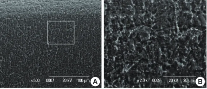
×500 and ×2,000. The isotropic microstructure consisted of sharp, pointed long ridges and v-shaped valleys (Fig. 3).
Test groups
Group 1 (100 mJ/pulse, 10 Hz) showed some partial changes after the two-minute irradiation treatment, but did not show significant changes in the specific ridge and valley structures (Figs. 4-6). After the laser energy was increased to 140 mJ/
pulse, the sharpness of the ridges tended to be reduced (Figs.
7-9). As a result of the collapse of the ridges the microstruc- ture of ridges and valleys had been gradually flattened with increased laser energy and irradiation time.
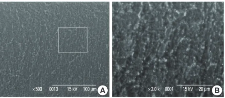
This flattening was more significant with the energy set- ting at 180 mJ/pulse, which flattened ridges that had still been present after only one minute of irradiation treatment (Figs.
10-12).
The change of microstructure can be easily noted in the oblique view of the surface (Fig. 13).
Surface roughness measurement
The mean surface roughness of the control implant was 0.423±0.039 µm. The amount of change in roughness be- tween before and after the laser irradiation was -0.119 µm, -0.027 µm, -0.173 µm, -0.038 µm, -0.028 µm, -0.013 µm, Figure 1. Specimen assignment depending on laser irradiation conditions.
Figure 2. The area of treatment and measurement. SEM, scanning electron microscopy.
3rd valley 6th valley 9th valley
The area of laser irradiation (2×2 mm2) The area of surface roughness measurement and SEM evaluation
Figure 3. Control specimen: double acid-etched implant surface without any conditioning. (A) ×500, (B) ×2,000. A microstructure consisting of ridges and valleys can be seen.
×500 0007 20 kV 100 μm A ×2.0 k 0009 20 kV 20 μm B Double acid-etched implants
(n=10)
Control group without laser irradiation
(n=1)
Test groups with laser irradiation
(n=9)
No. 2 (1.5 min)
No. 3 (2 min) No. 1
(1 min)
No. 5 (1.5 min)
No. 6 (2 min) No. 4
(1 min)
No. 8 (1.5 min)
No. 9 (2 min) No. 7
(1 min) Group 1
with 100 mJ/pulse, 10 Hz (n=3)
Group 3 with 180 mJ/pulse, 10 Hz (n=3) Group 2
with 140 mJ/pulse, 10 Hz (n=3)
-0.002 µm, -0.044 µm, and -0.003 µm, for experimental group implants from No. 1 to No. 9 in order. All the average
roughness values were reduced, regardless of the irradiation energy and time. However, these changes were not statisti- Figure 4. Double acid-etched implant surface irradiated under 100
mJ/pulse for 1 minute. (A) ×500, (B) ×2,000. No remarkable changes are visible.
×500 0003 20 kV 100 μm A ×2.0 k 0010 20 kV 20 μm B
Figure 10. Double acid-etched implant surface irradiated under 180 mJ/pulse for 1 minute. (A) ×500, (B) ×2,000. The loss of the ridges is visible.
×500 0007 15 kV 100 μm A ×2.0 k 0011 15 kV 20 μm B
Figure 7. Double acid-etched implant surface irradiated under 140 mJ/pulse for 1 minute. (A) ×500, (B) ×2,000. The sharpness of ridges is reduced.
×500 0003 20 kV 100 μm A ×2.0 k 0004 20 kV 20 μm B Figure 5. Double acid-etched implant surface irradiated under 100 mJ/pulse for 1.5 minutes. (A) ×500, (B) ×2,000. No remarkable changes can be seen.
×500 0001 20 kV 100 μm A ×2.0 k 0002 20 kV 20 μm B
Figure 11. Double acid-etched implant surface irradiated under 180 mJ/pulse for 1.5 minutes. (A) ×500, (B) ×2,000. The change is exten- sive.
×500 0012 15 kV 100 μm A ×2.0 k 0008 15 kV 20 μm B Figure 8. Double acid-etched implant surface irradiated under 140 mJ/pulse for 1.5 minutes. (A) ×500, (B) ×2,000. The change is exten- sive.
×500 0021 20 kV 100 μm A ×2.0 k 0016 20 kV 20 μm B
Figure 6. Double acid-etched implant surface irradiated under 100 mJ/pulse for 2 minutes. (A) ×500, (B) ×2000. No remarkable chang- es can be seen.
×500 0001 20 kV 100 μm A ×2.0 k 0002 20 kV 20 μm B
Figure 9. Double acid-etched implant surface irradiated under 140 mJ/pulse for 2 minutes. (A) ×500, (B) ×2,000. The change is exten- sive.
×500 0002 15 kV 100 μm A ×2.0 k 0020 20 kV 20 μm B
cally significant (P>0.05; Table 1).
DISCUSSION
To achieve successful implant treatment, a large number of surface modifications including mechanical, topographic, and physicochemical properties have been introduced.
Among these properties, surface roughness, which is a topo- graphic property, has been the main focus of dental implant studies for more than a decade [14]. Beginning in the mid- 1990s, the general trend related to the surface roughness has changed from using a minimally rough surface to a moder- ately rough one. Previous experiments demonstrated that rough surfaces promoted both bone anchoring and biome- chanical stability, and facilitated better osseointegration when compared to machined [15,16] or very rough surfaces [17,18].
The two theoretical bases are as follows: the biological ad- vantage of increased contact area with blood cells, adherence of more platelets [18,19], and migration of osteogenic cells result in increased contact osteogenesis [20,21]; and the me- chanical advantage of enhanced mechanical interlocking be- tween the macromolecules of the implant surface and the bone, which increases bone-to-implant contact, result in greater resistance to compression, tension, and shear stress
[22,23]. However, consideration of the negative effects of sur- face roughness is also necessary. Some implant surface prop- erties promote early plaque formation, resulting in inflam- matory reactions around implants, which can in turn result in implant failure [24]. In general, a higher degree of bacterial adhesion has been observed on rougher surfaces, which can hinder effective access for cleaning infected implants [25,26].
To achieve the successful treatment of a peri-implantitis le- sion, it is necessary to not only surgically remove the inflamed tissue and provide antimicrobial therapy, but also provide an efficient procedure for reconstituting the contaminated im- plant surface without modifying the surface, to allow for healthy tissue remodeling at the implant interface [27]. Al- though it is reported that some mechanical and chemical methods are inappropriate, in that they cause surface dam- age, it can be mostly prevented by using controlled methods including some types of dental lasers [28,29].
Among the various laser systems, the Er:YAG laser has been recognized as the most promising laser system for obtaining excellent tissue ablation with high bactericidal [30] and de- toxification effects [31,32]. Higher energy levels to achieve even more potent effects, as mentioned above, can lead to rapid heat generation, which creates tissue and implant sur- face damage [33]. However, during irradiation the specific wavelengths of an Er:YAG laser will not result in a significant increase in implant body temperature because they are poor- ly absorbed by titanium. In addition, due to the implant irra- diation occurring under sufficient water cooling, the Er:YAG laser affects only the superficial layer without affecting the deep regions of a substrate, since the applied energy is trans- ferred into a thermal reaction leading to the evaporation of the coolant. Depending on the different implant surfaces and laser irradiation conditions, the effects of an Er:YAG laser can vary and these have been evaluated in previous series studies [34-36].
The present study was performed to evaluate the effects of Er:YAG laser irradiation on the double acid-etched implant surface microstructure, according to the laser energy and the Figure 12. Double acid-etched implant surface irradiated under 180
mJ/pulse for 2 minutes. (A) ×500, (B) ×2,000. The change is exten- sive.
×500 0013 15 kV 100 μm A ×2.0 k 0001 15 kV 20 μm B
Figure 13. The comparison from the oblique perspective ×2,000 (A) control (B) 180 mJ/pulse, 10 Hz, 1.5 minutes.
×2.0 k 0001 15 kV 20 μm
A B
Table 1. Surface roughness values measured 3 valleys before and after surface detoxification by laser treatment (mean±SD).
Pulse energy (time) Ra-pre (m) Ra-post (m) P-value No.1 100 mJ/pulse, 1 min 0.467±0.135 0.348±0.011 0.109 No.2 100 mJ/pulse, 1.5 min 0.382±0.059 0.355±0.029 0.285 No.3 100 mJ/pulse, 2 min 0.506±0.091 0.333±0.051 0.109 No.4 140 mJ/pulse, 1 min 0.432±0.05 0.394±0.036 0.593 No.5 140 mJ/pulse, 1.5 min 0.409±0.082 0.381±0.07 0.593 No.6 140 mJ/pulse, 2 min 0.420±0.102 0.407±0.075 0.593 No.7 180 mJ/pulse, 1 min 0.410±0.007 0.408±0.068 1 No.8 180 mJ/pulse, 1.5 min 0.419±0.026 0.375±0.001 0.109 No.9 180 mJ/pulse, 2 min 0.399±0.045 0.396±0.015 1 Wilcoxon’s signed rank sum test (P>0.05).
protocol. The energy setting and irradiation exposure dura- tion was determined based on the conditions that do not al- low harmful heat generation or surface alteration, but allow effective cytotoxicity and biocompatibility; the method of ir- radiation such as the irradiation angle, the distance from the implant surface, and the course of tip movement were deter- mined by considering clinical situations.
The SEM image evaluation of this study showed that the original structure of the double acid-etched implant was not changed by the treatment condition of 100 mJ/pulse, at 10 Hz, with applied durations of 1 minute, 1.5 minutes, and 2 min- utes. Increased laser energy of 140 mJ/pulse and 180 mJ/pulse caused surface alteration that resulted in reduced sharpness of ridge structures. However, neither melting nor cracking was observed, and the subtractive surface treatment may have higher stability than additive surface treatment, specifically laser irradiation. In additive surface treatment such as plas- ma spray-coating of hydroxyapatite particles, titanium beads, or physical or chemical vapor deposition, deposited particles may have a tendency to cause resorption or separation of the coating. On the other hand, the acid-etching and blasting methods, which are subtractive surface treatments, did not show significant surface change and endured higher energy conditions.
The surface roughness evaluation revealed that the rough- ness value of the non-laser irradiated double acid-etched implant used in this study was as low as that of the machined implant. That is, except one specimen (No. 3), all implants had a low roughness value (Ra<0.5 µm) that could be classi- fied as a smooth surface according to the surface topography characterization advanced by Albrektsson and Wennerberg [14]. Although the kinds of measurement methods and im- plant surface area are not identical, the results are different from other experiments, and this showed that the double ac- id-etched implant system (Osseotite, Biomet, Inc.) had mini- mal roughness values (0.5 µm<Sa<1.0 µm) similar to those of turned implants (Table 2). To explain this discordance, un- derstanding of roughness parameters is necessary.
The two-dimensional (Ra) and three-dimensional (Sa) pa-
quite stable and insensitive to occasional high peaks or deep valleys, and are the most commonly used parameters in eval- uating implant surface roughness [37]. However, 3-D topo- graphic analysis using an optical interferometer is superior to 2-D analysis because the former (Sa) is more accurate, rep- resentative, and flexible. Mechanical contact profilometer disadvantages are that there is no precise information about which regions were actually measured and it cannot be used for non-destructive evaluations of implants because the tip cannot evaluate threaded parts. Therefore, the use of 3-D evaluations is recommended for implant surface evaluation.
As the surface roughness in the screw-type implant varies at different locations, the roughness of all these regions must be considered when surfaces of oral implants are evaluated.
It is widely accepted that the nine measurements on each screw (three tops, three valleys, and three flanks) of three im- plants are sufficient to obtain a reliable mean value [38]. In this study, we evaluated surface roughness on only three val- leys using a mechanical contact profilometer. The reason only three valleys were measured was to evaluate the rough- ness changes on the same area where SEM evaluation was done. Therefore, it must be noted that the roughness value of the double acid-etched implant in this study does not rep- resent the overall surface, but a restricted valley area. In other words, we cannot claim that this implant system had a smooth surface nor that the overall surface roughness was not changed significantly by laser irradiation. Compared with a previous study [39] conducted with the same protocol of this study, ex- cept for the detoxification method (rubbing with 50 mg/mL tetracycline-HCl) and the evaluation method of surface roughness (optical interferometer), the mechanical contact profilometer in this study showed more reproducible and consistent results than that of the optical interferometer.
This suggests that mechanical contact profilometer use is a possible method for some areas of implant dentistry (e.g., in confined, rather than flat surface measurement).
It is necessary to consider some limitations to this study.
First, although SEM provides high-quality images, it is more suitable for morphologic than topographic description. Top- ographic characterization is almost exclusively used as a com- parative method and is therefore prone to subjective inter- pretations. There was also an ambiguous interpretation in this study concerning whether or not the change was exam- ined (Fig. 4). Therefore, a more objective parameter is need- ed. Second, in this study, roughness measurement was evalu- ated by a mechanical contact profilometer on valleys. In or- der to evaluate the entire implant surface, a topographic sur- face analysis must be used, including three thread tops, three thread valleys, and three flank areas using an optical interfer- Table 2. The comparison of mean roughness value of double acid-
etched implants (Osseotite, Biomet, Inc., Palm Beach Gardens, FL, USA).
Roughness mean value (Sa, µm)a)
Abrahamsson et al. 2001 [34] 0.94b)
Albrektsson and Wennerberg 2004 [14] 0.5-1.0b) Sul et al. 2006 [35], 2008 [36] 0.72b)
a)All roughness value was evaluated by the optical interferometer.
b)Roughness mean value was obtained on tops, valleys, and flanks.
all measuring systems, irrespective of being 2-D or 3-D, have vertical and lateral limitations in terms of measuring range and resolution; the true surface topography will never be ob- tained due to surface deformation in the mechanical contact mode techniques and because of optical artifacts in optical instruments. Fourth, integral roughness parameters are of very limited value in describing the complex surface struc- tures presented on surface-treated titanium implants. In ad- dition, though surface roughness has been regarded as a predominant factor in cell adhesion and osteogenesis during the implant healing phases, no correlation was reported be- tween the profilometry data and bone contact percentage.
Double acid-etched implants have been recognized as hav- ing a minimally rough surface, have been reported to induce more bone contact, and have a 3.5-fold increase in mechani- cal pull-out force compared to untreated implants, possibly as a result of better fibrin clot retention and growth factor enhancement. Overall, some other factor dictating cell adhe- sion and bone apposition in direct contact with the implant surface, other than roughness, should be considered. Fifth, only the topographic characterization was studied in relation to the microstructure change contained in this study; there- fore, additional research is needed to evaluate the effect of the Er:YAG laser on cell attachment and bacteria removal on the implant surface. Simply roughening the implant surface may not result in a large change in bone conductivity and bi- ologic response; there may be more critical parameters of biocompatibility than surface roughness.
Standardization is needed for peri-implantitis treatment to determine under which conditions the Er:YAG laser can be used without altering the implant surface. In conclusion, Er:YAG laser irradiation on a double acid-etched implant surface is recommended to be set below 100 mJ/pulse, at 10 Hz, and with an applied duration of less than two minutes for detoxification of the implant surface in order to prevent surface alteration.
CONFLICT OF INTEREST
No potential conflict of interest relevant to this article was reported.
REFERENCES
1. Albrektsson TO, Johansson CB, Sennerby L. Biological as- pects of implant dentistry: osseointegration. Periodontol 2000 1994;4:58-73.
2. Mombelli A, Lang NP. The diagnosis and treatment of peri-implantitis. Periodontol 2000 1998;17:63-76.
of 589 consecutive implants supporting complete fixed prostheses. Part I: Periodontal aspects. J Prosthet Dent 1992;68:655-63.
4. Isidor F. Loss of osseointegration caused by occlusal load of oral implants. A clinical and radiographic study in monkeys. Clin Oral Implants Res 1996;7:143-52.
5. Pongnarisorn NJ, Gemmell E, Tan AE, Henry PJ, Marshall RI, Seymour GJ. Inflammation associated with implants with different surface types. Clin Oral Implants Res 2007;
18:114-25.
6. Meffert RM, Langer B, Fritz ME. Dental implants: a review.
J Periodontol 1992;63:859-70.
7. Mombelli A. Microbiology and antimicrobial therapy of peri-implantitis. Periodontol 2000 2002;28:177-89.
8. Krozer A, Hall J, Ericsson I. Chemical treatment of ma- chined titanium surfaces. An in vitro study. Clin Oral Im- plants Res 1999;10:204-11.
9. Schwarz F, Putz N, Georg T, Reich E. Effect of an Er:YAG laser on periodontally involved root surfaces: an in vivo and in vitro SEM comparison. Lasers Surg Med 2001;29:
328-35.
10. Schwarz F, Sculean A, Berakdar M, Szathmari L, Georg T, Becker J. In vivo and in vitro effects of an Er:YAG laser, a GaAlAs diode laser, and scaling and root planing on peri- odontally diseased root surfaces: a comparative histologic study. Lasers Surg Med 2003;32:359-66.
11. Ando Y, Aoki A, Watanabe H, Ishikawa I. Bactericidal effect of erbium YAG laser on periodontopathic bacteria. Lasers Surg Med 1996;19:190-200.
12. Yamaguchi H, Kobayashi K, Osada R, Sakuraba E, Nomura T, Arai T, et al. Effects of irradiation of an erbium:YAG la- ser on root surfaces. J Periodontol 1997;68:1151-5.
13. Matsuyama T, Aoki A, Oda S, Yoneyama T, Ishikawa I. Ef- fects of the Er:YAG laser irradiation on titanium implant materials and contaminated implant abutment surfaces. J Clin Laser Med Surg 2003;21:7-17.
14. Albrektsson T, Wennerberg A. Oral implant surfaces: Part 1--review focusing on topographic and chemical proper- ties of different surfaces and in vivo responses to them.
Int J Prosthodont 2004;17:536-43.
15. De Leonardis D, Garg AK, Pecora GE. Osseointegration of rough acid-etched titanium implants: 5-year follow-up of 100 minimatic implants. Int J Oral Maxillofac Implants 1999;14:384-91.
16. Wennerberg A, Ektessabi A, Albrektsson T, Johansson C, Andersson B. A 1-year follow-up of implants of differing surface roughness placed in rabbit bone. Int J Oral Maxil- lofac Implants 1997;12:486-94.
17. Lazzara RJ, Testori T, Trisi P, Porter SS, Weinstein RL. A hu-
faces using implants with 2 opposing surfaces. Int J Peri- odontics Restorative Dent 1999;19:117-29.
18. London RM, Roberts FA, Baker DA, Rohrer MD, O’Neal RB. Histologic comparison of a thermal dual-etched im- plant surface to machined, TPS, and HA surfaces: bone contact in vivo in rabbits. Int J Oral Maxillofac Implants 2002;17:369-76.
19. Park JY, Davies JE. Red blood cell and platelet interactions with titanium implant surfaces. Clin Oral Implants Res 2000;11:530-9.
20. Bowers KT, Keller JC, Randolph BA, Wick DG, Michaels CM. Optimization of surface micromorphology for en- hanced osteoblast responses in vitro. Int J Oral Maxillofac Implants 1992;7:302-10.
21. Ivanoff CJ, Widmark G, Johansson C, Wennerberg A. His- tologic evaluation of bone response to oxidized and turned titanium micro-implants in human jawbone. Int J Oral Maxillofac Implants 2003;18:341-8.
22. Hsu SH, Liu BS, Lin WH, Chiang HC, Huang SC, Cheng SS. Characterization and biocompatibility of a titanium dental implant with a laser irradiated and dual-acid etched surface. Biomed Mater Eng 2007;17:53-68.
23. Trisi P, Rao W, Rebaudi A. A histometric comparison of smooth and rough titanium implants in human low-den- sity jawbone. Int J Oral Maxillofac Implants 1999;14:689-98.
24. Grossner-Schreiber B, Teichmann J, Hannig M, Dorfer C, Wenderoth DF, Ott SJ. Modified implant surfaces show different biofilm compositions under in vivo conditions.
Clin Oral Implants Res 2009;20:817-26.
25. Drake DR, Paul J, Keller JC. Primary bacterial colonization of implant surfaces. Int J Oral Maxillofac Implants 1999;14:
226-32.
26. Renvert S, Roos-Jansaker AM, Claffey N. Non-surgical treatment of peri-implant mucositis and peri-implantitis:
a literature review. J Clin Periodontol 2008;35(8 Suppl):
305-15.
27. Maximo MB, de Mendonca AC, Renata Santos V, Figueire- do LC, Feres M, Duarte PM. Short-term clinical and mi- crobiological evaluations of peri-implant diseases before and after mechanical anti-infective therapies. Clin Oral Implants Res 2009;20:99-108.
28. Kreisler M, Gotz H, Duschner H. Effect of Nd:YAG, Ho:
YAG, Er:YAG, CO2, and GaAIAs laser irradiation on sur- face properties of endosseous dental implants. Int J Oral Maxillofac Implants 2002;17:202-11.
Jansen B, et al. Bactericidal effect of the Er:YAG laser on dental implant surfaces: an in vitro study. J Periodontol 2002;73:1292-8.
30. Folwaczny M, Mehl A, Aggstaller H, Hickel R. Antimicro- bial effects of 2.94 microm Er:YAG laser radiation on root surfaces: an in vitro study. J Clin Periodontol 2002;29:73-8.
31. Friedmann A, Antic L, Bernimoulin JP, Purucker P. In vitro attachment of osteoblasts on contaminated rough titani- um surfaces treated by Er:YAG laser. J Biomed Mater Res A 2006;79:53-60.
32. Schwarz F, Aoki A, Sculean A, Georg T, Scherbaum W, Becker J. In vivo effects of an Er:YAG laser, an ultrasonic system and scaling and root planing on the biocompati- bility of periodontally diseased root surfaces in cultures of human PDL fibroblasts. Lasers Surg Med 2003;33:140-7.
33. Kreisler M, Kohnen W, Christoffers AB, Gotz H, Jansen B, Duschner H, et al. In vitro evaluation of the biocompati- bility of contaminated implant surfaces treated with an Er: YAG laser and an air powder system. Clin Oral Im- plants Res 2005;16:36-43.
34. Abrahamsson I, Zitzmann NU, Berglundh T, Wennerberg A, Lindhe J. Bone and soft tissue integration to titanium implants with different surface topography: an experi- mental study in the dog. Int J Oral Maxillofac Implants 2001;16:323-32.
35. Sul YT, Johansson C, Albrektsson T. Which surface proper- ties enhance bone response to implants? Comparison of oxidized magnesium, TiUnite, and Osseotite implant sur- faces. Int J Prosthodont 2006;19:319-28.
36. Sul YT, Byon E, Wennerberg A. Surface characteristics of electrochemically oxidized implants and acid-etched im- plants: surface chemistry, morphology, pore configura- tions, oxide thickness, crystal structure, and roughness. Int J Oral Maxillofac Implants 2008;23:631-40.
37. Lang NP, Jepsen S; Working Group 4. Implant surfaces and design (Working Group 4). Clin Oral Implants Res 2009;20(Suppl 4):228-31.
38. Wennerberg A, Albrektsson T, Andersson B. Bone tissue response to commercially pure titanium implants blasted with fine and coarse particles of aluminum oxide. Int J Oral Maxillofac Implants 1996;11:38-45.
39. Jeong DM, Park JB, Kwon YH, Herr Y, Chung JH. The in- fluence of tetracycline-HCl for micromorphology of ther- mal dual acid etched surface implants. J Korean Acad Peri- odontol 2007;37:265-75.