This work was supported by a grant from the Korean Ministry of Health and Welfare, Republic of Korea (No. 01-PJ3-PG6-01GN04-003).
Address for correspondence : Choon-Sik Park, M.D.
1174 Jung-Dong, Wonmi-Gu, Bucheon-Si, Gyeonggi-Do, Seoul 420-767, Korea Phone : +82-32-621-5105
Fax : +82-32-621-5018 E-mail : [email protected] Received : Jun. 16. 2005 Accepted : Jul. 12. 2005
림프종 1예
순천향대학교 의과대학 내과학교실, 해부병리학교실1, 진단방사선과교실2
배준용, 나현식, 최재성, 최준호, 박성규, 장안수, 고은석1, 백상현2, 박재성2, 박춘식
A Case of Primary Mucosa-Associated Lymphoid Tissue Lymphoma of the Trachea and Colon
Jun Yong Bae, M.D., Hyun Sik Na, M.D., Jae Sung Choi, M.D., Jun Ho Choi, M.D., Sung Gyu Park, M.D., An Soo Jang, M.D., Eun Suk Ko, M.D.1, Sang Hyun Paik, M.D.2, Jae Sung Park, M.D.2, Choon Sik Park, M.D.
Department of Internal Medicine, Anatomical Pathology1, Diagnostic Radiology2, Soonchunhyang University, School of Medicine, Bucheon, Korea
The 64-year-old female patient with cough and intermittent hemoptysis of six months duration visited our hospital.
On chest computed tomography, a small, ovoid, 1.3cm sized and well enhanced lesion was detected on the distal trachea. Two multiple lobulated lesions on the sigmoid and transverse colon were revealed on the colonoscopy. The histological findings showed small and medium sized lymphocytes infiltration, CD20 and CD79a positive staining and multiple lymphoepithelial lesions on the distal trachea and colon tissues. Herein, a case of primary MALT lymphoma, with involvement of the trachea and colon, which was treated with rituximab (CD20 anti-monoclonal antibody), cyclophosphamide, adriamycin, vincristine and prednisolone (CHOP regimen), is reported.
(Tuberc Respir Dis 2005; 59: 193-197)
Key words : Mucosa-associated lymphoid tissue, Trachea, Colon
서 론
Mucosa-associated lymphoid tissue는 안구, 침샘, 갑 상선, 폐, 위, 소장, 대장, 방광, 여성 생식기 등에서 존재 하는 림프 조직으로 림프절과 형태와 기능 면에서 매우 비슷하다. Mucosa-associated lymphoid tissue 림프종 (이하 MALT 림프종)은 일반적인 림프종이 림프절에 서 면역세포 중 한 종류가 악성 변화를 가져오는 것과 비슷하게 mucosa-associated lymphoid tissue에서 면역세포 중 한 종류가 악성 변화를 가지는 것이 다1. 위장관의 경우에 있어서 MALT 림프종은 역 학, 병리 기전 및 치료가 잘 정립 되어 있지만 다른
장기의 MALT 림프종의 경우 매우 드물기 때문에 비교적 예후는 좋은 편이라고는 하지만 이에 대한 자세한 임상경과, 치료 및 예후가 드물게 보고되 어 있다. 이에 저자 등은 기관과 대장에 동시에 발 생한 MALT 림프종을 경험하였기에 보고하는 바 이다.
증 례
환 자 : 64세 여자
주 소 : 기침, 간헐적 객혈
현병력 : 내원 6개월 전부터 발생한 기침과 간헐적으 로 가래에 피가 묻어 나오는 정도의 객혈을 주소로 내 원함.
과거력 : 특이사항 없었고 흡연력도 없었음.
가족력 : 특이사항 없음.
진찰 소견 : 생체 징후는 130/80 mmHg, 체온 36.5℃
호흡수 20회/분, 맥박수 90회/분 이었으며 흉부청진에 서 양측 전 폐야에서 비교적 깨끗한 호흡음 들렸음.
림프절 종대나 간비종대는 관찰되지 않았다.
검사실 소견 : 일반 혈액 검사 및 혈청 화학 검사에서
Figure 2. Bronchoscopy shows smooth rounded lesion with normal mucosa on distal trachea.
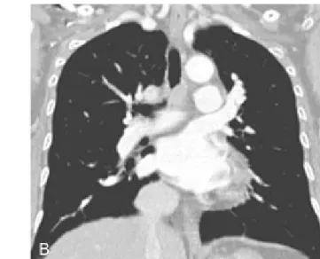
Figure 1. Computed tomography images (A. Axial scan, B. Coronal scan) show well enhanced, small ovoid lesion (about 1.3cm) on distal trachea.
특이 소견 없었다. β2-microglobulin의 증가나 다른 종양 표지자, HIV 감염이나 자가면역질환의 증거도 없었다. 폐기능 검사에서 FVC 3.36L(예측치의 123%), FEV1 2.10L(예측치의 106%), FEV1/FVC 62%를 보 였으며 노력성 호기류-용적 곡선에서 흉곽 내 폐쇄 소견을 보였다.
방사선 소견 : 단순 흉부 촬영에서는 특이 소견 없었으 나 흉부 전산화 단층 촬영에서 원위부 기관의 비교적 조영이 잘되는 1.3cm의 난원형 병변(Fig. 1)이 관찰이 되었다. 기관지 내시경으로 관찰 하였을 때 원위부 기관 에 주위와 비교하여 점막의 변화는 없었으나 기관 내로 표면이 부드러운 융기형 병변(Fig. 2)이 관찰 되었다.
조직학적 소견 : 경식 기관지 내시경하에서 조직 생검 을 시행 하였으며 조직 검사에서 작고 중간크기의 림 프구의 침윤을 보이며 기관 상피는 잘 유지되었다. 면 역화학염색에서 CD20과 CD79a에 양성소견을 보였으 며 Anti-kappa와 lamda light chain 염색에서는 단클 론성을 보이지 않는 mucosa-associated lymphoid tissue 림프종 소견이 관찰 되었다 (Fig. 3).
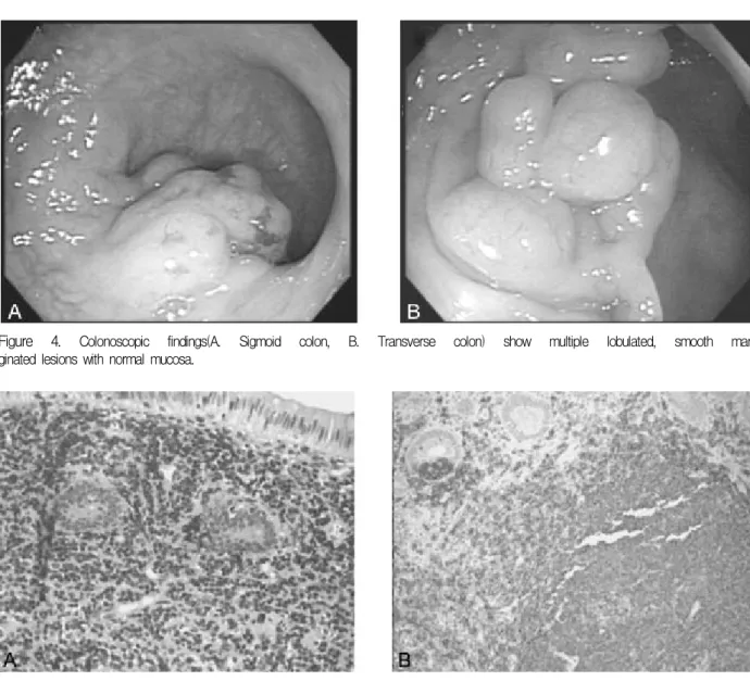
치료 및 경과 :환자의 기관지 MALT 림프종이 전신 적인 질환이 아님을 증명하기 위해 골수조직검사, 두 부 및 경부와 복부 컴퓨터 단층촬영, 위와 대장 내시 경, 혈액 및 뇨의 단백질 전기영동을 시행 하였다. 대 장 내시경에서 S자 대장과 횡행 대장의 소엽의 용종
모양을 보이는 병변(Fig. 4)이 관찰 되었다. 대장 조직 검사에서도 작은 림프구의 침윤과 함께 CD20과 CD79a의 양성을 보였으며 림프상피성 병변이 관찰 되었다 (Fig. 5).환자는 MALT 림프종으로 병기는 횡 경막 위아래의 양측에 병변을 가지는 IIIE로서 CD20 단클론항체인 rituximab과 CHOP (cyclophosphamide, Doxorubicin, Vincristine, Prednisolone)화학요법을 시 행하였고 추적관찰 중이다.
Figure 5 Histologic findings of the colonoscopic specimens (A. Haematoxyline-eosin staining, B. CD20, CD79a co- staining) show the small and medium sized lymphocytes with CD20, CD79a positive staining, lymphoepithelial lesions.
Figure 4. Colonoscopic findings(A. Sigmoid colon, B. Transverse colon) show multiple lobulated, smooth mar
ginated lesions with normal mucosa.
Figure 3. Histologic findings of the tracheal specimens (A. Haematoxyline-eosin staining, B. CD20, CD79a co-stain
ing) show the small and medium sized lymphocytes with CD20, CD79a positive staining.
고 찰
MALT 림프종은 Isaacson과 Wright가1 1983년 처 음으로 위장관의 저급 B 세포 림프종으로 보고 하였 다. MALT 림프종은 침범부위가 위장관인 경우가 대 부분이고 안구, 침샘, 방광, 소장, 대장, 폐, 갑상선, 여 성 생식기 등도 발생하는 것으로 알려져 있다. MALT 림프종의 조직학적 소견은 활성화된 배중심(germinal center)형성, 불규칙한 핵과 투명한 세포질을 가지는 중심세포(centrocyte-like cell)모양의 세포, 림프 상피 성 병변을 만드는 임파구의 상피침윤을 보이며 일부 에서 대세포(large cell)를 포함한다. 그리고 면역조직 화학 검사에서 특징적으로 단클론의 확장을 보이는 B cell 림프종의 형태를 보이며 B 세포 표지자인 CD20, CD19, CD79a에 양성을 보인다. 대부분의 MALT 림 프종은 외과적 수술 및 방사선 치료 등의 국소 치료에 우수한 효과 및 예후를 보이는 것으로 알려져 있다.
이것은 MALT 림프종에서 임파선의 국소 또는 원격 전이가 드물며 골수전이가 잘 일어나지 않기 때문이다.
원발성 기관 종양이 드물기는 하지만 Grillo과 Ma
thisen은2 대부분이 편평 세포 암종과 선양 낭성 암종 (adenoid cystic carcinoma)으로 보고하였다. MALT 림프종의 기관에서의 빈도는 Zinzani 등3의 비 위장관 MALT 림프종의 연구결과를 보면 75예 중 1예가 보 고되어 매우 드문 것으로 생각된다. 또한 기관을 침범 한 MALT 림프종의 경우 국소치료에 대한 효과가 있 는 것으로 보고하고 있으나 장기간의 예후에 대한 통 계는 거의 없다4,5. 1981년 Maeda 등6이 기관지 림프종 이 외과적 절제로만 치료하여 5년 이상의 생존율을 보 고 하였다. Okubo 등7은 외과적 절제만으로 53개월간 재발이 없었음을 보고하였다.
위장관의 MALT 림프종은 약 90%의 저급 위 MALT 림프종이 stage I 이며 80-95%의 5년 생존율을 가진 다. 조기에 발견된 경우 Helicobacter pylori 치료만으 로 대부분이 충분한 치료 효과를 볼 수 있었으며8-10, Lugano staging system을 이용하여 stage I 과 stage II의 경우에 방사선 치료(30CGy)는 100% 완전관해, 100%에서 재발 없음을 보고했다11. Ann Arbor stage IE 17명과 stage IVE 7명을 대상으로 단일 제제
(cyclophosphamide 또는 chlorambucil) 경구 화학요 법은 75%의 완전관해와 75%의 5년 생존율을 가져 왔 다.12 대장과 갑상선 에서도 MALT 림프종은 외과적 절제, 방사선치료, 화학요법이 좋은 결과를 가져 오는 것으로 알려져 있다.
MALT 림프종에서 여러 장기 침범을 보인 경우가 있는데 Thieblemont 등13은 MALT 림프종 108예 중 35예(32%)에서 다 장기 침범을 보고 하였다. Yoshino 등14이 7년간 MALT 림프종 304예를 추적관찰하여 7 예 에서 MALT 림프종이 2개 이상 장기를 침범한 경 우가 있었고, 7예 중 6예가 장의 침범을 포함하고 있 었다. 이와 같이 MALT 림프종은 대략 2%에서 32%
까지 다양한 다 장기 침범이 있으며 다 장기 침범의 경우에서 장관을 침범한 경우가 많음을 알 수 있다.
MALT 림프종의 불량한 예후가 대세포(large cell) 구성 비 , 다 장기 침범, 전신적인 증상이 연관이 있을 것이라 생각을 하지만 아직 정확한 보고는 없다.
따라서 저자 등은 MALT 림프종에서 매우 드문 기관 과 대장을 침범한 MALT 림프종을 가진 1예를 CD20의 단클론 항체인 rituximab과 CHOP의 화학요법으로 치료하고 추적관찰 중인 1예를 보고하는 바이다.
요 약
MALT 림프종은 안구, 침샘, 감상선, 폐, 위, 소장, 대장, 방광, 여성 생식기에 발생하며, 대부분이 단일 기관을 침범하고 국소 치료와 화학요법에 좋은 예후 를 보인다. MALT 림프종이 기관을 침범하는 경우는 거의 보고된 경우가 없으며 국내에선 폐의 MALT 림 프종이 보고된 적이 있었다15. 이에 저자 등은 기관과 대장에 발생한 원발성 MALT 림프종 1예를 문헌 고 찰과 함께 보고하는 바이다.
참 고 문 헌
1. Isaacson P, Wright DH. Malignant lymphoma of mu
cosa-associated lymphoid tissue: a distinctive type of B-cell lymphoma. Cancer 1983;52:1410-6.
2. Grillo HC, Mathisen DJ. Primary tracheal tumors:
treatment and results. Ann thorac Surg 1990;49:69- 77.
3. Zinzani PL, Magagnoli M, Galieni P, Martelli M, Poletti V, Zaja F, et al. Nongastrointestinal low-grade mu
cosa-associated lymphoid tissue lymphoma: analysis of 75 patients. J Clin Oncol 1999;17:1254.
4. Kaplan MA, Pettit CL, Zukerberg LR, Harris NL.
Primary lymphoma of the trachea with morphologic and immunophenotypic characteristics of low-grade B-cell lymphoma of mucosa-associated lymphoid ti
ssue. Am J Surg Pathol 1992;16:71-5.
5. Wiggins J, Shefield E, Green M. Primary B cell mali
gnant lymphoma of the trachea. Thorax 1988;43:497-8.
6. Maeda M, Kotake Y, Monden Y, Nakahara K, Kawa
shima Y, Kitamura H. Primary malignant lymphoma of the trachea: report of a case successfully treated by primary end to end anastomosis after circumferential resection of the trachea. J Thorac Cardiovasc Surg 1981;81:835-9.
7. Okubo K, Miyamoto N, Komaki C. Primary mucosa- associated lymphoid tissue (MALT) lymphoma of the trachea: a case of surgical resection and long term survival. Thorax 2005;60:82-3.
8. Steinbach G, Ford R, Glober G, Sample D, Hagemeister FB, Lynch PM, et al. Antibiotic treatment of gastric lymphoma of mucosa-associated lymphoid tissue: an uncontrolled trial. Ann Intern Med 1999;131:88-95.
9. Wotherspoon AC, Doglini C, Diss TC, Pan L, Moschini A, de Boni M, et al. Regression of primary low grade B-cell gastric lymphoma of mucosa-associated lymp
hoid tissue type after eradication of Helicobacter
pylori. Lancet 1993;342:575-7.
10. Roggero E, Zucca E, Pinotti G, Pascarella A, Capella C, Savia A, et al. Eradication of Helicobacter pylori infection in primary low-grade gastric lymphoma of mucosa-associated lymphoid tissue. Ann Intern Med 1995;122:767-9.
11. Schechter NR, Portlok CS, Yahalom J. Treatment of mucosa-associated lymphoid tissue lymphoma of the stomach with radiation alone. J Clin Oncol 1998;16:
1916-21.
12. Hammel P, Haioun C, Chaumette MT, Gaulard P, Divine M, Reyes F, et al. Efficacy of single agent chemotherapy in low grade B-cell mucosa-associa
ted lymphoid tissue lymphoma with prominent gastric expression. J Clin Oncol 1995;13:2524-9.
13. Thieblemont C, Bastion Y, Berger F, Rieux C, Salles G, Dumontet C, et al. Mucosa-associated lymphoid tissue gastrointestinal and nongastrointestinal lym
phoma behavior: analysis of 108 patients. J Clin Oncol 1997;15:1624-30.
14. Yoshino T, Ichimura K, Mannami T, Takase S, Ohara N, Okada H, et al. Multiple organ mucosa-associated lymphoid tissue lymphomas often involve the intestine.
Cancer 2001;91:346-53.
15. Han MS, Kang DW, Choi GY, Lee YD, Cho YS. A case of primary extranodal marginal zone B-cell lymph
oma of the MALT type. Tuberc Respir Dis 2003;54:
635-9.