• 검색 결과가 없습니다.

Primary Pulmonary Malignant Melanoma: An Unexpected Tumor

N/A
N/A
Protected

Academic year: 2021

Share "Primary Pulmonary Malignant Melanoma: An Unexpected Tumor"

Copied!
4
0
0

로드 중.... (전체 텍스트 보기)

전체 글

(1)

Primary Pulmonary Malignant Melanoma:

An Unexpected Tumor

Kyo-Bum Hwang, M.D.

1

, Ki-Eun Hwang, M.D.

1

, Jae-Wan Jung, M.D.

1

, Su-Jin Oh, M.D.

1

, Mi-Jeong Park, M.D.

1

, Young-Hoon Jeong, M.D.

1

, Keum-Ha Choi, M.D.

2

,

Eun-Taik Jeong, M.D.

1

and Hak-Ryul Kim, M.D.

1

Departments of

1

Internal Medicine and

2

Pathology, Institute of Wonkwang Medical Science, Wonkwang University School of Medicine, Iksan, Korea

Malignant melanoma occurs most frequently on the skin. However, it can also arise in other organs and tissues of the body. Primary pulmonary malignant melanoma is a very rare non-epithelial neoplasm accounting for 0.01% of all primary pulmonary tumors. The treatment of choice is surgical resection of the tumor with an oncologically adequate margin as in lobectomy or pneumonectomy. The prognosis of this condition is rather poor. Based on previous data, its 5-year survival is at least 10%. Here, we report a case of an 82-year-old woman whose primary pulmonary melanoma was detected incidentally.

Keywords: Melanoma; Lung

describe this unusual case and review the relevant literature.

Case Report

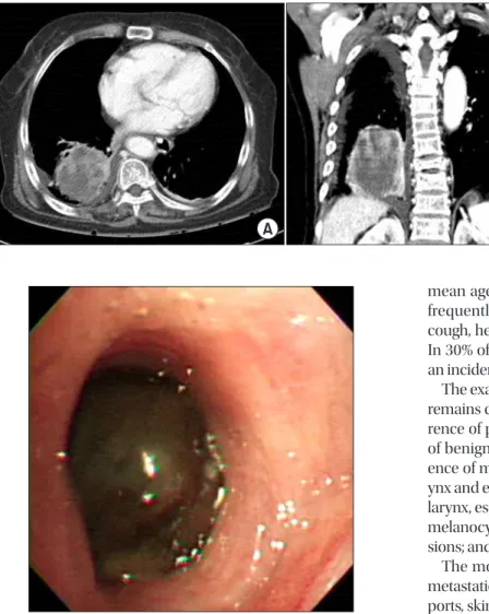
A solitary mass was detected incidentally in the right lower lobe of the lung in an 82-year-old woman. The results of physi- cal examinations and laboratory tests were within normal limits. Chest computed tomography (CT) showed a hetero- geneous enhancing mass lesion of approximately 8 cm in the right lower lobe of the lung (Figure 1). Bronchoscopic exami- nation showed a black-pigmented mass in the posterobasal segment of the right lower lobe, and biopsy was performed (Figure 2). Histopathological examination of the biopsy speci- men showed melanoma cells containing melanin granules and “nesting” of melanoma cells just beneath the bronchial epithelium. The melanoma cells were round or spindle shaped, with melanin pigmentation, and these cells were posi- tive for human melanoma black 45 (HMB-45) and vimentin and focally positive for antibodies to S-100 protein (Figure 3).

Accordingly, the patient was diagnosed with malignant mela- noma.

To exclude the possibility of metastasis, an extensive exami- nation was carried out. Positron emission tomography/CT showed a hypermetabolic lesion (standardized uptake value, Copyright © 2015

The Korean Academy of Tuberculosis and Respiratory Diseases.

All rights reserved.

Introduction

Malignant melanoma is a tumor arising from pigment- producing cells in the deeper layers of the skin. It accounts for 1.5% of all reported cancers. It occurs most frequently on the skin, but also occurs in other organs and tissues of the body.

However, melanoma of the lung without evidence of extra- pulmonary disease, i.e., primary pulmonary melanoma, is very rare

1

.

Here, we report the case of an 82-year-old woman in whom primary pulmonary melanoma was detected incidentally. We

CASE REPORT

http://dx.doi.org/10.4046/trd.2015.78.3.272

ISSN: 1738-3536(Print)/2005-6184(Online) • Tuberc Respir Dis 2015;78:272-275

272

Address for correspondence: Hak-Ryul Kim, M.D.

Department of Internal Medicine, Wonkwang University School of Medicine, 460 Iksan-daero, Iksan 570-974, Korea

Phone: 82-63-859-2583, Fax: 82-63-855-2025 E-mail: kshryj@wonkwang.ac.kr

Received: Aug. 14, 2014 Revised: Dec. 8, 2014 Accepted: Dec. 9, 2014

cc

It is identical to the Creative Commons Attribution Non-Commercial

License (http://creativecommons.org/licenses/by-nc/4.0/).

(2)

Primary pulmonary malignant melanoma

http://dx.doi.org/10.4046/trd.2015.78.3.272 273

www.e-trd.org

14.5) in the right lower lobe of the lung, with no evidence of metastatic disease (Figure 4). The patient had no history of skin lesions, and did not have any skin, ear, or ocular lesions.

Gastrointestinal endoscopy, colonoscopy, and gynecologi- cal examinations were performed, and no possible primary tumor was detected. The final diagnosis was primary pulmo- nary melanoma. The patient refused to undergo treatment because of old age and poor general health.

Discussion

Malignant melanoma mainly occurs on the skin, but has also been described at other mucosal sites and organs, in- cluding the paranasal sinuses, oral cavity, esophagus, larynx, vagina, anorectal region, and liver

2,3

. Primary pulmonary malignant melanoma is a very rare non-epithelial neoplasm, accounting for 0.01% of all primary pulmonary tumors

4

. The

mean age at diagnosis is 57 years (range, 41–82 years). It is frequently endobronchial and manifests with symptoms of cough, hemoptysis, pneumonia, lobar collapse, or atelectasis.

In 30% of cases, primary pulmonary malignant melanoma is an incidental finding on chest radiography

2

.

The exact pathogenesis of pulmonary malignant melanoma remains controversial. Some likely explanations for the occur- rence of pulmonary melanoma are as follows

5

: (1) migration of benign melanocytes during embryogenesis; (2) the pres- ence of melanocytes and melanocytic proliferation in the lar- ynx and esophagus; (3) a common embryological origin of the larynx, esophagus, and lungs, suggesting possible migration of melanocytes; (4) spontaneous regression of previous skin le- sions; and (5) melanogenic metaplasia in the submucosa.

The most important consideration is the exclusion of the metastatic origin of the tumor, because according to some re- ports, skin melanomas may spontaneously disappear without any trace after they have already metastasized

6

. A recent com- prehensive systemic review of the existing literature showed that the incidence of malignant melanoma with an unknown primary site is estimated to be 3.2%

7

. The final diagnosis of a primary pulmonary malignant melanoma is established based on clinical, radiological, and pathological findings.

Therefore, Allen and Drash

8

proposed the following diagnos- tic criteria for primary pulmonary malignant melanoma

9

: (1) no history suggestive of a previous melanoma; (2) no demon- strable melanoma in any other organ at the time of surgery; (3) a solitary tumor in the surgical specimen from the lung; (4) tu- mor morphology compatible with that of a primary tumor; (5) no evidence at autopsy of a primary melanoma elsewhere; (6) obvious melanoma cells confirmed by immunohistochemi- cal staining for S-100 and HMB-45, and possibly by electron microscopy; (7) evidence of junctional change; (8) “nesting” of cells beneath the bronchial epithelium; and (9) invasion of the intact bronchial epithelium by melanoma cells. Our patient had a solitary mass and fulfilled the diagnostic criteria. More- over, pathological examination showed “nesting” of mela- noma cells just beneath the bronchial epithelium, a suggested histopathological criterion for primary pulmonary melanoma.

Figure 1. (A, B) Chest computed tomog- raphy showing a heterogeneous enhanc- ing mass of 8 cm containing an inner low attenuating portion in the right lower lobe.

Figure 2. Bronchoscopy showing a black-pigmented mass in the

posterobasal segment of the right lower lobe.

(3)

KB Hwang et al.

274 Tuberc Respir Dis 2015;78:272-275 www.e-trd.org

Therefore, she was finally diagnosed as having primary malig- nant melanoma according to the proposed criteria.

The treatment of choice is surgical resection of the tumor with an oncologically adequate margin, as is lobectomy or pneumonectomy. Some studies have shown that patients un- dergoing surgical resection have a better prognosis than those treated non-surgically

10

. In inoperable cases, chemotherapy with dacarbazine and immunotherapy with interleukin-2 or interferon are also considered. The prognosis of this condition is rather poor, but existing data are not sufficient to draw a de- finitive conclusion. Based on previous data, 5-year survival is at least 10%

2

. In addition, follow-ups were inadequate in 15%

of the patients who were still alive when the case reports were published.

In conclusion, although rare, primary pulmonary malignant melanoma should be considered in the differential diagnosis of primary bronchial tumors of the lung as an extremely rare possibility.

Conflicts of Interest

No potential conflict of interest relevant to this article was reported.

Acknowledgements

This study was supported by a grant from Wonkwang Uni- versity in 2013.

References

1. Scolyer RA, Bishop JF, Thompson JF. Primary melanoma of the lung. In: Raghavan D, Brecher ML, Johnson DH, Meropol NJ, Moots PL, Rose PG, et al., editors. Textbook of uncommon cancer. 3rd ed. Chichester: John Wiley & Sons Ltd; 2006. p.

Figure 4. (A, B) Positron emission tomog- raphy/computed tomography showing a hypermetabolic lesion (standardized uptake value, 14.5) in the right lower lobe without evidence of metastatic disease.

Figure 3. (A) Histopathological examination of the biopsy specimen showing melanoma cells containing melanin granules (H&E stain, ×200).

(B) Histopathological features of “nesting” of melanoma cells just beneath the bronchial epithelium (H&E stain, ×400). (C) The melanoma

cells are round or spindle shaped with melanin pigmentation and positive for human melanoma black 45 (×200).

(4)

Primary pulmonary malignant melanoma

http://dx.doi.org/10.4046/trd.2015.78.3.272 275

www.e-trd.org 293-8.

2. Ost D, Joseph C, Sogoloff H, Menezes G. Primary pulmonary melanoma: case report and literature review. Mayo Clin Proc 1999;74:62-6.

3. Gong L, Li YH, Zhao JY, Wang XX, Zhu SJ, Zhang W. Primary malignant melanoma of the liver: a case report. World J Gas- troenterol 2008;14:4968-71.

4. Dountsis A, Zisis C, Karagianni E, Dahabreh J. Primary malig- nant melanoma of the lung: a case report. World J Surg Oncol 2003;1:26.

5. Jennings TA, Axiotis CA, Kress Y, Carter D. Primary malignant melanoma of the lower respiratory tract: report of a case and literature review. Am J Clin Pathol 1990;94:649-55.

6. Smith JL Jr, Stehlin JS Jr. Spontaneous regression of primary

malignant melanomas with regional metastases. Cancer 1965;18:1399-415.

7. Kamposioras K, Pentheroudakis G, Pectasides D, Pavlidis N.

Malignant melanoma of unknown primary site. To make the long story short. A systematic review of the literature. Crit Rev Oncol Hematol 2011;78:112-26.

8. Allen MS Jr, Drash EC. Primary melanoma of the lung. Cancer 1968;21:154-9.

9. Jensen OA, Egedorf J. Primary malignant melanoma of the lung. Scand J Respir Dis 1967;48:127-35.

10. Seitelman E, Donenfeld P, Kay K, Takabe K, Andaz S, Fox S.

Successful treatment of primary pulmonary melanoma. J

Thorac Dis 2011;3:207-8.

수치

Figure 1. (A, B) Chest computed tomog- tomog-raphy showing a heterogeneous  enhanc-ing mass of 8 cm containenhanc-ing an inner low  attenuating portion in the right lower  lobe.
Figure 4. (A, B) Positron emission tomog- tomog-raphy/computed tomography showing  a hypermetabolic lesion (standardized  uptake value, 14.5) in the right lower lobe  without evidence of metastatic disease.

참조

관련 문서

공동 번영과 세계 평화를 위해 힘을 모으고 있습니다..

Climate Change Close-Up Climate Change Policy Climate Change Science Climate Change News... Climate Change Close-Up Climate Change Policy Climate Change Science

Primary hepatocytes were incubated with T090 for 24 h following Res pretreatment (1 h). Cells were fixed and stained with Oil red O and observed under a microscope. The Oil

The melanoma cell line WM793 was employed for the Fascin and MST2 knock-down and then analyzed by the Western blot assay, and the melanoma xenograft followed by Fascin

As the observation elements have been changed at the stations 201 ~ 295, the following data are no longer available since August 1, 2000: number of days for weather conditions

As the observation elements have been changed at the stations 201 ~ 295, the following data are no longer available since August 1, 2000: number of days for weather conditions

As the observation elements have been changed at the stations 201 ~ 295, the following data are no longer available since August 1, 2000: number of days for weather conditions

 Kernel 1, which has an arithmetic intensity of 0.5 FLOPs/byte, is limited by memory bandwidth to no more than 8 GFLOPS/sec.  Kernel 2, which has an arithmetic intensity of