Primary Gastric Malignant Melanoma Mimicking Adenocarcinoma
Jun-Min Cho, Chang Min Lee, You-Jin Jang, Sung-Soo Park, Seong-Heum Park, Seung-Joo Kim, Young-Jae Mok, Chong-Suk Kim, Ju-Han Lee1, and Jong-Han Kim
Departments of Surgery and 1Pathology, Korea University College of Medicine, Seoul, Korea
We report a case of primary gastric malignant melanoma that was diagnosed after curative resection but initially misdiagnosed as adeno- carcinoma. A 68-year-old woman was referred to our department for surgery for gastric adenocarcinoma presenting as a polypoid lesion with central ulceration located in the upper body of the stomach. The preoperative diagnosis was confirmed by endoscopic biopsy. We performed laparoscopic total gastrectomy, and the final pathologic evaluation led to the diagnosis of primary gastric malignant melanoma without a primary lesion detected in the body. To the best of our knowledge, primary gastric malignant melanoma is extremely rare, and this is the first case reported in our country. According to the literature, it has aggressive biologic activity compared with adenocarcino- ma, and curative resection is the only promising treatment strategy. In our case, the patient received an early diagnosis and underwent curative gastrectomy with radical lymphadenectomy, and no recurrence was noted for about two years.
Key Words: Melanoma; Stomach; Primary malignant
Correspondence to: Jong-Han Kim
Department of Surgery, Korea University Ansan Hospital, 123 Jeokgeum-ro, Danwon-gu, Ansan 425-707, Korea
Tel: +82-31-412-5957, Fax: +82-31-413-4829 E-mail: [email protected]
Received August 7, 2014 Revised September 17, 2014 Accepted September 18, 2014
Copyrights © 2014 by The Korean Gastric Cancer Association www.jgc-online.org
This is an open-access article distributed under the terms of the Creative Commons Attribution Non-Commercial License (http://creativecommons.org/
licenses/by-nc/3.0) which permits unrestricted noncommercial use, distribution, and reproduction in any medium, provided the original work is properly cited.
Introduction
Malignant melanoma is a tumor of the melanocytes with high malignant potential. Malignant melanoma arises usually in the skin or less frequently in the eyes. Occasionally, it is found in the gastro- intestinal (GI) tract. However, malignant melanomas of the GI tract are mostly metastases from a primary cutaneous tumor. Primary malignant tumors in the GI tract are usually adenocarcinomas, and melanomas are rare. Specifically, a melanoma located primarily in the gastric mucosa is extremely rare. We report an extremely rare case of primary malignant melanoma mimicking adenocarcinoma arising in the upper body of the stomach.
Case Report
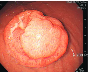
A 68-year-old woman was referred to our department after gastroscopy, which showed a mass in the stomach. She had under- gone total abdominal hysterectomy with bilateral salpingo-oopho- rectomy because of an ovarian serous carcinoma (stage IIIa) nine years ago. On routine workup, gastroscopy showed a round mass, approximately 3 cm in diameter, in the upper body on the greater curvature side of the stomach (Fig. 1).
A biopsy was performed and histologic examination indicated a poorly differentiated adenocarcinoma. Tumor markers includ- ing carcinoembryonic antigen and carbohydrate antigen 19-9 were within the normal ranges. Contrast-enhanced computed tomog- raphy showed focal, eccentric, enhancing wall thickening in the greater curvature of the stomach body and no significantly enlarged perigastric lymph nodes (Fig. 2). We diagnosed the patient with gastric adenocarcinoma and performed a laparoscopic-assisted to- tal gastrectomy with D2 lymphadenectomy. No ascites or peritoneal lesion was observed. Reconstruction was performed with a Roux-
en-Y anastomosis.
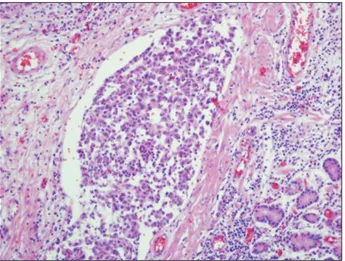
The resected specimen contained a polypoid tumor with cen- tral ulceration 3 cm in diameter (Fig. 3). Tumor cells had spread through the submucosal layer of the stomach with clear resection margins (Fig. 4). None of the 44 resected lymph nodes were posi- tive for metastasis, but lymphatic tumor emboli were detected by immunostaining with CD-31 and D2-40 antibodies (Fig. 5). The tumor cells showed atypical hyperchromatic nuclei (Fig. 6) and ac- companying melanin distribution within the tumor nest (Fig. 7).
Immunohistochemical examination revealed a positive reaction to HMB-45 antibodies and S-100 proteins and a negative reaction to cytokeratin antibodies (Fig. 8).
Based on these findings, we diagnosed the patient with malig- nant melanoma. In the pathologic examination, the depth of tumor involvement was to the submucosal level and no metastasis showed in 44 lymph nodes. Therefore, the final pathologic tumor’s TNM Fig. 1. Grossly, the tumor was a round, polypoid lesion with central
ulceration on gastroscopy.
Fig. 2. Computed tomography showed a focal enhancing wall thicken- ing in the greater curvature of the upper body of the stomach. Enlarged perigastric lymph nodes were not noted.
Fig. 3. Photograph of the gross specimen that contained a polypoid tumor with central ulceration 3 cm in diameter located in the greater curvature of the upper body of the stomach.
A B
Fig. 4. Microscopic findings showed that tumor cells were spreading through the submucosal layer of the stomach with a clear resection margin (A:
H&E, ×20; B: H&E, ×200).
stage was pT1bN0M0. Postoperatively, further examinations were performed to identify the primary tumor or other metastatic foci.
Ophthalmologic, dermatologic, and oral examinations showed neg- ative findings. Positron emission tomography/computed tomogra- phy was performed, but no hot spots were detected (Fig. 9). These results indicated a final diagnosis of primary malignant melanoma of the stomach. After total gastrectomy, a follow-up study was performed with abdominal computed tomography scans at 3, 6, 12, 18, and 24 months (5 times) over the course of 2 years and there was no recurrence. The TNM tumor stage was early (pT1bN0M0);
therefore, we did not perform any adjuvant chemotherapy, but the patient has taken oral immunotherapeutic medication for 4 months.
Discussion
Malignant melanomas of the GI tract are rare. Most of them are metastases from cutaneous lesions and are reported in up to 4% of patients who are alive and in up to 60% of patients at autopsy.1-4 Primary GI melanomas are very rare and have been reported to arise in the esophagus, anorectum, and small intestine.5 Primary gastric melanoma has also been reported sporadically.6-10 An au- topsy series of patients with melanoma revealed gastric metastasis rates of more than 22%.4 The median overall survival associated with primary malignant melanoma of the GI tract is reportedly 17 months, whereas that associated with primary malignant melanoma of the stomach is only 5 months.11 Criteria for the diagnosis of Fig. 5. Findings of immunohistochemical staining: lymphatic tumor
emboli were noted by immunostaining with CD-31 and D2-40 anti- body (×200).
Fig. 6. The tumor cells contained atypical hyperchromatic nuclei on H&E staining (×400).
Fig. 7. The tumor nest was accompanied by melanin on H&E staining (×400).
Fig. 8. Immunohistochemical examination revealed a positive reaction with HMB-45 antibodies and S-100 proteins and a negative reaction with cytokeratin antibodies (immunostaining, ×200).
primary malignant melanoma of the GI tract include the absence of other primary site melanomas and no history of melanoma or atypical melanocytic lesion removal from the skin or other or- gans.6 On diagnosis, the computed tomography scan is commonly used to detect intra-abdominal lesions. Also, the positron emis- sion tomography/computed tomography scan has been shown to have higher sensitivity and accuracy in the detection of visceral metastases, including extra-intestinal metastatic disease.12 Since there are no melanocytes in the stomach, gastric melanoma may originate from ectopic melanocytes that migrate to the GI tract during embryogenesis or differentiation of amine precursor up- take and decarboxylation cells to melanocytes.13 Surgical resection is the only identifiable treatment modality for which independent predictive prognostic values have been demonstrated.11 Systemic chemotherapy regimens show no benefit in overall survival and are not recommended for the treatment of malignant GI melanomas.14 In this case, we initially misdiagnosed the mass as adenocarcinoma of the stomach in the biopsied tumor cell specimens obtained dur- ing endoscopic examination. We supposed that the initial biopsied tumor cells might have been so scanty that they resembled poorly
differentiated adenocarcinoma in the pathologic view. The patient had a history of tubular adenoma with high-grade dysplasia in the angular portion of the stomach three years ago and had undergone endoscopic mucosal resection. Therefore, we mistook the mass as adenocarcinoma from the intestinal metaplastic precancerous le- sion, and the grossly protruding tumor resembled Borrmann type I or early gastric cancer type I. However, this lesion was located away from the previous site of dysplasia and was not associated with it.
During the pathologic examination of the resected specimen, the tumor appearance differed from ordinary adenocarcinoma.
Therefore, we performed immunohistochemical staining with sev- eral markers, and the results were positive for HMB45, S-100, and vimentin and negative for cytokeratin, CAM5.2, chromogranin, and synaptophysin. This patient was finally diagnosed with primary malignant melanoma based on the immunohistochemical staining results. After curative laparoscopic total gastrectomy, the patient received routine follow-up, and there was no recurrence for two years.
Fig. 9. Positron emission tomography/
computed tomography scan showed no other hot spots except the stomach cancer portion.
References
1. Elsayed AM, Albahra M, Nzeako UC, Sobin LH. Malignant melanomas in the small intestine: a study of 103 patients. Am J Gastroenterol 1996;91:1001-1006.
2. Woollons A, Derrick EK, Price ML, Darley CR. Gastrointesti- nal malignant melanoma. Int J Dermatol 1997;36:129-131.
3. Liang KV, Sanderson SO, Nowakowski GS, Arora AS. Meta- static malignant melanoma of the gastrointestinal tract. Mayo Clin Proc 2006;81:511-516.
4. Patel JK, Didolkar MS, Pickren JW, Moore RH. Metastatic pat- tern of malignant melanoma. A study of 216 autopsy cases.
Am J Surg 1978;135:807-810.
5. Lagoudianakis EE, Genetzakis M, Tsekouras DK, Papadima A, Kafiri G, Toutouzas K, et al. Primary gastric melanoma: a case report. World J Gastroenterol 2006;12:4425-4427.
6. Yamamura K, Kondo K, Moritani S. Primary malignant mela- noma of the stomach: report of a case. Surg Today 2012;42:195- 199.
7. Goral V, Ucmak F, Yildirim S, Barutcu S, Ileri S, Aslan I, et al.
Malignant melanoma of the stomach presenting in a woman: a case report. J Med Case Rep 2011;5:94.
8. Alazmi WM, Nehme OS, Regalado JJ, Rogers AI. Primary gas- tric melanoma presenting as a nonhealing ulcer. Gastrointest
Endosc 2003;57:431-433.
9. Germano D, Rosati G, Romano R, Vita G, Lepore G, De Sanc- tis D, et al. Primary gastric melanoma presenting as a double ulcer. J Clin Gastroenterol 2004;38:828.
10. Jelincic Z, Jakic-Razumovic J, Petrovic I, Cavcic AM, Unusic J, Trotic R. Primary malignant melanoma of the stomach. Tu- mori 2005;91:201-203.
11. Cheung MC, Perez EA, Molina MA, Jin X, Gutierrez JC, Fran- ceschi D, et al. Defining the role of surgery for primary gastro- intestinal tract melanoma. J Gastrointest Surg 2008;12:731-738.
12. Marsden JR, Newton-Bishop JA, Burrows L, Cook M, Cor- rie PG, Cox NH, et al; British Association of Dermatologists Clinical Standards Unit. Revised U.K. guidelines for the management of cutaneous melanoma 2010. Br J Dermatol 2010;163:238-256.
13. Krausz MM, Ariel I, Behar AJ. Primary malignant melanoma of the small intestine and the APUD cell concept. J Surg Oncol 1978;10:283-288.
14. Verma S, Petrella T, Hamm C, Bak K, Charette M; Melanoma Disease Site Group of Cancer Care Ontario's Program in Evidence-based Care. Biochemotherapy for the treatment of metastatic malignant melanoma: a clinical practice guideline.
Curr Oncol 2008;15:85-89.