PRIMARY MALIGNANT MELANOMA OF THE UTERINE CERVIX: A CASE REPORT
Nae Ri Yun, MD 1 , Jung Woo Park, MD 1 , Jee Hyun Park, MD, PhD 1 , Suk Jin Choi, MD, PhD 2 , Sung Ook Hwang, MD, PhD 1
Departments
1Obstetrics and Gynecology,
2Pathology, Inha University College of Medicine, Incheon, Korea
After the discovery of melanocytes in the cervix in 1959, it was recognized that primary malignant melanoma of the cervix exists as a separate entity. A 74-year-old woman visited hospital for vaginal bleeding from a black colored cervical mass. The pathology of cervical punch biopsy showed a malignant melanoma with positive immnunohistochemical stainings for S100 protein and HMB- 45 antibody. Abdominal radical hysterectomy with pelvic and paraaortic lymphadenectomy was performed. The fi nal pathology was a malignant melanoma of the cervix with metastases for both external iliac lymph nodes and tumor involvement in the margin of vaginal resection. She received cisplatin based concurrent chemoradiotherapy postoperatively. But 6 months later, she received another chemotherapy with dacarbazine and cisplatin for recurrence. We report a case of a 74-year-old patient with a malignant melanoma of the uterine cervix with a brief review.
Keywords: Malignant melanoma; Uterine cervix; Vaginal bleeding
Received: 2012.2.4. Accepted: 2012.4.1.
Corresponding author: Sung Ook Hwang, MD, PhD Department of Obstetrics and Gynecology, Inha University Hospital, Inha University College of Medicine, 27 Inhang-ro, Jung-gu, Incheon 400-711, Korea
Tel:+82-32-890-2270 Fax: +82-32-890-3097 E-mail: [email protected]
Th is is an Open Access article distributed under the terms of the Creative Commons Attribution Non-Commercial License (http://creativecommons.org/licenses/
by-nc/3.0/) which permits unrestricted non-commercial use, distribution, and reproduction in any medium, provided the original work is properly cited.
Copyright © 2012. Korean Society of Obstetrics and Gynecology
Since melanocytes were demonstrated in the cervical epithelium in as many as 3.5% of women [1], malignant melanomas of the cer- vix were often reported. Approximately 3%–7% of malignant mela- nomas in women develop within genital tract [2]. The vast majority of these cases occur in vulva and vagina. Cervix is a rare site for melanomas with about 60 cases of malignant melanomas of the cervix have been described as primary tumors within the cervix [3-5].
Malignant melanomas present clinically as an advanced stage and the diagnosis is confi rmed by histological examination using special staining and by immunohistochemical study. Radical hysterectomy with regional lymphadenectomy are generally advocated.
Primary malignant melanoma of the cervix has a poor outcome as a consequence of delayed diagnosis and lack of standardized treatment. There is lack of evidence on the effi cacy of postopera- tive radiation or chemotherapy. Radiation therapy may play a role in the treatment of patients with close resection margins, regional nodal metastases or unresectable tumors. Diagnosis is confi rmed by immunohistochemical methods and by exclusion of other pri- mary sites of melanomas.
Case Report
A 74-year-old, multiparous woman was admitted to the Depart-
ment of Obstetrics and Gynecology at Inha University Hospital for vaginal bleeding for several months. A elongated, large sausage like, soft black colored mass with active bleeding and blood clots was noted on the anterior lip of the cervix with suspicious both upper vagina and parametrial invasion. A biopsy from the lesion was taken. The emergency radiation therapy for active cervical bleeding was performed under the impression of cervical squa- mous cell carcinoma (SCC), stage 1B2, or stage 2B less likely.
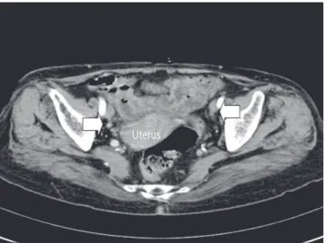
CA-125 and SCC were normal. The abdominopelvic computed tomography (CT) revealed cervical cancer with upper vaginal in- volvement and suspicious parametrial invasion, and multiple tiny http://dx.doi.org/10.5468/KJOG.2012.55.5.343
pISSN 2233-5188 · eISSN 2233-5196
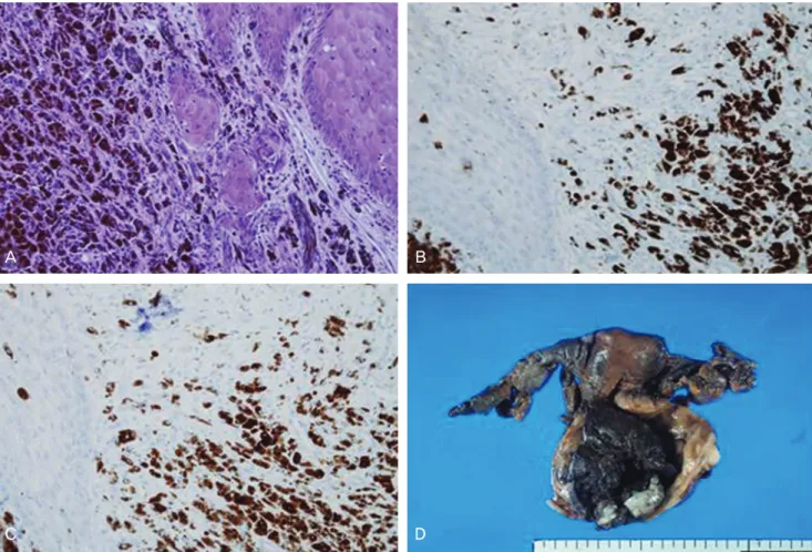
small lymph nodes in both external iliac chain (Fig. 1). Whole body positron emission tomography-CT showed malignancy in the uter- ine cervix and tiny pulmonary nodule in right medial lobe of lung (Fig. 2). The pathology of cervical punch biopsy showed malignant melanoma. The immunohistochemical staining for S100 protein and HMB-45 antibody were positive, but negative for mixed cyto- keratin and leukocyte common antigen (Fig. 3).
An extensive search for a melanotic lesion including skin and eye was performed to verify the primary site of melanoma, but we could not fi nd any primary site of melanoma. We discussed what
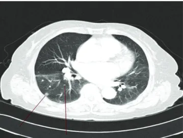
is the optimal treatment for this patient with Radio-oncologist and concluded that we would do the surgery because primary mela- nomas of cervix were poor responders to radiation. We performed the radical hysterectomy with pelvic and paraaortic lymphadenec- tomy (Fig. 3D). The frozen biopsies of posterior vaginal resection margins were positive tumor involvement in spite of repeated three times of resection in the vaginal cuff. The final pathologic report showed a malignant melanoma, cervix with tumor size 6.0 × 4.7 × 2.0 cm, depth of invasion/thickness of cervix, 2.4 cm/2.5 cm, and tumor involvement of vaginal resection margin, metastatic tumors in 6 out of 38 lymph nodes, positive parame- trial, both external iliac and right common iliac lymph nodes. She received postoperative cisplatin based concurrent chemoradio- therapy. Postoperatively 6 months later, her pelvic examination and vaginal stump were clear with good general condition. She had a regular follow-up PET-CT, because there is no tumor markers in malignant melanoma of the uterine cervix. The PET-CT showed metastasis in right adrenal gland and several new pulmonary nod- ules in right lung (Fig. 4). The chest CT revealed newly appeared multiple well-defi ned various size pulmonary nodules in right lung and lower lobe of left lung that suspected pulmonary metastasis (Fig. 5). She had a chemotherapy with dacarbazine and cisplatin.
Discussion
Primary melanoma of the cervix is very rare. Melanoma in the uter- ine cervix may be melanotic or amelanotic [3,6]. About half of the melanomas are amelanotic. Diagnosis of amelanotic melanomas may be diffi cult due to the absence of pigment.
Patients with cervical melanoma have ranged in age from 19 to 83 years, although the majority have been between 60 to 70 years [3,7,8]. Vaginal bleeding or discharge is the usual presenting com- plaints. Some patients have been asymptomatic. Patients may re- main asymptomatic until the lesions were ulcerated and infected, after then the lesions were easy to bleed. Clinical examination has usually revealed an exophytic polypoid pigmented cervical mass with variable sizes. About half of the cases, brown to blue-black pigmentation of the tumors have been noted. Cervical melanoma is seldom diagnosed by Pap smear in absence of typical pigmented polypoid growth [9]. Morphological features of primary cervical melanoma in Pap smear have been reported, showing bizarre and abnormal cells containing pigment with the hope of early diagno- sis [9].
Diagnosis is usually based on pelvic examination and histopathol- Fig. 1. Computed tomography findings of preoperation; upper margin
of mild enhancing cervical mass. The multiple small lymph nodes in both external iliac chains were enlarged.
Uterus
Fig. 2. Positron emission tomography findings of abnormal fluoro-D-
glucose uptake in whole cervix mass.
Fig. 3. Histologic fi ndings are showing proliferation of malignant melanin-producing melanocytes (A). Most of the atypical cells are positive for HMB-45 (B) and S100 (C) (A: H&E, ×20; B: Immunohistochemical stain, ×20; C: Immunohistochemical stain for S100 protein after melanin bleaching, ×20). The uterus was enlarged with a huge dark brown ulcerofungating tumor arising in the exocervix and endocervix, extending to the vagina with near full-thickness invasion of the cervical wall (D).
A B
C D
Fig. 4. About 6 months after radical hysterectomy, newly appeared multiple well-defi ned various size pulmonary nodule in right lung and left lower lobe that suspected pulmonary metastasis. Computed tomography fi ndings of round shape well-defi ned 0.6 cm and 0.4 cm sized pulmonary nodules in right lower lobe (allow).
A B
ogy. Though histological examination in primary cervical melano- ma displays predominantly eosinophilic nucleoli, diagnosis should always be confi rmed by immunohistochemical staining. A combi- nation of S100 protein (more sensitive) or HMB-45 (more specifi c) is the best combination for an accurate diagnosis, and is useful in distinguishing amelanotic melanoma from anaplastic carcinoma, high grade lymphoma and sarcoma [7].
Primary cervical melanoma must be differentiated from second- ary metastasis of melanoma of other sites in the body, including skin and eye. Norris and Taylor [10] have suggested the diagnostic criterias for primary malignant melanoma: the presence of mela- nin in the normal cervical epithelium, the absence of malanoma elsewhere in the body, the demonstration of junctional change in the cervix, the metastases according to the pattern of cervical car- cinoma.
Histologically the tumors do not differ significantly from mela- nomas in other sites, being composed of nests of polygonal to spindle-shaped cells, containing mitotically active pleomorphic nuclei. Primary melanoma of the cervix with a prominent spindle cell component should be distinguished from leiomyosarcoma and many benign melanotic lesions, including blue nevus [11]. Primary cervical melanoma has the presence of melanin, junctional activity, immunoreactivity for S100 and HMB-45 antigens, and absence of immunoactivity for markers of smooth muscle differentiation.
There is no consensus on optimal management of the primary malignant melanoma, because of rarity of the lesion. Radical hys- terectomy with pelvic and paraaortic lymphadenectomy usually is the most common procedure [12]. There is lack of evidence on the
effi cacy of postoperative radiation or chemotherapy [13]. Radio- therapy can be used for the palliation of an inoperable patient or as an adjuvant therapy [14]. Dacarbazine which has been shown to reduce tumor in patients with cutaneous malignant melanoma may be useful for cervical malignant melanoma [4,12]. A combi- nation chemotherapy with cisplatin, vinblastin and bleomycin may produce a better response than a single agent dacarbazine [3,12].
The prognosis of the primary cervical melanoma is generally poor, because diagnosis is usually made at advanced stage and the tu- mor is highly aggressive as both local recurrence and widespread metastases [6]. The 5-year survival rate after radical hysterectomy is not exceeding 40% in stage 1 and reaching only 14% in stage 2 [7]. The reported survival time ranges from 6 months to 14 years, 90% of the reported patients with follow-up data have been died of their diseases, within 2-3 years of presentation [12].
In conclusion, primary malignant melanoma of the cervix should be considered in the differential diagnosis of cervical malignan- cies. The diagnosis should be confi rmed using special stains and immunohistochemistry. This is essential since cervical melanoma is incurable even with the currently available therapies and hence needs to be diagnosed early.
References
1. Cid JM. Melanoid pigmentation of the endocervix: a neuro- genic visceral argument. Ann Anat Pathol (Paris) 1959;4:617- 28.
2. Podczaski E, Abt A, Kaminski P, Larson J, Sorosky J, DeGeest K, et al. A patient with multiple, malignant melanomas of the lower genital tract. Gynecol Oncol 1990;37:422-6.
3. Gupta R, Singh S, Mandal AK. Primary malignant melanoma of cervix - a case report. Indian J Cancer 2005;42:201-4.
4. Ma SQ, Bai CM, Zhong S, Yu XH, Lang JH. Clinical analysis of primary malignant melanoma of the cervix. Chin Med Sci J 2005;20:257-60.
5. Mousavi AS, Fakor F, Nazari Z, Ghaemmaghami F, Hashemi FA, Jamali M. Primary malignant melanoma of the uterine cervix:
case report and review of the literature. J Low Genit Tract Dis 2006;10:258-63.
6. DeMatos P, Tyler D, Seigler HF. Mucosal melanoma of the fe- male genitalia: a clinicopathologic study of forty-three cases at Duke University Medical Center. Surgery 1998;124:38-48.
7. Clark KC, Butz WR, Hapke MR. Primary malignant melanoma of the uterine cervix: case report with world literature review.
Fig. 5. Positron emission tomography-computed tomography imaging.
Abnormal FDG uptake in newly appeared 2 cm sized right adrenal gland
nodule 6 month after radical hysterectomy.
Int J Gynecol Pathol 1999;18:265-73.
8. Mordel N, Mor-Yosef S, Ben-Baruch N, Anteby SO. Malignant melanoma of the uterine cervix: case report and review of the literature. Gynecol Oncol 1989;32:375-80.
9. Deshpande AH, Munshi MM. Primary malignant melanoma of the uterine cervix: report of a case diagnosed by cervical scrape cytology and review of the literature. Diagn Cytopathol 2001;25:108-11.
10. Norris HJ, Taylor HB. Melanomas of the vagina. Am J Clin Pathol 1966;46:420-6.
11. Hytiroglou P, Domingo J. Development of melanosis of uterine cervix after cryotherapy for epithelial dysplasia. A case report
and brief review of the literature on pigmented lesions of the cervix. Am J Clin Pathol 1990;93:802-5.
12. Kristiansen SB, Anderson R, Cohen DM. Primary malignant melanoma of the cervix and review of the literature. Gynecol Oncol 1992;47:398-403.
13. Cantuaria G, Angioli R, Nahmias J, Estape R, Penalver M. Pri- mary malignant melanoma of the uterine cervix: case report and review of the literature. Gynecol Oncol 1999;75:170-4.
14. Herbert SH, Solin LJ, Rate WR, Schultz DJ, Hanks GE. The effect of palliative radiation therapy on epidural compression due to metastatic malignant melanoma. Cancer 1991;67:2472-6.
자궁경부의 일차성 악성 흑색종의 1예
인하대학교 의과대학 1산부인과학교실, 2병리학교실 윤내리1, 박정우1, 박지현1, 최석진2, 황성욱1
1959년 자궁경부의 멜라닌 세포 발견 이후, 자궁경부의 일차성 악성 흑색종은 별개의 독립체로 존재했다. 본 증례는 74세 여자환자가 자궁경부의 검은색 덩이로부터의 출혈을 주소로 입원하여 자궁경부 생검에서 악성 흑색종이 발견되었고 면역조직학적 검사에서 S100단 백과 HMB-45 항체 양성으로 나왔다. 골반림프절, 대동맥 주위 림프절 절제를 포함한 근치적 자궁 절제 수술이 시행 후 최종 병리결과는 외장골 림프절 부위와 질 원개에 전이가 있는 자궁경부의 악성 흑색종이었다. 수술 후 시스플라틴을 사용하여 동시 항암화학방사선 요법 을 시행하였으나 6개월 후 재발하여 다카르바진과 시스플라틴을 사용한 항암화학요법을 시행하였다. 이에 저자들은 74세 여자에서 생긴 자궁경부의 악성 흑색종의 증례에 대하여 간단한 문헌고찰과 함께 보고하는 바이다.
중심단어: 악성 흑색종, 자궁경부, 질출혈